. 2023 Apr 25;120(17):e2220982120.
doi: 10.1073/pnas.2220982120. Epub 2023 Apr 19.

Fragmentation landscape of cell-free DNA revealed by deconvolutional analysis of end motifs

Affiliations

Fragmentation landscape of cell-free DNA revealed by deconvolutional analysis of end motifs

Ze Zhou et al. Proc Natl Acad Sci U S A. .

Abstract

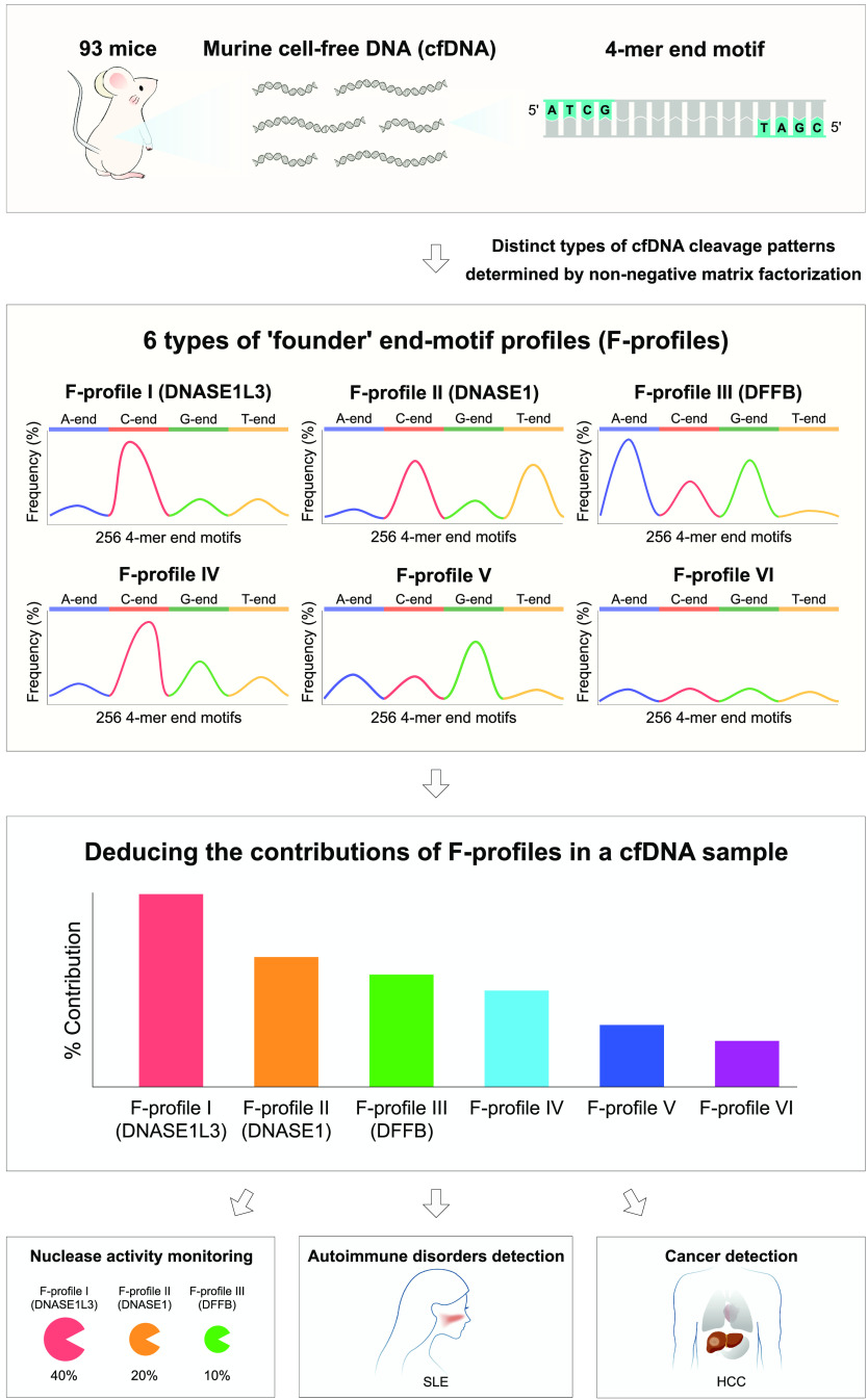
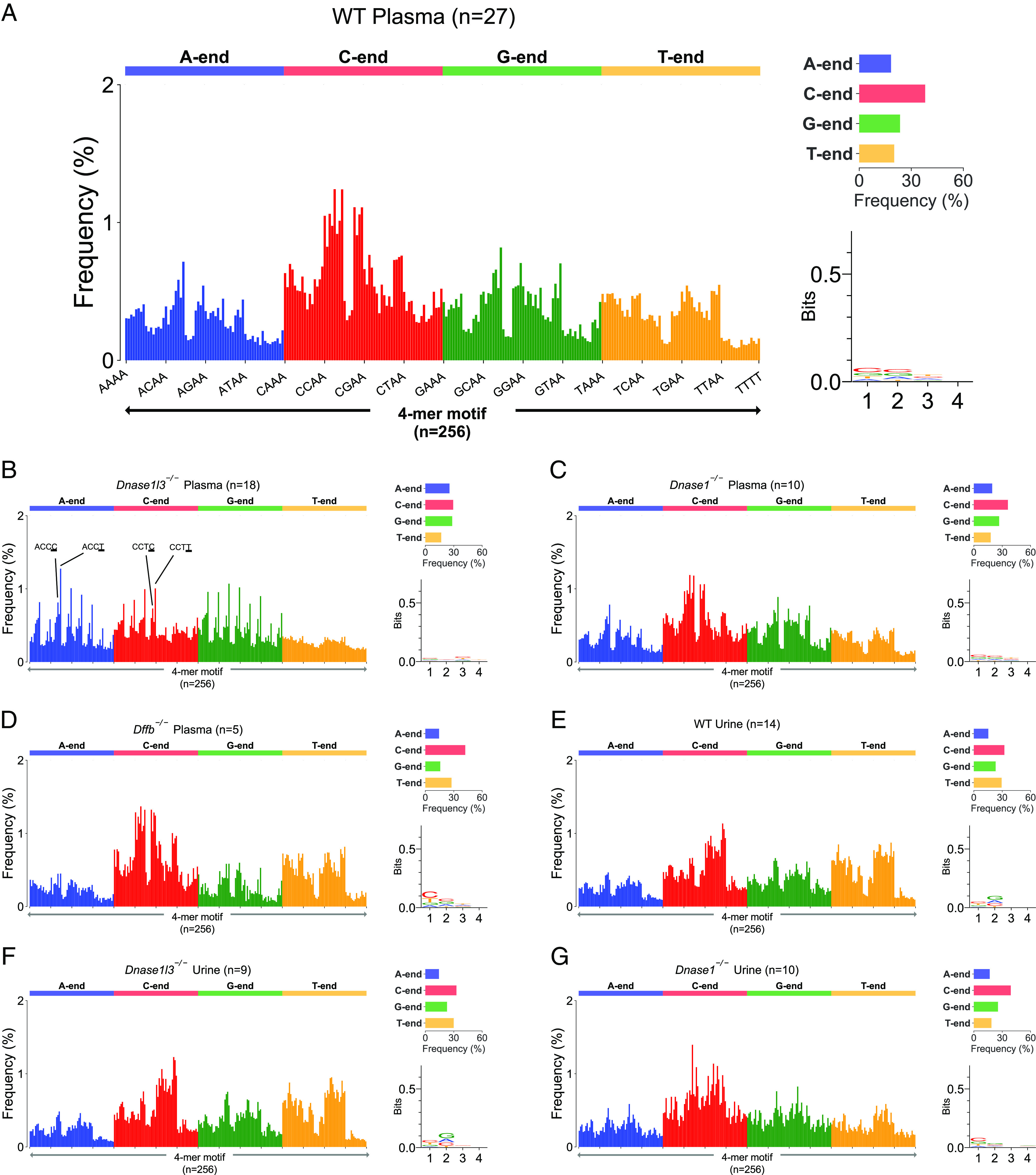
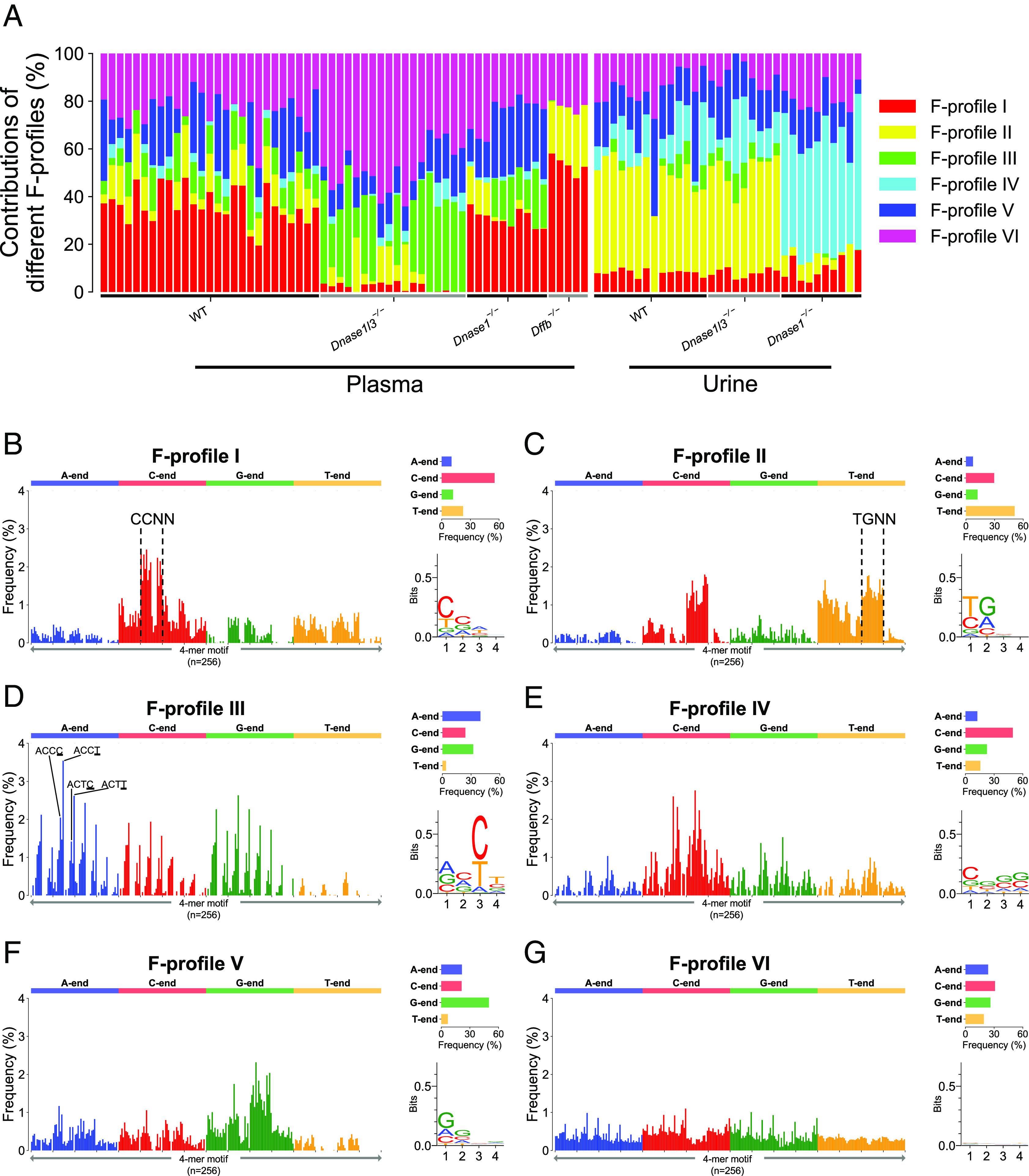
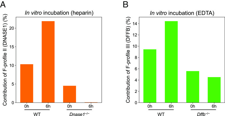
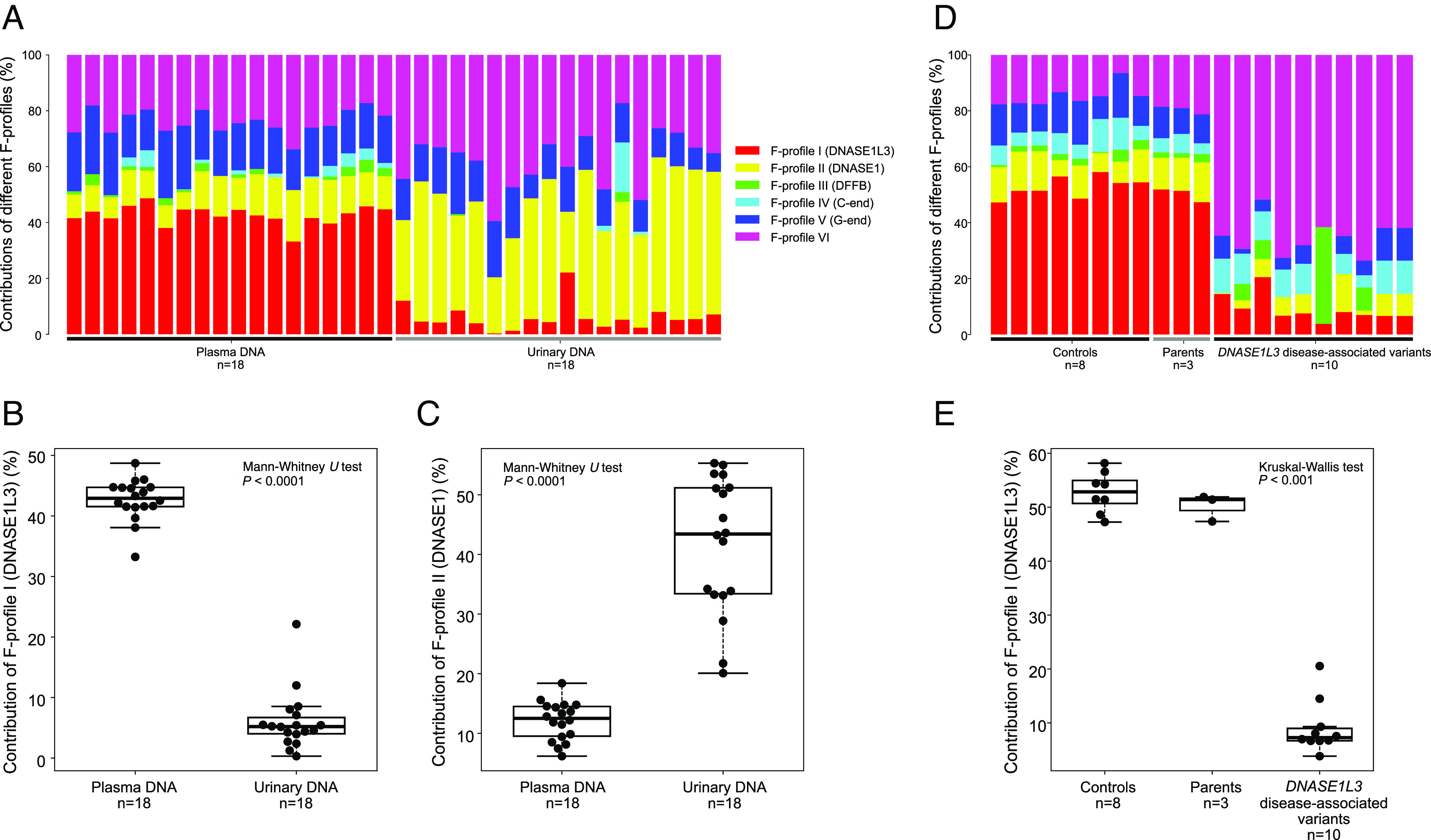
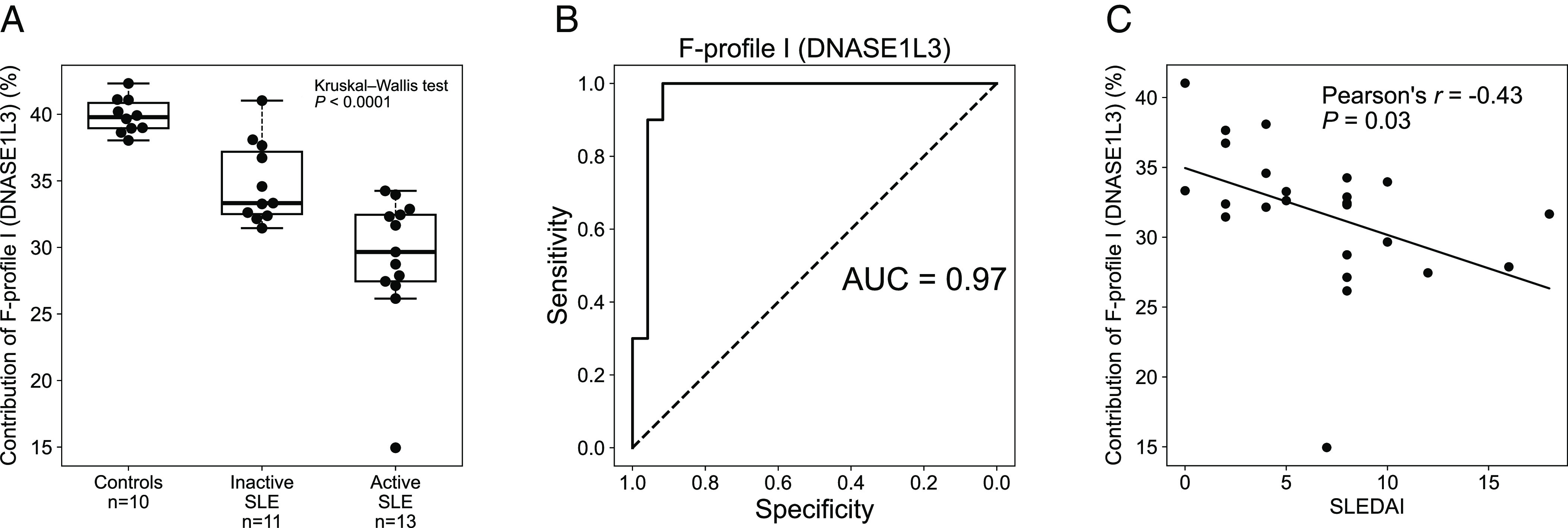
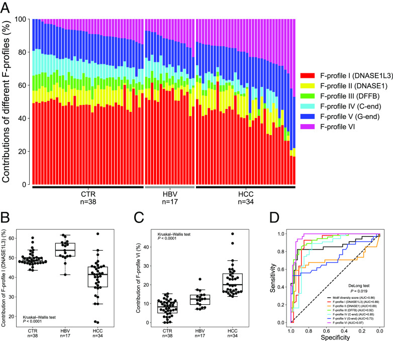
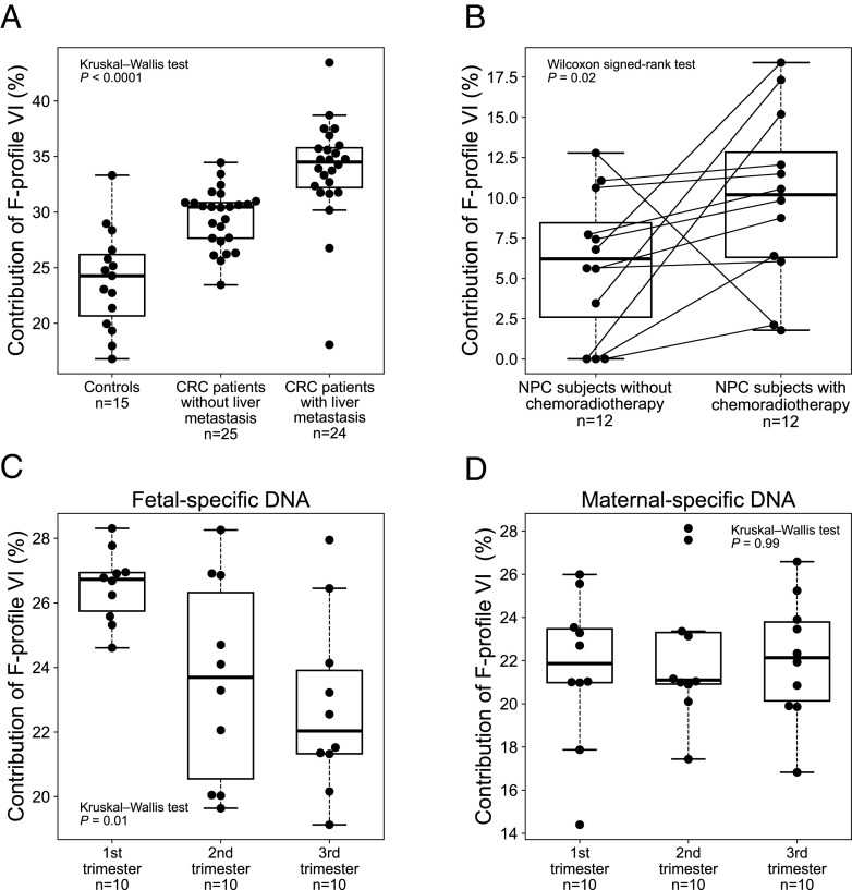
Cell-free DNA (cfDNA) fragmentation is nonrandom, at least partially mediated by various DNA nucleases, forming characteristic cfDNA end motifs. However, there is a paucity of tools for deciphering the relative contributions of cfDNA cleavage patterns related to underlying fragmentation factors. In this study, through non-negative matrix factorization algorithm, we used 256 5' 4-mer end motifs to identify distinct types of cfDNA cleavage patterns, referred to as "founder" end-motif profiles (F-profiles). F-profiles were associated with different DNA nucleases based on whether such patterns were disrupted in nuclease-knockout mouse models. Contributions of individual F-profiles in a cfDNA sample could be determined by deconvolutional analysis. We analyzed 93 murine cfDNA samples of different nuclease-deficient mice and identified six types of F-profiles. F-profiles I, II, and III were linked to deoxyribonuclease 1 like 3 (DNASE1L3), deoxyribonuclease 1 (DNASE1), and DNA fragmentation factor subunit beta (DFFB), respectively. We revealed that 42.9% of plasma cfDNA molecules were attributed to DNASE1L3-mediated fragmentation, whereas 43.4% of urinary cfDNA molecules involved DNASE1-mediated fragmentation. We further demonstrated that the relative contributions of F-profiles were useful to inform pathological states, such as autoimmune disorders and cancer. Among the six F-profiles, the use of F-profile I could inform the human patients with systemic lupus erythematosus. F-profile VI could be used to detect individuals with hepatocellular carcinoma, with an area under the receiver operating characteristic curve of 0.97. F-profile VI was more prominent in patients with nasopharyngeal carcinoma undergoing chemoradiotherapy. We proposed that this profile might be related to oxidative stress.

Keywords: cancer detection; fragmentomics; liquid biopsy; non-negative matrix factorization; oxidative stress.


Conflict of interest statement

The authors have organizational affiliations to disclose, K.C.A.C. and Y.M.D.L. hold leadership positions in Centre for Novostics. R.W.K.C., K.C.A.C., and Y.M.D.L. hold equities in Take2. Z.Z., M.-J.L.M., K.C.A.C., Y.M.D.L., and P.J. have filed a patent application on the described technology.

Figures

Fig. 1.
Fig. 1.
Schematic of distinct types of cfDNA cleavage analysis for cfDNA molecules. The terminal 4 nucleotides at each of the 5′ fragment ends (i.e., 4-mer end motifs; n = 256) were determined from 93 murine cfDNA samples, including WT mice and nuclease-deficient mice. Six categories of distinct types of cfDNA cleavage patterns were found, referred to as "founder" end-motif profiles (i.e., F-profiles), by applying NMF analysis to the 4-mer end-motif profiles. F-profiles I, II, and III were associated with the cutting preference of DNASE1L3, DNASE1, and DFFB, respectively. The distinct types of cfDNA cleavage patterns learned from murine cfDNA could be extrapolated to human cfDNA for informing the proportional contributions of F-profiles in both mouse and human cfDNA samples (referred to as deconvolutional analysis of end motifs), allowing the detection of immune diseases and cancers.
Fig. 2.
Fig. 2.
The observed end-motif profiles of mouse plasma and urinary cfDNA molecules. The observed end-motif frequencies of plasma cfDNA from (A) WT mice, (B) Dnase1l3−/− mice, (C) Dnase1−/− mice, and (D) Dffb−/− mice, respectively. The observed end-motif frequencies of urinary cfDNA from (E) WT mice, (F) Dnase1l3−/− mice, and (G) Dnase1−/− mice, respectively.
Fig. 3.
Fig. 3.
Six F-profiles deduced from mouse plasma and urinary cfDNA using NMF analysis. (A) Proportional contribution of each F-profile in murine cfDNA samples with different knockout genotypes. (B–G) Plots for the six F-profiles.
Fig. 4.
Fig. 4.
Deconvolutional analysis of end motifs of mouse plasma cfDNA samples that were subjected to whole blood in vitro incubation. (A) F-profile II (DNASE1) levels in plasma cfDNA from WT mice before and after 6 h incubation in heparin-contained tube, and from Dnase1−/− mouse before and after 6 h heparin incubation. (B) F-profile III (DFFB) levels in plasma cfDNA from WT mice before and after 6 h incubation in EDTA-contained tube, and from Dffb−/− mice before and after 6 h EDTA incubation.
Fig. 5.
Fig. 5.
Deconvolutional analysis of end motifs in paired human plasma and urinary cfDNA samples, and human plasma cfDNA of subjects with and without DNASE1L3 deficiency. (A) Bar chart of results of deconvolutional analysis between human plasma and urinary cfDNA. (B–C) Boxplots of F-profile I and II levels between human plasma and urinary cfDNA. (D) Bar chart of results of deconvolutional analysis in plasma cfDNA between subjects with and without DNASE1L3 disease-associated variants. (E) Boxplot of F-profile I levels for healthy subjects, patients with DNASE1L3 deficiency, and parents of the patients.
Fig. 6.
Fig. 6.
Deconvolutional analysis of end motifs in plasma cfDNA of human subjects with and without SLE. (A) Boxplot of F-profile I levels (DNASE1L3) in plasma cfDNA across healthy control subjects, patients with inactive SLE, and patients with active SLE. (B) Area under the receiver operating characteristic (ROC) curve (AUC) for differentiation between patients with and without SLE using F-profile I. (C) Correlation between the SLEDAI and F-profile I levels in patients with SLE.
Fig. 7.
Fig. 7.
The distinct types of cfDNA cleavage analysis in plasma cfDNA of human subjects with and without HCC. (A) Bar chart of F-profile levels in plasma cfDNA of patients with and without HCC. Boxplots of F-profile (B) I and (C) VI levels in plasma cfDNA of patients with and without HCC. (D) ROC curves for the differentiation between non-HCC and HCC groups using different metrics, including motif diversity score and six F-profiles.
Fig. 8.
Fig. 8.
The F-profile VI levels in plasma cfDNA of human subjects under different oxidative stress. (A) Boxplot of F-profile VI contributions in healthy control subjects, CRC patients without and with liver metastasis. (B) Boxplot of F-profile VI contributions in patients with NPC who were subjected to chemoradiotherapy or not. Boxplots of F-profile VI contributions in the (C) fetal- and (D) maternal-specific DNA in plasma cfDNA of pregnant women across first, second and third trimesters.

References

    1. Sun K., et al. , Plasma DNA tissue mapping by genome-wide methylation sequencing for noninvasive prenatal, cancer, and transplantation assessments. Proc. Natl. Acad. Sci. U.S.A. 112, E5503–E5512 (2015). - PMC - PubMed
    1. Li W., et al. , CancerDetector: Ultrasensitive and non-invasive cancer detection at the resolution of individual reads using cell-free DNA methylation sequencing data. Nucleic Acids Res. 46, e89 (2018). - PMC - PubMed
    1. Lo Y. M. D., et al. , Maternal plasma DNA sequencing reveals the genome-wide genetic and mutational profile of the fetus. Sci. Transl. Med. 2, 61ra91 (2010). - PubMed
    1. Markus H., et al. , Analysis of recurrently protected genomic regions in cell-free DNA found in urine. Sci. Transl. Med. 13, eaaz3088 (2021). - PMC - PubMed
    1. Lo Y. M. D., Han D. S. C., Jiang P., Chiu R. W. K., Epigenetics, fragmentomics, and topology of cell-free DNA in liquid biopsies. Science 372, eaaw3616 (2021). - PubMed

Publication types

LinkOut - more resources