. 2023 Apr 21;9(16):eadf4888.
doi: 10.1126/sciadv.adf4888. Epub 2023 Apr 19.

BBB opening with focused ultrasound in nonhuman primates and Parkinson's disease patients: Targeted AAV vector delivery and PET imaging

Affiliations

BBB opening with focused ultrasound in nonhuman primates and Parkinson's disease patients: Targeted AAV vector delivery and PET imaging

Javier Blesa et al. Sci Adv. .

Abstract

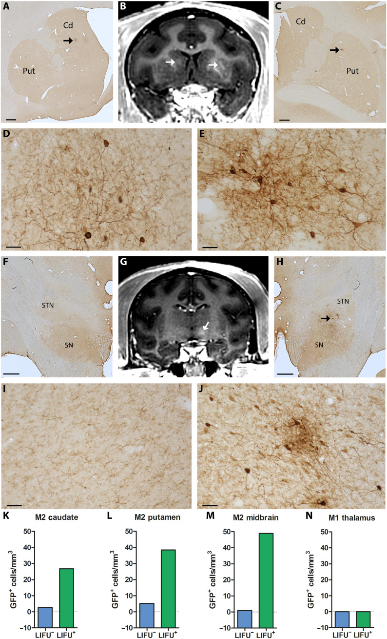
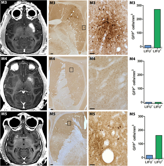
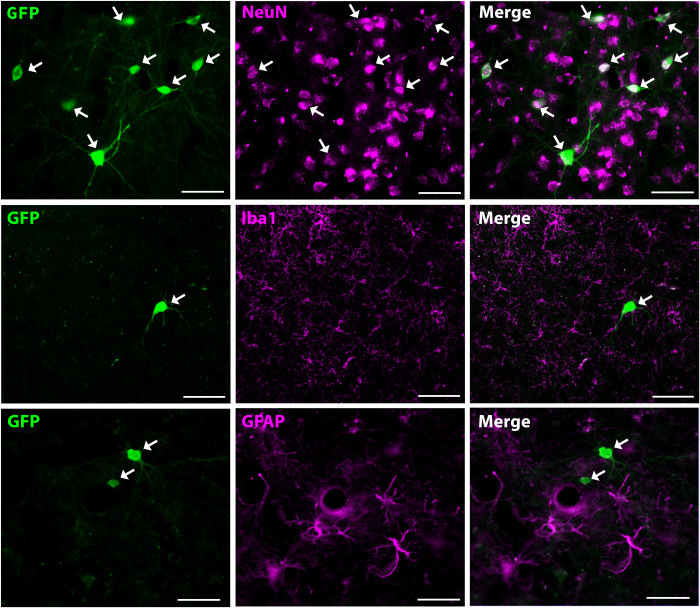
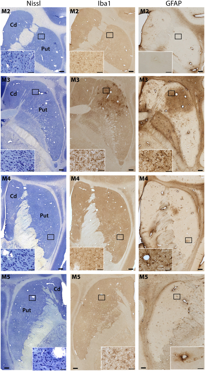
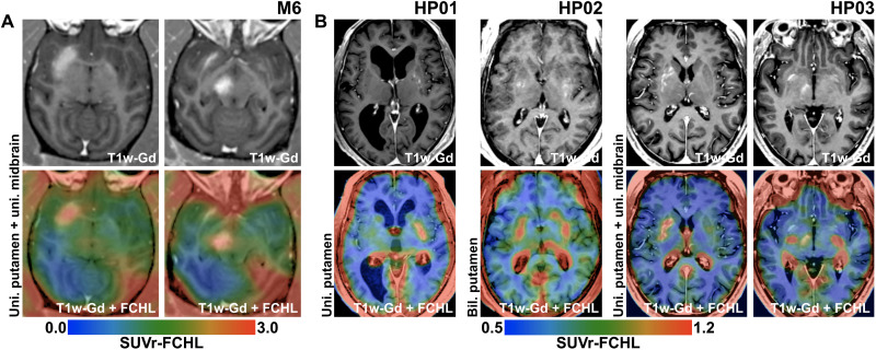
Intracerebral vector delivery in nonhuman primates has been a major challenge. We report successful blood-brain barrier opening and focal delivery of adeno-associated virus serotype 9 vectors into brain regions involved in Parkinson's disease using low-intensity focus ultrasound in adult macaque monkeys. Openings were well tolerated with generally no associated abnormal magnetic resonance imaging signals. Neuronal green fluorescent protein expression was observed specifically in regions with confirmed blood-brain barrier opening. Similar blood-brain barrier openings were safely demonstrated in three patients with Parkinson's disease. In these patients and in one monkey, blood-brain barrier opening was followed by 18F-Choline uptake in the putamen and midbrain regions based on positron emission tomography. This indicates focal and cellular binding of molecules that otherwise would not enter the brain parenchyma. The less-invasive nature of this methodology could facilitate focal viral vector delivery for gene therapy and might allow early and repeated interventions to treat neurodegenerative disorders.


Figures

Fig. 1.
Fig. 1.. Initial BBB openings in NHPs targeting brain regions relevant to PD.
Successful BBB openings were demonstrated in two monkeys (M1/M2) by delivery of an MR contrast agent (Gd) that does not normally extravasate in the brain. Openings were achieved in the targeted regions with relative accuracy. (A) Axial contrast-enhanced T1-weighted MRI image shows BBB disruption in the PMC (indicated by the red circle; M1). (B) Coronal contrast-enhanced MRI shows BBB disruption in the posterior ventrolateral thalamic region (M1). (C) Axial contrast-enhanced MRI shows BBB disruption in the striatum (both the head of the caudate nucleus and the posterior putamen on one side and the anterior putamen on the other; M2). (D) Coronal contrast-enhanced MRI shows BBB disruption in the midbrain (M2). All the enhancements were largely restricted to the targeted regions. Although the same approach was used for each sonication, the size and magnitude of BBB disruption varied depending on the targeted region.
Fig. 2.
Fig. 2.. Safe and large-volume BBB openings in the putamen of normal and parkinsonian NHPs by LIFU.
Axial contrast-enhanced T1-weighted images show homogeneous Gd extravasation in the putamen in M3, M4, and parkinsonian monkey M5 immediately after treatment (left column) and 30 days later (right column). The areas of enhancement almost perfectly overlapped the targeted regions.
Fig. 3.
Fig. 3.. After immediate and after 30-day safety assessment of large-volume BBB openings in the putamen of normal and parkinsonian NHPs.
Follow-up MRI including susceptibility-weighted (SWI), T2*-weighted (T2-FSE), and FLAIR (T2-FLAIR) images in M3, M4, and parkinsonian monkey M5. All images are shown in axial orientation in a single slice corresponding to the largest BBB disruption area, as demonstrated by Gd extravasation. Follow-up sessions were performed immediately and 30 days after treatment.
Fig. 4.
Fig. 4.. Multitargeted focal delivery of modified AAV9 vector in the striatum and midbrain.
In M2, the distribution areas of GFP+ cells in the caudate nucleus [A, low magnification; D, higher magnification taken from the site specified by the arrow in (A)] and the putamen [C, low magnification; E, higher magnification taken from the site specified by the arrow in (C)] overlap the regions of Gd extravasation observed immediately after sonication (arrows in B). Similarly, GFP+ cells in the midbrain [H, low magnification; J, higher magnification taken from the site specified by the arrows in (H)] are distributed within the hyperintense BBB-opened area [arrow in (G)]. The same region on the contralateral side served as a negative control showing no hyperintense signal (G) or GFP expression (F and I, low and higher magnification, respectively). Number of GFP+ cells in the BBB-opened regions (LIFU+) and their contralateral non-opened regions (LIFU) in the caudate nucleus (K), putamen (L), and midbrain (M) of M2 and in the thalamus of M1 (with NAbs) (N). Cd, caudate nucleus; Put, putamen; SN, substantia nigra; STN, subthalamic nucleus. Scale bars, 1 mm (A, C, F, and H) and 50 μm (D, E, I, and J).
Fig. 5.
Fig. 5.. Delivery of modified AAV9 vectors after large-volume BBB openings in the putamen of normal and parkinsonian NHPs.
Representative images of distributions of Gd extravasation (first column) and GFP+ cells (low and higher magnifications in second and third columns, respectively) in the putamen of M3, M4, and parkinsonian monkey M5. Note that the distribution areas of GFP+ cells overlap the regions of Gd extravasation observed immediately after sonication, except for monkey M4 which had NAbs against AAV and, therefore, displayed no GFP expression. Fourth column shows the number of GFP+ cells in the BBB-opened regions (LIFU+) and their contralateral non-opened regions (LIFU) for each monkey. Scale bars, 1 mm (for low magnifications in second column) and 50 μm (for higher magnifications in third column).
Fig. 6.
Fig. 6.. Neuron specificity of AAV vector–induced transgene expression.
Colocalization with the neuronal marker NeuN (top row), but not with the microglial marker Iba1 (middle row) or astroglial marker GFAP (bottom row) in GFP+ cells indicates that GFP is expressed specifically in neuronal cells. Scale bars, 50 μm.
Fig. 7.
Fig. 7.. Tissue assessment following LIFU BBB openings and AAV vector delivery.
Three adjacent sections were processed for Nissl staining (left column) and immunostaining for Iba1 (central column) and GFAP (right column) following LIFU BBB openings and AAV vector delivery in M2, M3, M4, and parkinsonian M5. No marked histopathological changes were observed, except for M3 who exhibited minor focal inflammation but no tissue distortion in the BBB-opened area. Scale bars, 1 mm and 50 μm (insets).
Fig. 8.
Fig. 8.. In vivo PET imaging of BBB permeability using 18F-Choline in NHPs and patients with PD.
Data obtained in monkey M6 (A) and three patients with PD (B) are included. The top row shows targeted regions on T1-weighted images with Gd extravasation after BBB opening sessions. The bottom row depicts colored overlays of 18F-Choline PET images and the T1-weighted images. PET images are represented in blue-green-red colormaps, where red indicates the highest uptake value. Increased 18F-Choline uptake is seen only in the regions where net BBB opening occurred, such as the putamen and midbrain in M6 (A) and unilateral putamen (HP01), bilateral putamen (HP02), and the unilateral putamen and midbrain (HP03) in patients with PD (B).

Comment in

References

    1. G. Deuschl, E. Beghi, F. Fazekas, T. Varga, K. A. Christoforidi, E. Sipido, C. L. Bassetti, T. Vos, V. L. Feigin, The burden of neurological diseases in Europe: An analysis for the Global Burden of Disease Study 2017. Lancet Public Health 5, e551–e567 (2020). - PubMed
    1. R. Daneman, A. Prat, The blood–brain barrier. Cold Spring Harb. Perspect. Biol. 7, a020412 (2015). - PMC - PubMed
    1. A. Akhtar, A. Andleeb, T. S. Waris, M. Bazzar, A.-R. Moradi, N. R. Awan, M. Yar, Neurodegenerative diseases and effective drug delivery: A review of challenges and novel therapeutics. J. Control. Release 330, 1152–1167 (2021). - PubMed
    1. V. Sudhakar, R. M. Richardson, Gene therapy for neurodegenerative diseases. Neurotherapeutics 16, 166–175 (2019). - PMC - PubMed
    1. C. N. Bedbrook, B. E. Deverman, V. Gradinaru, Viral strategies for targeting the central and peripheral nervous systems. Annu. Rev. Neurosci. 41, 323–348 (2018). - PubMed