

Case Reports
doi: 10.1093/jnen/nlad032.
An unusual case of metastatic diffuse midline glioma in an adult
Affiliations
- PMID: 37075315
- PMCID: PMC10501466
- DOI: 10.1093/jnen/nlad032
Item in Clipboard
Case Reports
An unusual case of metastatic diffuse midline glioma in an adult
J Neuropathol Exp Neurol.
.
No abstract available
Conflict of interest statement
The authors have no duality or conflicts of interest to declare.
Figures
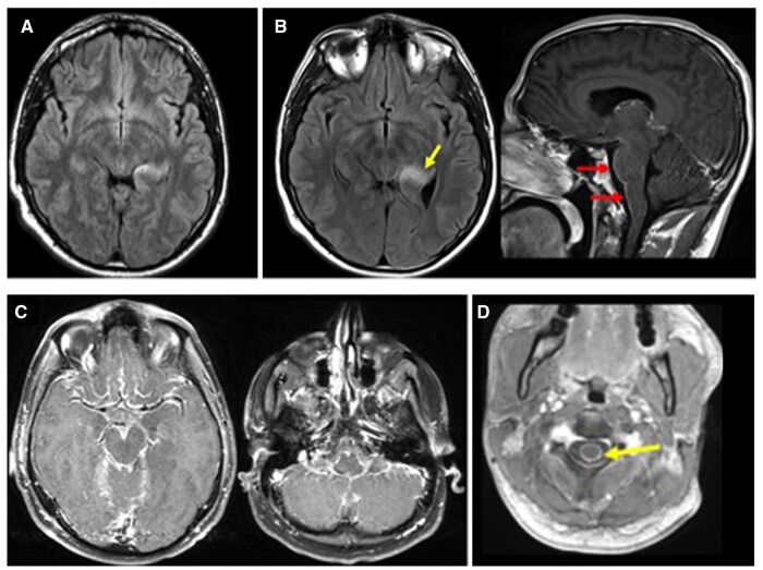
(A) MRI at initial presentation showing a 2 × 1 cm nonenhancing T2/FLAIR hyperintense lesion in the posteromedial left temporal lobe and thalamus; (B) follow-up MRI 3 months later showing mild increase in size and expansion of the component in the posteromedial left temporal lobe (yellow arrow) and new subtle leptomeningeal enhancement along the ventral surface of the brainstem (red arrows); (C) MRI an additional month later showing marked progression of leptomeningeal enhancement in the supratentorial compartment; (D) spine MRI showing leptomeningeal enhancement encasing the spinal cord (yellow arrow).
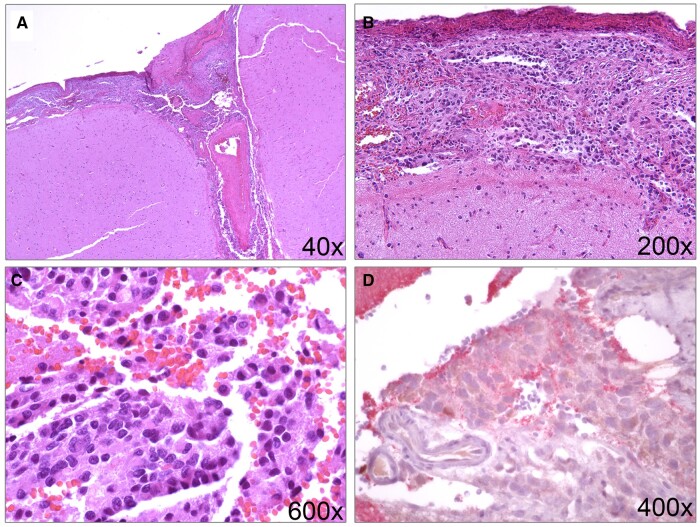
(A–C) H&E images of the temporal lobe biopsy in region of meningeal enhancement depicting tumor cells localized to the subarachnoid space. (D) Synaptophysin stain highlights a few tumor cells.
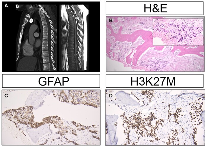
(A) CT showing widespread patchy sclerosis throughout the spine and sternum, corresponding to diffuse hypointensity on T1-weighted MRI consistent with diffuse marrow replacement. (B) H&E of bone depicting metastatic glioma (inset demonstrating cytology at higher power). (C, D) GFAP and H3K27M immunostains highlighting metastatic glial cells.
Similar articles
-
An adult case of diffuse midline glioma with H3 K27M mutation.Neuropathology. 2020 Dec;40(6):627-631. doi: 10.1111/neup.12689. Epub 2020 Sep 20. Neuropathology. 2020. PMID: 32954563
-
Indolent presentation of a diffuse midline glioma, H3 K27-altered.Childs Nerv Syst. 2023 Mar;39(3):833-835. doi: 10.1007/s00381-022-05668-4. Epub 2022 Sep 12. Childs Nerv Syst. 2023. PMID: 36094605
-
[H3K27 positive diffuse midline glioma. Report of one case].Rev Med Chil. 2019 Nov;147(11):1487-1490. doi: 10.4067/S0034-98872019001101487. Rev Med Chil. 2019. PMID: 32186609 Spanish. No abstract available.
-
Molecular and clinical characterization of H3 K27M-mutant "non-midline" glioblastoma: A case report and literature review.Neurocirugia (Engl Ed). 2022 Nov-Dec;33(6):356-360. doi: 10.1016/j.neucie.2021.06.008. Neurocirugia (Engl Ed). 2022. PMID: 36333093 Review.
-
Adult diffuse midline gliomas H3 K27-altered: review of a redefined entity.J Neurooncol. 2022 Jul;158(3):369-378. doi: 10.1007/s11060-022-04024-5. Epub 2022 May 14. J Neurooncol. 2022. PMID: 35567713 Review.
Cited by
-
Diffuse midline glioma H3K27-altered with thoracic epidural metastasis: illustrative case.J Neurosurg Case Lessons. 2025 Jul 14;10(2):CASE25249. doi: 10.3171/CASE25249. Print 2025 Jul 14. J Neurosurg Case Lessons. 2025. PMID: 40658994 Free PMC article.
References
-
- WHO, ed. Central Nervous System Tumours. 5th ed. Lyon: International Agency for Research on Cancer; World Health Organization Classification of tumours; 2021:568
-
- Korshunov A, Schrimpf D, Ryzhova M, et al.H3-/IDH-wild type pediatric glioblastoma is comprised of molecularly and prognostically distinct subtypes with associated oncogenic drivers. Acta Neuropathol 2017;134:507–16 - PubMed
-
- Hsu E, Keene D, Ventureyra E, et al. Bone marrow metastasis in astrocytic gliomata. J Neurooncol 1998;37:285–93 - PubMed
Publication types
MeSH terms
Grants and funding
LinkOut - more resources
Full Text Sources
Medical
