. 2023 Jul;15(4):451-472.
doi: 10.4168/aair.2023.15.4.451. Epub 2023 Feb 28.

Tissue Inhibitor of Metalloproteinase-1 Enhances Eosinophilic Airway Inflammation in Severe Asthma

Affiliations

Tissue Inhibitor of Metalloproteinase-1 Enhances Eosinophilic Airway Inflammation in Severe Asthma

Thi Bich Tra Cao et al. Allergy Asthma Immunol Res. 2023 Jul.

Abstract

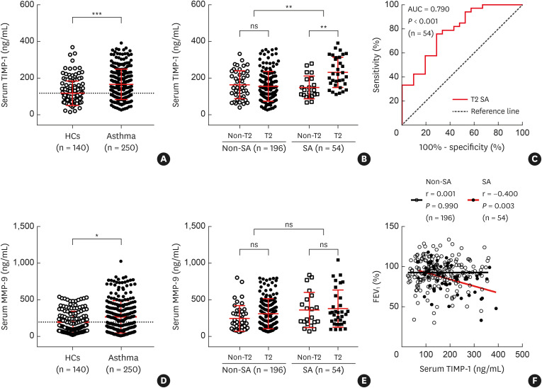
Purpose: Severe asthma (SA) is characterized by persistent airway inflammation and remodeling, followed by lung function decline. The present study aimed to evaluate the role of tissue inhibitor of metalloproteinase-1 (TIMP-1) in the pathogenesis of SA.

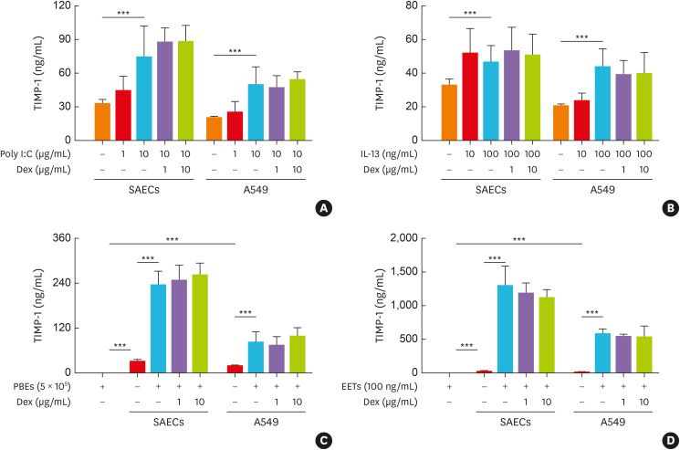
Methods: We enrolled 250 adult asthmatics (54 with SA and 196 with non-SA) and 140 healthy controls (HCs). Serum TIMP-1 levels were determined by enzyme-linked immunosorbent assay. The release of TIMP-1 from airway epithelial cells (AECs) in response to stimuli as well as the effects of TIMP-1 on the activations of eosinophils and macrophages were evaluated in vitro and in vivo.

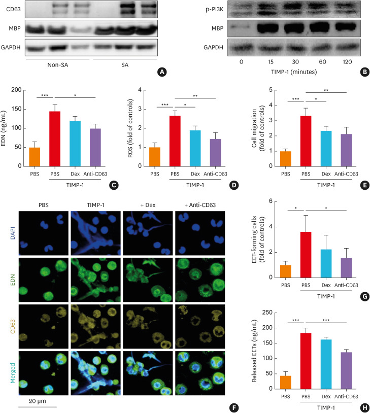
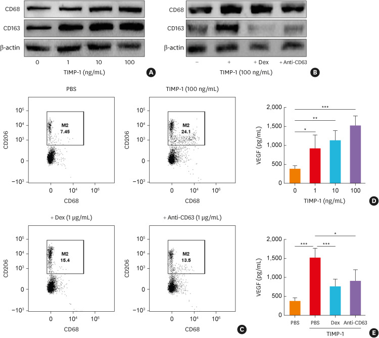
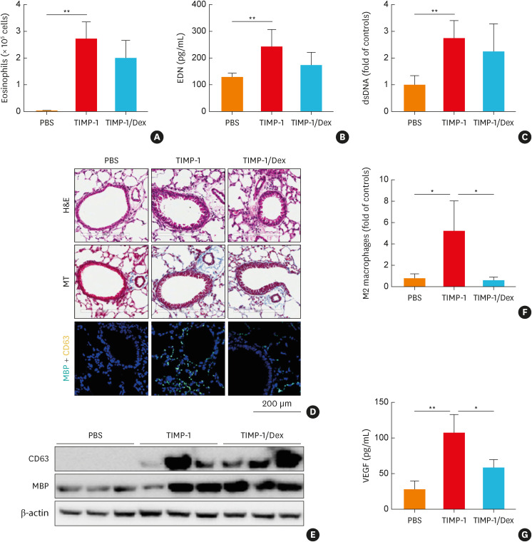
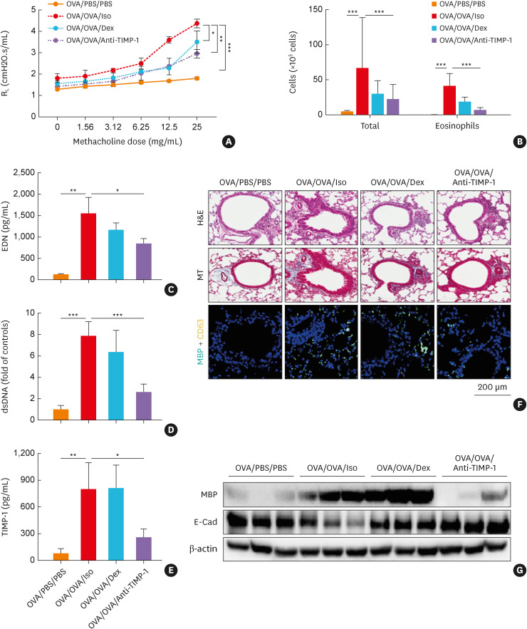
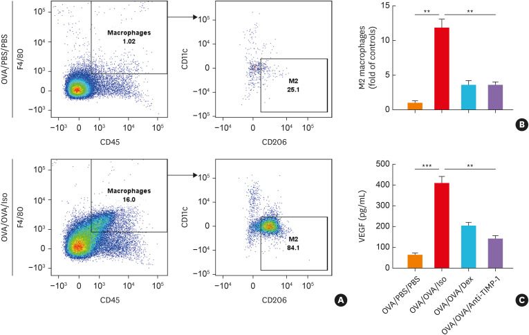
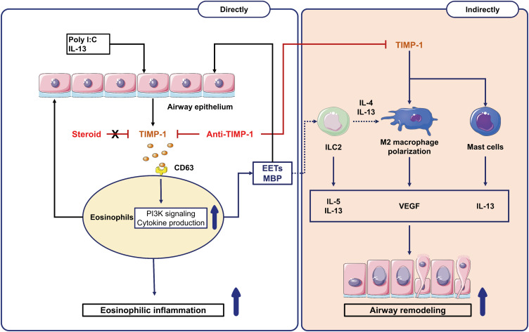
Results: Significantly higher levels of serum TIMP-1 were noted in asthmatics than in HCs, in the SA group than in non-SA group, and in the type 2 SA group than in non-type 2 SA group (P < 0.01 for all). A negative correlation between serum TIMP-1 and FEV1% values (r = -0.400, P = 0.003) was noted in the SA group. In vitro study demonstrated that TIMP-1 was released from AECs in response to poly I:C, IL-13, eosinophil extracellular traps (EETs) and in coculture with eosinophils. TIMP-1-stimulated mice showed eosinophilic airway inflammation, which was not completely suppressed by steroid treatment. In vitro and in vivo functional studies showed that TIMP-1 directly activated eosinophils and macrophages, and induced the release of EETs and macrophages to polarize toward M2 subset, which was suppressed by anti-TIMP-1 antibody.

Conclusions: These findings suggest that TIMP-1 enhances eosinophilic airway inflammation and that serum TIMP-1 may be a potential biomarker and/or therapeutic target for type 2 SA.

Keywords: Asthma; TIMP-1; airway remodeling; eosinophils; epithelial cells; inflammation; macrophages.


Conflict of interest statement

There are no financial or other issues that might lead to conflict of interests.

Figures

Fig. 1
Fig. 1. Increased levels of serum TIMP-1 in patients with SA. Comparisons of serum TIMP-1 levels between (A) HCs (n = 140) and asthmatic patients (n = 250), between (B) patients with SA and those with non-SA, and between T2 and non-T2. Asthmatics were grouped into non-T2 non-SA (n = 45), T2 non-SA (n = 151), non-T2 SA (n = 21), and T2 SA (n = 33). (C) The receiver operating characteristic curve of serum TIMP-1 levels for discriminating the patients with T2 SA from those with non-T2 SA. Comparisons of serum MMP-9 levels between (D) HCs and asthmatic patients as well as (E) patients with SA and those with non-SA. Data are presented as median with interquartile range. (F) Correlations between serum TIMP-1 levels and FEV1% in patients with SA or non-SA. Data are presented as Spearman correlation coefficient r.
SA, severe asthma; HCs, healthy controls; T2, type 2; ns, not significant; FEV1, forced expiratory volume in the first second; MMP-9, matrix metallopeptidase 9; TIMP-1, tissue inhibitor of metalloproteinase-1; AUC, area under the curve. *P < 0.05, **P < 0.01, and ***P < 0.001 by Mann-Whitney U test or Kruskal-Wallis test with Dunn’s post hoc test.
Fig. 2
Fig. 2. The production of TIMP-1 from human AECs. The production of TIMP-1 from AECs (SAECs and A549) in response to (A) poly I:C, (B) IL-13, and in coculture with (C) PBEs as well as (D) in response to EETs (n = 6 for each group). Data are presented as mean ± standard deviation.
AECs, airway epithelial cells; A549, type II alveolar epithelial cell line; IL, interleukin; Dex, dexamethasone; EETs, eosinophil extracellular traps; PBEs, peripheral blood eosinophils; SAECs, primary small airway epithelial cells; TIMP-1, tissue inhibitor of metalloproteinase-1. ***P < 0.001 by one-way analysis of variance with the Bonferroni post hoc test.
Fig. 3
Fig. 3. The effects of TIMP-1 on human PBEs. (A) The protein expressions of CD63 and MBP in PBEs from asthmatics according to asthma severity. (B) The effect of TIMP-1 on the phosphorylation of PI3K in a time-dependent manner. TIMP-1 induced the release of (C) EDN and (D) ROS with (E) eosinophil recruitment (n = 6 for each group). (F, G) The levels of EETs were evaluated using confocal microscopy. Cells were stained with EDN (green), CD63 (yellow), and DAPI (blue). Scale bar, 20 µm. (H) The levels of released EETs were evaluated by measuring dsDNA concentrations using PicoGreen assay (n = 6 for each group). Data are presented as mean ± standard deviation.
PBEs, peripheral blood eosinophils; DAPI, 4’,6-diamidino-2-phenylindole; Dex, dexamethasone; EETs, eosinophil extracellular traps; GAPDH, glyceraldehyde 3-phosphate dehydrogenase; MBP, major basic protein; PI3K, phosphatidylinositol 3-kinases; ROS, reactive oxygen species; SA, severe asthma; TIMP-1, tissue inhibitor of metalloproteinase-1; EDN, eosinophil-derived neurotoxin. *P < 0.05, **P < 0.01, and ***P < 0.001 by one-way analysis of variance with the Bonferroni post hoc test.
Fig. 4
Fig. 4. The effect of TIMP-1 on human macrophages. (A) The protein expressions of CD68 (macrophage maturation marker) and CD163 (M2 macrophage marker) from macrophages in response to TIMP-1 in a dose-dependent manner. (B) The effects of anti-CD63 antibody treatment on TIMP-1-stimulated macrophages. (C) Macrophage polarization was evaluated by flow cytometry. The levels of VEGF from macrophages in response to TIMP-1 in (D) a dose-dependent manner or with (E) the presence of anti-CD63 treatment (n = 6 for each group). Data are presented as mean ± standard deviation.
Dex, dexamethasone; TIMP-1, tissue inhibitor of metalloproteinase-1; VEGF, vascular endothelial growth factor; PBS, phosphate buffered saline. *P < 0.05, **P < 0.01, and ***P < 0.001 by one-way analysis of variance with the Bonferroni post hoc test.
Fig. 5
Fig. 5. The effects of TIMP-1 on airway inflammation in vivo. (A) Eosinophils in the BALF. The levels of (B) EDN and (C) dsDNA in the BALF. (D) Lung histology stained with H&E and MT staining. Immunofluorescence staining for DAPI (nuclear, blue), MBP (turquoise), and CD63 (yellow). Scale bar, 200 µm. (E) The expressions of CD63 and MBP in the lung tissues. (F) M2 macrophage counts in the lung tissues were obtained by flow cytometry. (G) The levels of VEGF in the BALF (n = 5 for each group). Data are presented as mean ± standard deviation.
BALF, bronchoalveolar lavage fluid; DAPI, 4’,6-diamidino-2-phenylindole; Dex, dexamethasone; EDN, eosinophil-derived neurotoxin; H&E, hematoxylin and eosin; MT, Masson’s trichrome; MBP, major basic protein; TIMP-1, tissue inhibitor of metalloproteinase-1; VEGF, vascular endothelial growth factor; PBS, phosphate buffered saline. *P < 0.05 and **P < 0.01 by one-way analysis of variance with the Bonferroni post hoc test.
Fig. 6
Fig. 6. The effects of anti-TIMP-1 antibody treatment in a mouse model of chronic allergic asthma. (A) Airway hyperresponsiveness. (B) Total cells and eosinophils in the BALF. The levels of (C) EDN, (D) dsDNA, and (E) TIMP-1 in the BALF (n = 5 for each group). Data are presented as mean ± standard deviation. (F) Lung histology stained with H&E and MT staining. Immunofluorescence staining for DAPI (nuclear, blue), MBP (turquoise), and CD63 (yellow). Scale bar, 200 µm. (G) The expressions of MBP and E-Cad in the lung tissues.
BALF, bronchoalveolar lavage fluid; DAPI, 4’,6-diamidino-2-phenylindole; Dex, dexamethasone; EDN, eosinophil-derived neurotoxin; H&E, hematoxylin and eosin; MT, Masson’s trichrome; MBP, major basic protein; RL, resistance to airflow across the lung; OVA, ovalbumin; TIMP-1, tissue inhibitor of metalloproteinase-1; E-Cad, E-cadherin; PBS, phosphate buffered saline. *P < 0.05, **P < 0.01, and ***P < 0.001 by one-way analysis of variance with the Bonferroni post hoc test.
Fig. 7
Fig. 7. The effects of anti-TIMP-1 antibody treatment on mouse macrophages. (A) The percentage of macrophage count (CD45+F4/80+ cells) and M2 macrophages (CD11cCD206+ macrophages) measured by flow cytometry. (B) The degree of M2 macrophage count in lung tissue. (C) The levels of VEGF in the BALF (n = 5 for each group). Data are presented as mean ± standard deviation.
BALF, bronchoalveolar lavage fluid; Dex, dexamethasone; OVA, ovalbumin; TIMP-1, tissue inhibitor of metalloproteinase-1; VEGF, vascular endothelial growth factor; PBS, phosphate buffered saline. **P < 0.01 and ***P < 0.001 by one-way analysis of variance with the Bonferroni post hoc test.
Fig. 8
Fig. 8. Available mechanisms of TIMP-1 in type 2 SA. The exogenous (poly I:C) and endogenous factors (IL-13, eosinophils, and EETs) stimulate airway epithelial cells to release TIMP-1. The left panel showed TIMP-1-induced eosinophilic inflammation through (1) directly activating eosinophils (MBP release) and (2) indirectly activating EET formation (through CD63/PI3K signaling) in SA, which were suppressed by anti-TIMP-1 antibody (not by steroid). The right panel showed TIMP-1-induced airway remodeling via releasing proinflammatory cytokines (IL-13 and VEGF) from activated ILC2, M2 macrophages, mast cells, and eosinophils.
EETs, eosinophil extracellular traps; ILC2, group 2 innate lymphoid cells; MBP, major basic protein; TIMP-1, tissue inhibitor of metalloproteinase-1; PI3K; phosphatidylinositol 3-kinases; VEGF, vascular endothelial growth factor; IL, interleukin; SA, severe asthma.

References

    1. Lee Y, Quoc QL, Park HS. Biomarkers for severe asthma: lessons from longitudinal cohort studies. Allergy Asthma Immunol Res. 2021;13:375–389. - PMC - PubMed
    1. Woo SD, Yang EM, Jang J, Lee Y, Shin YS, Ye YM, et al. Serum-free immunoglobulin E: a useful biomarker of atopy and type 2 asthma in adults with asthma. Ann Allergy Asthma Immunol. 2021;127:109–115.e1. - PubMed
    1. Nair P, Surette MG, Virchow JC. Neutrophilic asthma: misconception or misnomer? Lancet Respir Med. 2021;9:441–443. - PubMed
    1. Lee Y, Park Y, Kim C, Lee E, Lee HY, Woo SD, et al. Longitudinal outcomes of severe asthma: real-world evidence of multidimensional analyses. J Allergy Clin Immunol Pract. 2021;9:1285–1294.e6. - PubMed
    1. Park SY, Kang SY, Song WJ, Kim JH. Evolving concept of severe asthma: transition from diagnosis to treatable traits. Allergy Asthma Immunol Res. 2022;14:447–464. - PMC - PubMed