. 2023 Apr 19;14(1):2247.
doi: 10.1038/s41467-023-37986-9.

Allosteric role of the citrate synthase homology domain of ATP citrate lyase

Affiliations

Allosteric role of the citrate synthase homology domain of ATP citrate lyase

Xuepeng Wei et al. Nat Commun. .

Abstract

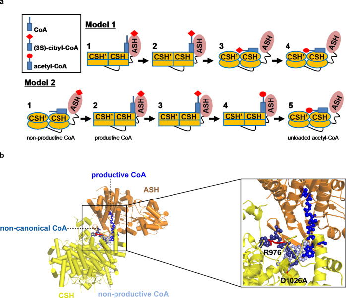
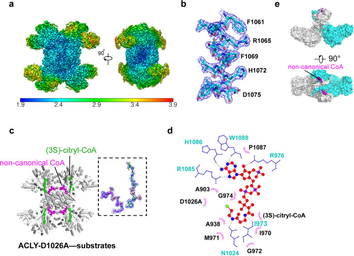
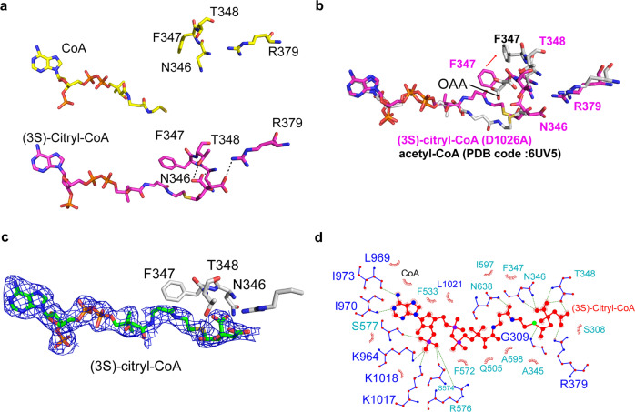
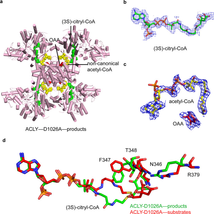
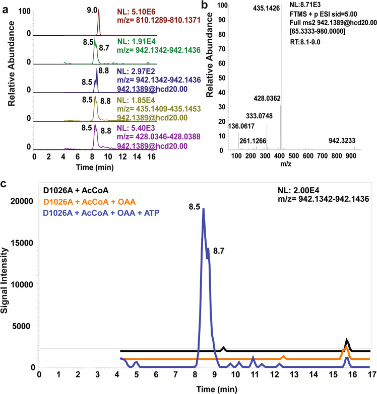
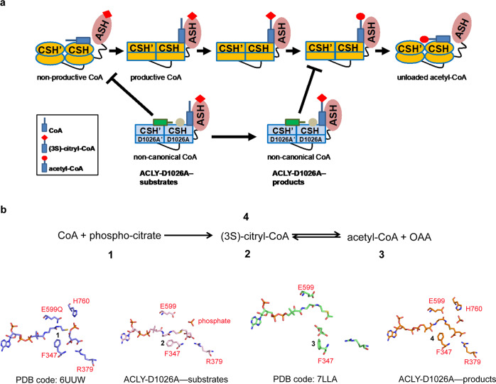
ATP citrate lyase (ACLY) is the predominant nucleocytosolic source of acetyl-CoA and is aberrantly regulated in many diseases making it an attractive therapeutic target. Structural studies of ACLY reveal a central homotetrameric core citrate synthase homology (CSH) module flanked by acyl-CoA synthetase homology (ASH) domains, with ATP and citrate binding the ASH domain and CoA binding the ASH-CSH interface to produce acetyl-CoA and oxaloacetate products. The specific catalytic role of the CSH module and an essential D1026A residue contained within it has been a matter of debate. Here, we report biochemical and structural analysis of an ACLY-D1026A mutant demonstrating that this mutant traps a (3S)-citryl-CoA intermediate in the ASH domain in a configuration that is incompatible with the formation of acetyl-CoA, is able to convert acetyl-CoA and OAA to (3S)-citryl-CoA in the ASH domain, and can load CoA and unload acetyl-CoA in the CSH module. Together, this data support an allosteric role for the CSH module in ACLY catalysis.


Conflict of interest statement

The authors declare no competing interests.

Figures

Fig. 1
Fig. 1. Models for ACLY catalysis and CoA binding modes.
a Model 1—ACLY-WT in complex 1 binds CoA through the ASH domain to form (3 S)-citryl-CoA in the ASH domain (complex 2). (3 S)-citryl-CoA then translocates to the CSH domain where catalysis to acetyl-CoA occurs (complex 4). Model 2—ACLY-WT in complex 1 loads CoA onto the CSH domain in a non-productive conformation with the ASH domain in an open conformation. An allosteric change in the CSH domain, coupled with a conformational change of the ASH domain to a closed conformation (complex 2), mediates the translocation of CoA to the ASH domain. Complex 3 initiates catalysis by forming (3S)-citryl-CoA in the ASH domain, followed by complex 4, that completes catalysis to acetyl-CoA in the ASH domain. An allosteric change in the CSH domain, coupled with a conformational change of the ASH domain to an open conformation (complex 5), mediates the translocation of acetyl-CoA to the CSH domain prior to product release. Different bound states of CoA and acetyl-CoA are annotated below the corresponding schematic diagrams. Residue D1026 is located in the CSH domain and interacts with non-productive CoA. Only one of the four ASH domains of the tetramer is shown, and the binding of ATP and OAA to the ASH domain is not shown for clarity. The red diamond and circle represent citrate and acetate, respectively. b Comparison of productive and non-productive CoA binding modes highlighting key residues highlighted in this study.
Fig. 2
Fig. 2. Structure of ACLY-D1026A with substrates.
a Cryo-EM map and local resolution estimation of ACLY-D1026A—substrates. b Representative side chain cryo-EM density of residues in the CSH domain. c Cartoon presentation of ACLY-D1026A—substrates highlighting bound non-canonical CoA (purple) and (3S)-citryl-CoA (green) molecules. d Ligplot highlighting interactions between the non-canonical CoA, two adjacent CSH domains, and (3 S)-citryl-CoA. e Surface representation of ACLY-D1026A—substrates structure highlighting how the non-canonical CoA molecule (magenta) interacts with the adjacent CSH domain; for clarity, only two subunits (cyan and gray) are shown.
Fig. 3
Fig. 3. (3S)-Citryl-CoA binding sites in the ASH domain of ACLY-D1026A with substrates.
a Comparison of CoA and (3S)-Citryl-CoA bound to the ASH domain of ACLY-WT bound to CoA (PDB 6UUZ) and ACLY-D1026A—substrates structures, respectively. Residues that change positions to accommodate the different ligands are highlighted. b Overlay of ACLY-D1026A—substrates and ACLY-WT bound to acetyl-CoA + OAA (PDB 6UV5) highlighting the conformational change of F347. c Cryo-EM density of (3 S)-citryl-CoA in the ASH domain is displayed at a contour level of 2.5 σ. d A ligplot highlighting the interactions between (3 S)-citryl-CoA and surrounding residues within the ASH domain.
Fig. 4
Fig. 4. Structure ACLY-D1026A with products.
a Cartoon presentation of ACLY-D1026A—products structure highlighting bound (3S)-citryl-CoA (green), non-canonical acetyl-CoA (yellow), and OAA (red) molecules. b Cryo-EM density of (3S)-citryl-CoA bound at the ASH domain. c Cryo-EM density of non-canonical acetyl-CoA and OAA bound at the CSH domain. d Overlay of (3S)-citryl-CoA from ACLY-D1026A—products and ACLY-D1026A—substrates structures.
Fig. 5
Fig. 5. Identification of citryl-CoA by LC-HRMS and LC-MS/MS produced by ACLY-D1026A.
a Unreacted acetyl-CoA (top, in red retention time 9.0, [M + H]+ ion at m/z 810.1330) and citryl-CoA (retention time 8.5-8.8, [M + H]+ ion at m/z 942.1389). Co-eluting MS/MS peaks corresponding to predominant acyl-CoA peaks at m/z 428.0362 and the [M-507 + H]+ neutral loss diagnostic for acyl-CoAs at 435.1426 support the identification of citryl-CoA. b MS/MS spectra for citryl-CoA. c LC-HRMS chromatograms of the citryl-CoA product from ACLY-D1026A + AcCoA (black), ACLY-D1026A + AcCoA + OAA (orange), and ACLY-D1026A + AcCoA + OAA + ATP (blue). An intense peak corresponding to citryl-CoA was identified in the only extract from ACLY-D1026A + AcCoA + OAA + ATP with the baseline offset for clarity.
Fig. 6
Fig. 6. Allosteric regulation of the CSH domain in ACLY catalysis and inhibition of product formation in ACLY-D1026A.
a Model 2 for ACLY catalysis, as illustrated in Fig. 1, with the mechanism of inhibition by the ACLY-D1026A mutant based on the structures of ACLY-D1026–substrates and ACLY-D1026A–products. Different bound states of CoA and acetyl-CoA are annotated below the corresponding schematic diagrams, and the position of the D1026A mutation is highlighted with a brown dot. The red diamond and circle represent citrate and acetate, respectively. b The reversible nature of the transformation between (3S)-citryl-CoA and acetyl-CoA and structurally captured states are indicated and shown, highlighting residues that undergo key conformational changes.

Similar articles

Cited by

References

    1. Shi L, Tu BP. Acetyl-CoA and the regulation of metabolism: mechanisms and consequences. Curr. Opin. Cell Biol. 2015;33:125–131. doi: 10.1016/j.ceb.2015.02.003. - DOI - PMC - PubMed
    1. Chypre M, Zaidi N, Smans K. ATP-citrate lyase: a mini-review. Biochem. Biophys. Res. Commun. 2012;422:1–4. doi: 10.1016/j.bbrc.2012.04.144. - DOI - PubMed
    1. Guo L, et al. Enhanced acetylation of ATP-citrate lyase promotes the progression of nonalcoholic fatty liver disease. J. Biol. Chem. 2019;294:11805–11816. doi: 10.1074/jbc.RA119.008708. - DOI - PMC - PubMed
    1. Ma TT, Huang C, Ni Y, Yang Y, Li J. ATP citrate lyase and LncRNA NONMMUT010685 play crucial role in nonalcoholic fatty liver disease based on analysis of microarray data. Cell Physiol. Biochem. 2018;51:871–885. doi: 10.1159/000495384. - DOI - PubMed
    1. Feng X, Zhang L, Xu S, Shen AZ. ATP-citrate lyase (ACLY) in lipid metabolism and atherosclerosis: an updated review. Prog. Lipid Res. 2019;77:101006. doi: 10.1016/j.plipres.2019.101006. - DOI - PubMed

Publication types

MeSH terms