. 2023 Apr 19;14(1):2233.
doi: 10.1038/s41467-023-37839-5.

Identification of D-arabinan-degrading enzymes in mycobacteria

Affiliations

Identification of D-arabinan-degrading enzymes in mycobacteria

Omar Al-Jourani et al. Nat Commun. .

Abstract

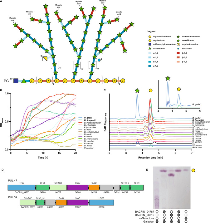
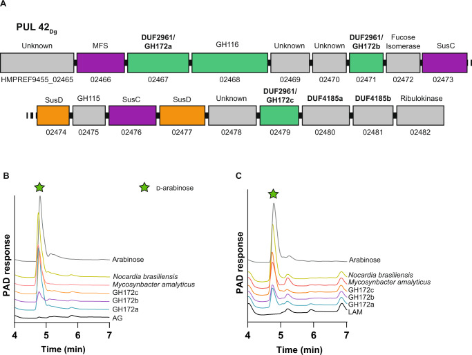
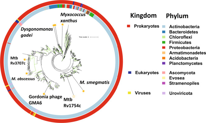
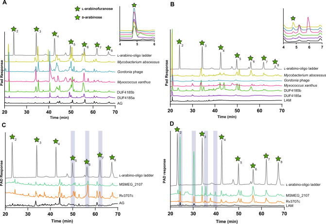
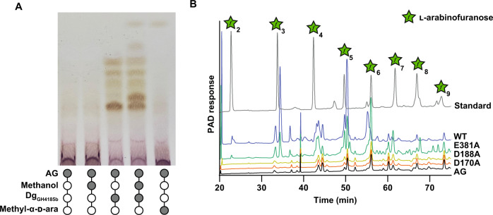
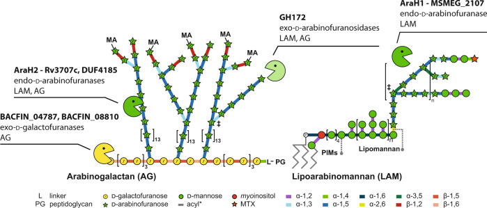
Bacterial cell growth and division require the coordinated action of enzymes that synthesize and degrade cell wall polymers. Here, we identify enzymes that cleave the D-arabinan core of arabinogalactan, an unusual component of the cell wall of Mycobacterium tuberculosis and other mycobacteria. We screened 14 human gut-derived Bacteroidetes for arabinogalactan-degrading activities and identified four families of glycoside hydrolases with activity against the D-arabinan or D-galactan components of arabinogalactan. Using one of these isolates with exo-D-galactofuranosidase activity, we generated enriched D-arabinan and used it to identify a strain of Dysgonomonas gadei as a D-arabinan degrader. This enabled the discovery of endo- and exo-acting enzymes that cleave D-arabinan, including members of the DUF2961 family (GH172) and a family of glycoside hydrolases (DUF4185/GH183) that display endo-D-arabinofuranase activity and are conserved in mycobacteria and other microbes. Mycobacterial genomes encode two conserved endo-D-arabinanases with different preferences for the D-arabinan-containing cell wall components arabinogalactan and lipoarabinomannan, suggesting they are important for cell wall modification and/or degradation. The discovery of these enzymes will support future studies into the structure and function of the mycobacterial cell wall.


Conflict of interest statement

Drs. Moynihan and Lowe are co-inventors on an unpublished patent application pertaining to some of the enzymes described in this manuscript. The remaining authors declare no competing interests.

Figures

Fig. 1
Fig. 1. Bacteroidetes growth on mycobacterial arabinogalactan.
A Schematic of the structure of mycobacterial mycolyl-arabinogalactan-peptidoglycan complex. Succinate (‡) and galactosamine (*) modifications are non-stoichiometric. B Growth of selected Bacteroidetes species on arabinogalactan as monitored by change in OD600nm. C Ion chromatography with pulsed amperometric detection (IC-PAD) analysis of culture supernatants for bacteria grown on mycobacterial AG as a sole-carbon source. Production of arabinose (green star) and galactose (yellow circle) are indicated and compared to standards. Data from D. gadei is also shown as an inset. D Schematics of PUL47 and 39 from B. finegoldii, whose homologs in B. cellulosilyticus were identified by RNAseq as being upregulated during growth on AG. E TLC analysis of endpoint reaction products of B. finegoldii enzymes with C. glutamicumubiA galactan. Filled circles indicate the presence of a given reaction component. The major product appears to be galactobiose suggesting endo-activity. A detailed analysis of this enzyme is beyond the scope of this study. Source data are provided in the Source Data file.
Fig. 2
Fig. 2. D. gadei DUF2961 genes encode GH172 exo-d-arabinofuranosidases with orthologs in diverse bacteria.
A Schematic of PUL42 as identified by proteomic analysis of D. gadei grown on d-arabinan. B DUF2961 enzymes (1 µM) vs 2 mg ml-1 AG and (C) LAM. Samples were analyzed by ion chromatography with pulsed amperometric detection (IC-PAD) on a Dionex ICS-6000 with CarboPac Pa300 column, 100 mM NaOH, 20 min isocratic elution followed by a 0-60% 500 mM sodium acetate gradient over 60 minutes. Green star = d-arabinose. Source data are provided in the Source Data file.
Fig. 3
Fig. 3. Phylogeny of the DUF4185 enzyme family.
Unrooted ML phylogeny (LG model with empirical base frequencies, invariable sites and the discrete gamma model) of DUF4185 family sequences. Branches with greater than 75% bootstrap support (100 replicates) are highlighted in green. Units for tree scale are inferred substitutions per amino acid residue. Colored rings indicate phylum (inner) and kingdom (outer) taxonomy information for sequences. Stars highlight sequences of interest and are filled for proteins that have been experimentally characterized in this work.
Fig. 4
Fig. 4. DUF4185 proteins are endo-d-arabinofuranases that cleave mycobacterial AG and LAM.
DUF4185 enzymes were incubated with 2 mg∙ml−1 AG (A and C) or 2 mg · ml−1 LAM (B and D) for 16 hours as described in Materials and Methods. Samples were analyzed by ion chromatography with pulsed amperometric detection (IC-PAD) on a Dionex ICS-6000w with CarboPac Pa300 column, 100 mM NaOH 20 min isocratic elution followed by a 0-60% 500 mM sodium acetate gradient over 60 minutes. A ladder of α-1,5-l-arabino-oligosaccharides (25 µM) derived from plant arabinan was used as a standard. In panel C the chromatogram of this ladder has been scaled on the y-axis by a factor of 0.2 for clarity of presentation. Source data are provided in the Source Data file.
Fig. 5
Fig. 5. Catalytic analysis of DUF4185 endo-d-arabinofuranases.
A Methanolysis of AG catalyzed by DgGH4185b. The production of methyl arabinosides in the presence of methanol indicates a retaining mechanism. B DgGH4185b-WT, DgGH4185b-D149A and DgGH4185b-D167A were incubated at 1 µM with 2 mg ml-1 AG for 16 hours and analyzed by ion chromatography with pulsed amperometric detection (IC-PAD). No activity was observed for the D188A or D170A mutants. Source data are provided in the Source Data file.
Fig. 6
Fig. 6. Mycobacterial arabinogalactan-degrading enzymes discovered in this study and their substrates.
Each enzyme or enzyme family is listed with its identified function and the mycobacterial cell wall component it acts upon. MA mycolic acids, L linker unit, PG peptidoglycan, PIMs phosphatidylinostol mannosides.

References

    1. Abrahams KA, Besra GS. Synthesis and recycling of the mycobacterial cell envelope. Curr. Opin. Microbiol. 2021;60:58–65. doi: 10.1016/j.mib.2021.01.012. - DOI - PMC - PubMed
    1. Dulberger CL, Rubin EJ, Boutte CC. The mycobacterial cell envelope - a moving target. Nat. Rev. Microbiol. 2020;18:47–59. doi: 10.1038/s41579-019-0273-7. - DOI - PubMed
    1. Daffe M, McNeil M, Brennan PJ. Major structural features of the cell wall arabinogalactans of Mycobacterium, Rhodococcus, and Nocardia spp. Carbohydr. Res. 1993;249:383–398. doi: 10.1016/0008-6215(93)84102-C. - DOI - PubMed
    1. Justen AM, et al. Polysaccharide length affects mycobacterial cell shape and antibiotic susceptibility. Sci. Adv. 2020;6:eaba4015. doi: 10.1126/sciadv.aba4015. - DOI - PMC - PubMed
    1. Safi H, Sayers B, Hazbon MH, Alland D. Transfer of embB codon 306 mutations into clinical Mycobacterium tuberculosis strains alters susceptibility to ethambutol, isoniazid, and rifampin. Antimicrob. Agents Chemother. 2008;52:2027–2034. doi: 10.1128/AAC.01486-07. - DOI - PMC - PubMed

Publication types

MeSH terms