

Qualitative study on diabetic cutaneous wound healing with radiation crosslinked bilayer collagen scaffold in rat model
- PMID: 37076561
- PMCID: PMC10115801
- DOI: 10.1038/s41598-023-33372-z
Qualitative study on diabetic cutaneous wound healing with radiation crosslinked bilayer collagen scaffold in rat model
Abstract
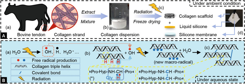
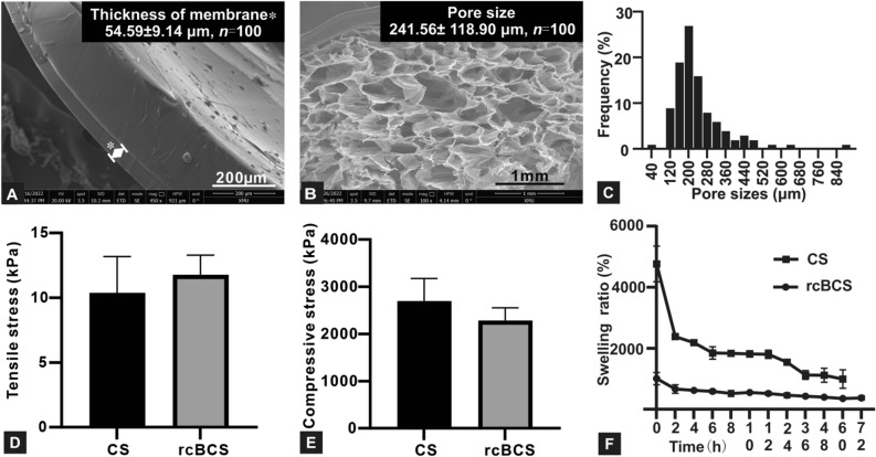
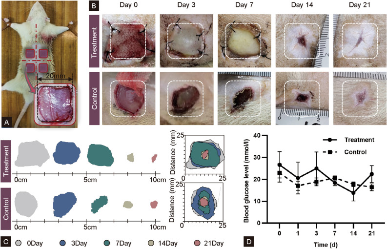
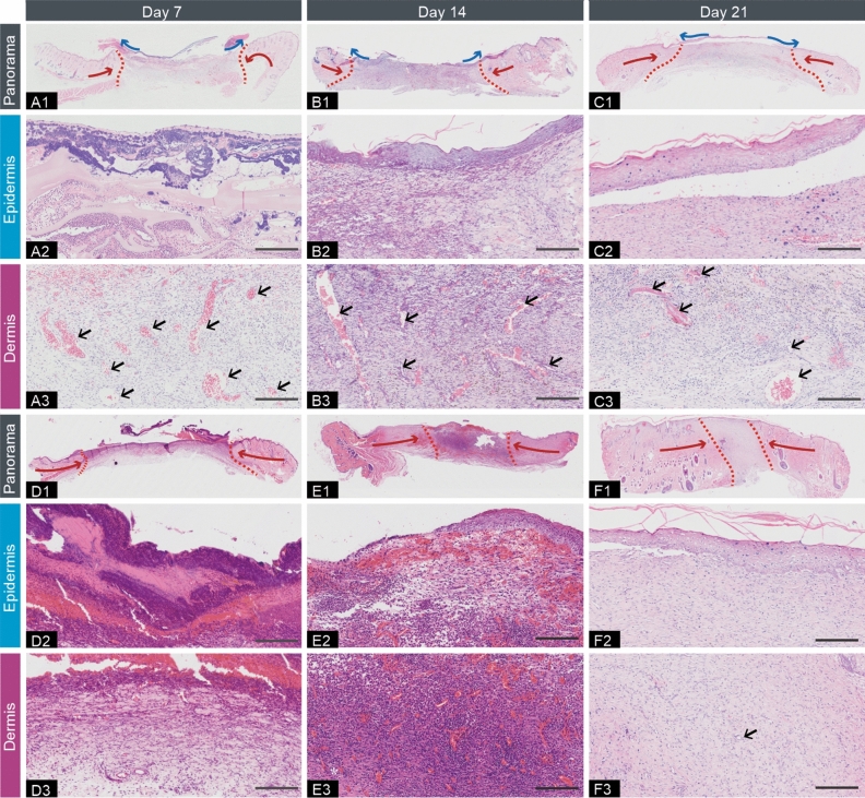
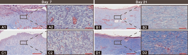
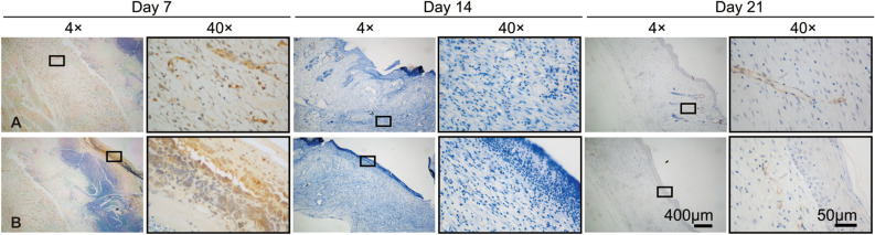
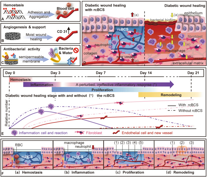
Diabetes may leave patients more prone to skin problems, and minor skin conditions can more easily turn into serious damage to the extracellular matrix, which further impairs the skin's mechanical properties and delays wound healing. Therefore, the aim of the work is to develop extracellular matrix substitution to remodel the mechanical properties of diabetic cutaneous wound and thus accelerate diabetic wound healing. A green fabrication approach was used to prepare radiation crosslinked bilayer collagen scaffold from collagen dispersion. The morphological, mechanical and swelling characteristics of radiation crosslinked bilayer collagen scaffold were assessed to be suitable for cutaneous wound remodeling. The feasibility of radiation crosslinked bilayer collagen scaffold was performed on full-skin defect of streptozotocin-induced diabetic rats. The tissue specimens were harvested after 7, 14, and 21 days. Histopathological analysis showed that radiation crosslinked bilayer collagen scaffold has beneficial effects on inducing skin regeneration and remodeling in diabetic rats. In addition, immunohistochemical staining further revealed that the radiation crosslinked bilayer collagen scaffold could not only significantly accelerate the diabetic wound healing, but also promote angiogenesis factor (CD31) production. Vascularization was observed as early as day 7. The work expands the therapeutic ideas for cutaneous wound healing in diabetes.
© 2023. The Author(s).
Conflict of interest statement
The authors declare no competing interests.
Figures
Similar articles
-
Controlled release of thymosin beta 4 using a collagen-chitosan sponge scaffold augments cutaneous wound healing and increases angiogenesis in diabetic rats with hindlimb ischemia.Tissue Eng Part A. 2015 Feb;21(3-4):541-9. doi: 10.1089/ten.TEA.2013.0750. Epub 2014 Oct 14. Tissue Eng Part A. 2015. PMID: 25204972
-
Accelerated wound healing in a diabetic rat model using decellularized dermal matrix and human umbilical cord perivascular cells.Acta Biomater. 2016 Nov;45:234-246. doi: 10.1016/j.actbio.2016.08.053. Epub 2016 Aug 31. Acta Biomater. 2016. PMID: 27591919 Free PMC article.
-
Promotion of diabetic wound healing by collagen scaffold with collagen-binding vascular endothelial growth factor in a diabetic rat model.J Tissue Eng Regen Med. 2014 Mar;8(3):195-201. doi: 10.1002/term.1513. Epub 2012 May 8. J Tissue Eng Regen Med. 2014. PMID: 22570298
-
The remarkable effect of menstrual blood stem cells seeded on bilayer scaffold composed of amniotic membrane and silk fibroin aiming to promote wound healing in diabetic mice.Int Immunopharmacol. 2022 Jan;102:108404. doi: 10.1016/j.intimp.2021.108404. Epub 2021 Dec 2. Int Immunopharmacol. 2022. PMID: 34863653
-
Translational Biochemistry of the Skin.Facial Plast Surg Clin North Am. 2023 Nov;31(4):443-452. doi: 10.1016/j.fsc.2023.06.009. Epub 2023 Aug 12. Facial Plast Surg Clin North Am. 2023. PMID: 37806678 Review.
Cited by
-
Scaffold-Mediated Drug Delivery for Enhanced Wound Healing: A Review.AAPS PharmSciTech. 2024 Jun 14;25(5):137. doi: 10.1208/s12249-024-02855-1. AAPS PharmSciTech. 2024. PMID: 38877197 Review.
-
The effect of chitosan/alginate hydrogel loaded quercetin on wound healing in diabetic rat model.J Mol Histol. 2025 Jul 12;56(4):225. doi: 10.1007/s10735-025-10508-1. J Mol Histol. 2025. PMID: 40650827
-
Recent Advances in the Local Drug Delivery Systems for Diabetic Wound Healing: A Comprehensive Review.AAPS PharmSciTech. 2025 Jul 1;26(6):177. doi: 10.1208/s12249-025-03172-x. AAPS PharmSciTech. 2025. PMID: 40593363 Review.
-
Alternative therapeutic strategies in diabetes management.World J Diabetes. 2024 Jun 15;15(6):1142-1161. doi: 10.4239/wjd.v15.i6.1142. World J Diabetes. 2024. PMID: 38983831 Free PMC article. Review.
-
Radiation cross-linked collagen scaffolds facilitate root coverage and keratinized gingival regeneration.Bioact Mater. 2025 May 11;51:138-149. doi: 10.1016/j.bioactmat.2025.04.023. eCollection 2025 Sep. Bioact Mater. 2025. PMID: 40475084 Free PMC article.
References
-
- Yuan Y, Fan D, Shen S, Ma X. An M2 macrophage-polarized anti-inflammatory hydrogel combined with mild heat stimulation for regulating chronic inflammation and impaired angiogenesis of diabetic wounds. Chem. Eng. J. 2022;433:133859–133867. doi: 10.1016/j.cej.2021.133859. - DOI
Publication types
MeSH terms
Substances
LinkOut - more resources
Full Text Sources
