Review
. 2023 Jun;22(6):449-475.
doi: 10.1038/s41573-023-00672-y. Epub 2023 Apr 19.

Therapeutic strategies for COVID-19: progress and lessons learned

Affiliations
Review

Therapeutic strategies for COVID-19: progress and lessons learned

Guangdi Li et al. Nat Rev Drug Discov. 2023 Jun.

Abstract

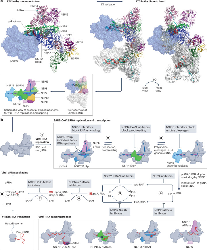
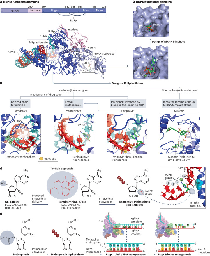
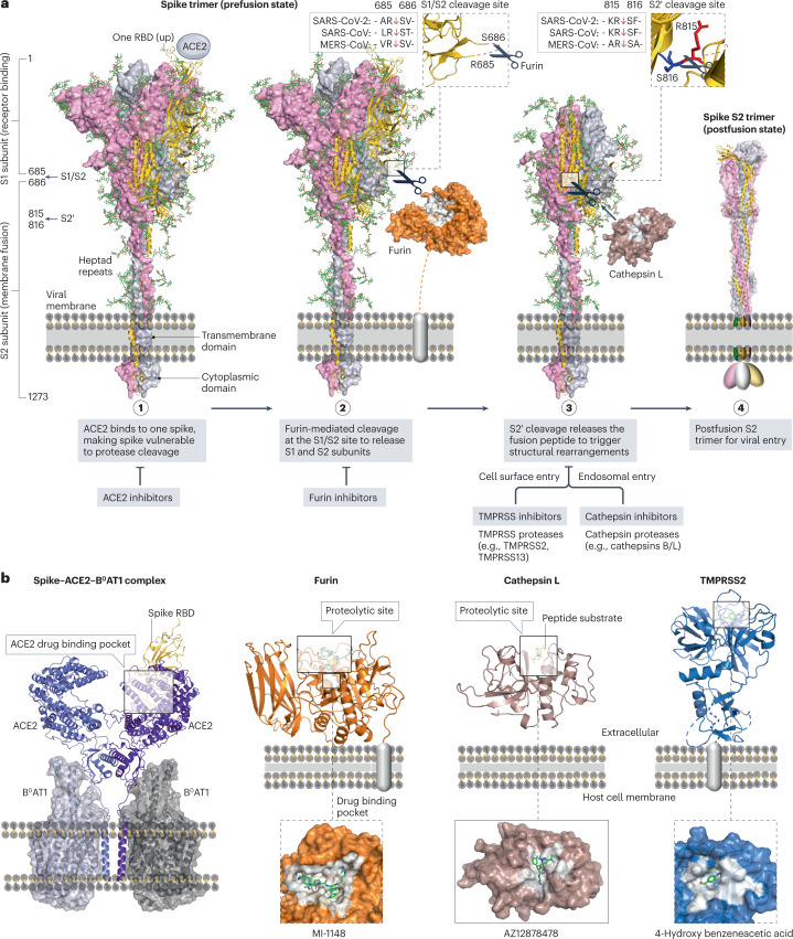
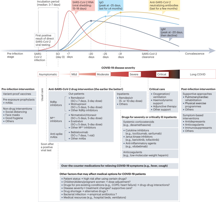
The coronavirus disease 2019 (COVID-19) pandemic has stimulated tremendous efforts to develop therapeutic strategies that target severe acute respiratory syndrome coronavirus 2 (SARS-CoV-2) and/or human proteins to control viral infection, encompassing hundreds of potential drugs and thousands of patients in clinical trials. So far, a few small-molecule antiviral drugs (nirmatrelvir-ritonavir, remdesivir and molnupiravir) and 11 monoclonal antibodies have been marketed for the treatment of COVID-19, mostly requiring administration within 10 days of symptom onset. In addition, hospitalized patients with severe or critical COVID-19 may benefit from treatment with previously approved immunomodulatory drugs, including glucocorticoids such as dexamethasone, cytokine antagonists such as tocilizumab and Janus kinase inhibitors such as baricitinib. Here, we summarize progress with COVID-19 drug discovery, based on accumulated findings since the pandemic began and a comprehensive list of clinical and preclinical inhibitors with anti-coronavirus activities. We also discuss the lessons learned from COVID-19 and other infectious diseases with regard to drug repurposing strategies, pan-coronavirus drug targets, in vitro assays and animal models, and platform trial design for the development of therapeutics to tackle COVID-19, long COVID and pathogenic coronaviruses in future outbreaks.


Conflict of interest statement

R.H. filed a patent application covering α-ketoamide Mpro inhibitors of the 13b family. He discloses a research collaboration with Atea Pharmaceuticals. Other authors declare no competing interests.

Figures

Fig. 1
Fig. 1. The SARS-CoV-2 RNA genome.
Antiviral drug targets are indicated beneath the genome map. Accessory proteins are not mapped. NiRAN, nidovirus RdRp-associated nucleotidyltransferase domain; NSP, non-structural protein; ORF, open reading frame; SARS-CoV-2, severe acute respiratory syndrome coronavirus 2.
Fig. 2
Fig. 2. The life cycle of SARS-CoV-2 and drug targets.
a, Entry of severe acute respiratory syndrome coronavirus 2 (SARS-CoV-2) into host cells. The viral spike protein binds to angiotensin-converting enzyme 2 (ACE2) (in complex with the sodium-dependent neutral-amino-acid transporter B0AT1) on the membrane surface. Spike is cleaved by furin at the S1/S2 cleavage site and subsequently cleaved at the S2′ site by transmembrane protease serine subfamily (TMPRSS) proteases in the cell surface entry pathway, or cathepsins in the endosomal entry pathway (see Fig. 7 for further details). b, Viral uncoating. The (+ss) genomic RNA is released from the viral particle into the host cell. The genomic RNA is translated into open reading frame 1a (ORF1a) and ORF1ab polyproteins (pp1a and pp1ab), which are subsequently cleaved by papain-like protease (PLpro) and the main protease (Mpro) to release 16 non-structural proteins (NSPs). c, Viral RNA synthesis. Several NSPs assemble into the replication–transcription complex (RTC) that replicates and translates viral genomic RNA in replication organelles. d, Viral mRNA translation. Structural proteins are sorted into the endoplasmic reticulum (ER) and Golgi apparatus for maturation. Accessory proteins modulate virus−host interactions and viral pathogenesis. e, Viral assembly. The genomic RNA is packed with viral nucleocapsid (N) for viral assembly, along with structural proteins. f, Viral release by exocytosis. g, Viral RNA triggers host immune signalling pathways, which involve activation of transcription factors to produce cytokines such as interleukin (IL)-6, chemokines such as C–C motif chemokine ligand 2 (CCL2) and C–X–C motif chemokine ligand 10 (CXCL10), and interferons such as interferon-α (IFNα). h, Excessive production and secretion may result in cytokine-induced damage, multiorgan failure, thrombosis or death (see reviews elsewhere,,). Immune cells also provide positive feedback to release more cytokines, chemokines and interferons. Ps in red circles denote phosphorylation sites. dsRNA, double-stranded RNA; E, envelope protein; IRF, interferon regulatory factor; ISG, interferon-stimulated gene; ISRE, interferon-stimulated response element; JAK, Janus kinase; M, membrane protein; NF-κB, nuclear factor-κB; ssRNA, single-stranded RNA; STAT, signal transducer and activator of transcription; UU...UU, polyuridines. The electron micrograph image of SARS-CoV-2 (contributed by C.S. Goldsmith and A. Tamin) was retrieved from the CDC Public Health Image Library.
Fig. 3
Fig. 3. Development of anti-spike monoclonal antibodies.
a, Screening of monoclonal antibodies (mAbs). Peripheral blood mononuclear cells (PBMCs) are collected from convalescent donors or humanized mice exposed to severe acute respiratory syndrome coronavirus 2 (SARS-CoV-2). Single-cell cultures of plasma or memory B cells are prepared to screen potent mAbs. b, Fragment crystallizable (Fc)-engineering approaches that modify amino acids in the Fc domain of mAbs to enhance the half-life and reduce the effector functions. Common modifications such as LALA, TM, LS, YTE and GAALIE are indicated, with their wild-type residues visualized using the structure of human IgG1 Fc (PDB: 4X4M), and their applications in selected mAbs are shown below. c, Structure of a mAb in complex with the prefusion spike trimer. Bamlanivimab (PDB: 7KMG) blocks the binding of angiotensin-converting enzyme 2 (ACE2) to the receptor-binding domain (RBD) of spike (PDB: 6M17). d, Four antibody classes can be defined based on their interactions with the spike RBD. Representative antibodies from each class are shown, including amubarvimab (class 1, PDB: 7CDI), bamlanivimab (class 2, PDB: 7KMG), S309 (class 3, PDB: 7TNO) and CR3022 (class 4, PDB: 6W41). e, Antibodies from multiple classes can be combined to inhibit SARS-CoV-2 variants. SARS-CoV-2 variants harbour amino acid substitutions in the RBD (for example, 15 mutations in the Omicron variant) that potentially hamper antibody potency. The timeline of the earliest documented samples provided by the WHO (see Related links) shows viral evolution from the SARS-CoV-2 original strain to more than ten variants to date, including Alpha (B.1.1.7), Beta (B.1.351), Gamma (P.1), Delta (B.1.617.2), Epsilon (B.1.427/B.1.429), Zeta (P.2), Eta (B.1.525), Theta (P.3), Iota (B.1.526), Kappa (B.1.617.1), Lambda (C.37), Mu (B.1.621) and Omicron (B.1.1.529). A three-antibody combination of Omi-18 from class 1, Omi-31 from class 2 and nanobody C1 from class 4 can simultaneously target one RBD of an Omicron variant (PDB: 7ZFB). Fab, fragment antigen binding.
Fig. 4
Fig. 4. Structure of the SARS-CoV-2 main protease and its drug-binding pocket.
a, The severe acute respiratory syndrome coronavirus 2 (SARS-CoV-2) main protease (Mpro) functional domains, protein structures and cleavage sequences. The Mpro homodimer is the active form (PDB: 7VU6). The cleavage sequence ‘AVLQ↓SGFR’ between adjacent NSP4 and NSP5 in the pp1a and pp1ab polyproteins is localized across the Mpro catalytic dyad formed by Cys145 and His41 (PDB: 7DVP). Mpro cleavage sequences from the reference genome are shown on the right. b, Mpro catalytic site in the pre-cleavage state with the NSP4−NSP5 cleavage sequence (PDB: 7DVP). Mpro inhibitors with P1′ warhead, P1, P2, P3 and P4 moieties can be developed to maximize the drug–receptor interactions at the S1′, S1, S2, S3 and S4 subsites of Mpro, respectively. c, Four classes of Mpro inhibitors and the drug-binding pockets of nirmatrelvir (PDB: 7VH8), 14c (PDB: 7T4B), ensitrelvir (PDB: 7VU6) and x1187 (PDB: 5RFA). Reversible covalent inhibition of nirmatrelvir via the catalytic dyad Cys145−His41 is also shown. The drug-binding pocket of x1187 is captured at the dimer interface (see panel a). d, Development of nirmatrelvir from PF-00835231 (ref. ). Nirmatrelvir and boceprevir share identical structures at the backbone and P2/P3 moieties. The half-maximal effective concentration (EC50) values of PF-00835231, nirmatrelvir and boceprevir against SARS-CoV-2 USA_WA1/2020 and oral bioavailability (oral F) in rats were obtained from the literature,.
Fig. 5
Fig. 5. The SARS-CoV-2 replication–transcription complex and its drug targets.
a, Model of the severe acute respiratory virus syndrome coronavirus 2 (SARS-CoV-2) replication–transcription complex (RTC) in complex with the product RNA (p-RNA) and template RNA (t-RNA), based on the superimposition of protein structures from the PDB codes 7EGQ, 7JYY, 7RDY and 7TQV. The exact structure of the SARS-CoV-2 RTC is yet to be discovered. The monomer is shown on the left, and two views of the active dimeric RTC are shown on the right. A schematic view of the essential RTC components is shown below. b, RTC activities and mechanisms of action of drugs that target it. Step 1: the RTC initiates viral RNA replication after unwinding the viral genomic RNA (gRNA). Step 2: non-structural protein 12 (NSP12) RNA-dependent RNA polymerase (RdRp) and NSP14 exoribonuclease (ExoN) mediate RNA synthesis and proofreading, respectively. Step 3: NSP15 cleaves uridines in the viral single-stranded RNA/double-stranded RNA (ssRNA/dsRNA), especially the long polyuridine tracts at the 5′-end of negative gRNA, to avoid host immune defences. The products of ss gRNA and mRNA undergo a four-step process (steps 4–8) to complete viral RNA capping for immune evasion,. Step 4: NSP13 ATPase hydrolyses and releases the γ-phosphate of the 5′-triphosphate of viral RNA. An alternative pathway is mediated by the RNAylated NSP9 (refs. ,). Step 5: NSP12 nidovirus RdRp-associated nucleotidyltransferase domain (NiRAN) transfers a covalently linked guanosine 5′-monophosphate to the 5′-diphosphate end of viral RNA. Step 6: the NSP14 guanine-N7-methyltransferase (N7-MTase) domain uses S-adenosylmethionine (SAM) as the methyl donor to produce the intermediate Cap-0-RNA structure (m7GpppA1-RNA). Step 7: NSP16 2′-O-methyltransferase (2′-O-MTase) uses SAM as the methyl donor to produce the cap-1-RNA structure (m7GpppA1m-RNA). Step 8: the capped RNA genome is translocated for viral packaging, while the other capped mRNAs are translocated to host ribosomes for translation. Schematic RTC models involved in the steps of the viral RNA capping process are shown at the bottom of the figure.
Fig. 6
Fig. 6. NSP12 protein structure and its drug-binding pockets.
a, Non-structural protein 12 (NSP12) functional domains (PDB: 7EIZ). NSP12 RNA-dependent RNA polymerase (RdRp) and nidovirus RdRp-associated nucleotidyltransferase domain (NiRAN) catalytic sites are highlighted with the template RNA (cyan) and product RNA (red). b, Drug-binding pocket at the catalytic site of the NiRAN domain. ADP (PDB: 7RDY) binds to the NiRAN active site. AT-9010 is the active 5′-triphosphate form of bemnifosbuvir that blocks the NiRAN active site (PDB: 7ED5). c, RdRp inhibitors include nucleos(t)ide analogues such as remdesivir triphosphate (PDB: 7B3C), molnupiravir triphosphate (PDB: 7OZU), favipiravir ribonucleoside triphosphate (PDB: 7AAP) and non-nucleos(t)ide analogues such as suramin (PDB: 7D4F). d, Development of remdesivir from GS-441524 using the ProTide approach. The post-translocation positions (+1, −1 to −4) of the nascent base pairs in the product viral RNA (p-RNA) are indicated, and the substrate active site of natural nucleoside triphosphates (NTPs) is located at the −1 position. Remdesivir triphosphate acts as a delayed chain terminator, and its cyano group clashes with S861 of RdRp to stall RNA synthesis. In vitro half-maximal effective concentration (EC50) and half-life values were obtained from the literature,. e, Lethal mutagenesis caused by molnupiravir, which is converted intracellularly into an active triphosphate form (EIDD-2061) that increases the frequency of G-to-A and C-to-U transition errors within genomic RNA (gRNA). RTC, replication–transcription complex.
Fig. 7
Fig. 7. Interactions between spike, ACE2 and cellular proteases as drug targets.
a, Structural rearrangements of the severe acute respiratory syndrome coronavirus 2 (SARS-CoV-2) spike trimer from the prefusion state (PDB: 7WEA) to the postfusion state (PDB: 6XRA). Viral entry is initiated via the binding of one prefusion spike with angiotensin-converting enzyme 2 (ACE2), rendering the spike cleavage sites vulnerable to host proteases. The SARS-CoV-2 spike undergoes extensive conformational changes from the closed (down) prefusion state to the open (up) fusion-prone state. Subsequently, furin and cellular proteases (such as cathepsins B/L and transmembrane protease serine subfamily (TMPRSS) 2/13) cleave the S1/S2 site and the S2′ site of the spike, respectively. b, Drug-binding pockets within the spike–ACE2–B0AT1 complex (PDB: 6M17), furin in complex with MI-1148 (PDB: 4RYD), cathepsin L in complex with the Gln-Leu-Ala peptide substrate (PDB: 3K24) and AZ12878478 (PDB: 3HHA), and TMPRSS2 in complex with 4-hydroxy benzeneacetic acid (PDB: 7MEQ), which is the active metabolite of camostat mesylate.
Fig. 8
Fig. 8. Therapeutic strategies for COVID-19 and future coronavirus outbreaks.
a, Therapeutic interventions at various stages of coronavirus disease 2019 (COVID-19). At the pre-infection stage, variant-proof vaccines, pre-exposure prophylaxis and nonpharmaceutical interventions can be considered. Once severe acute respiratory syndrome coronavirus 2 (SARS-CoV-2) infection is diagnosed, anti-SARS-CoV-2 therapies are ideally administered to outpatients as soon as possible so that the viral load is significantly reduced at an early stage. SARS-CoV-2 viral load usually peaks within the first week after symptom onset (SO) and SARS-CoV-2 RNA shedding in the upper respiratory tract has a mean duration of approximately 17 days. Levels of anti-SARS-CoV-2 immunoglobulin M (IgM), IgG and neutralizing antibodies peak at approximately 20, 25 and 31 days, respectively. At the advanced stage of COVID-19 progression, immunomodulators, anticoagulants, anti-inflammatory drugs and/or critical care can be considered for severely or critically ill inpatients under certain conditions (Box 1). Post-infection interventions might be needed for some survivors experiencing persistent symptoms after COVID-19 infection. COPD, chronic obstructive pulmonary disease; mAbs, monoclonal antibodies; Mpro, main protease; RdRp, RNA-dependent RNA polymerase.
Fig. 9
Fig. 9. Strategies to combat coronavirus outbreaks.
The early stage of a potential or declared outbreak requires immediate nonpharmaceutical interventions to prevent pathogen spread. Genomic sequencing can be used to identify coronavirus strains, which guide the clinical use and/or development of vaccines and antiviral drugs. Steps involved in antiviral development are shown, which typically progress from target and hit identification to preclinical development, clinical evaluation and drug authorization and marketing. ACE2, angiotensin-converting enzyme 2; APN, aminopeptidase N; CC50, half-maximal cytotoxic concentration; DPP4, dipeptidyl peptidase 4; EC50, half-maximal effective concentration; HCoV, human coronavirus; IC50, half-maximal inhibitory concentration; ICU, intensive care unit; RCT, randomized control trial.

Similar articles

Cited by

References

    1. Holmes EC, et al. The origins of SARS-CoV-2: a critical review. Cell. 2021;184:4848–4856. doi: 10.1016/j.cell.2021.08.017. - DOI - PMC - PubMed
    1. Zhu N, et al. A novel coronavirus from patients with pneumonia in China, 2019. N. Engl. J. Med. 2020;382:727–733. doi: 10.1056/NEJMoa2001017. - DOI - PMC - PubMed
    1. Dolgin E. Pan-coronavirus vaccine pipeline takes form. Nat. Rev. Drug Discov. 2022;21:324–326. doi: 10.1038/d41573-022-00074-6. - DOI - PubMed
    1. Chaudhary N, Weissman D, Whitehead KA. mRNA vaccines for infectious diseases: principles, delivery and clinical translation. Nat. Rev. Drug Discov. 2021;20:817–838. doi: 10.1038/s41573-021-00283-5. - DOI - PMC - PubMed
    1. De Clercq E, Li G. Approved antiviral drugs over the past 50 years. Clin. Microbiol. Rev. 2016;29:695–747. doi: 10.1128/CMR.00102-15. - DOI - PMC - PubMed

Publication types

Substances