. 2023 Apr;616(7958):843-848.
doi: 10.1038/s41586-023-05963-3. Epub 2023 Apr 19.

The Smc5/6 complex is a DNA loop-extruding motor

Affiliations

The Smc5/6 complex is a DNA loop-extruding motor

Biswajit Pradhan et al. Nature. 2023 Apr.

Abstract

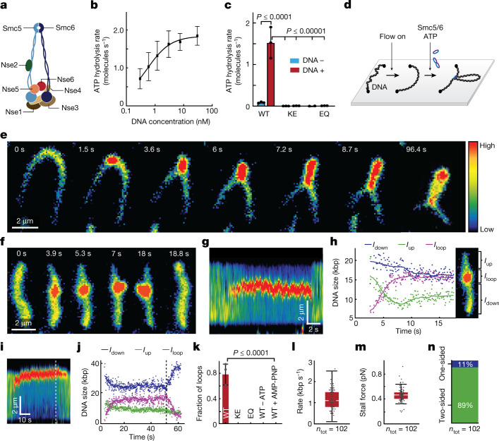
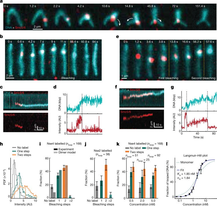
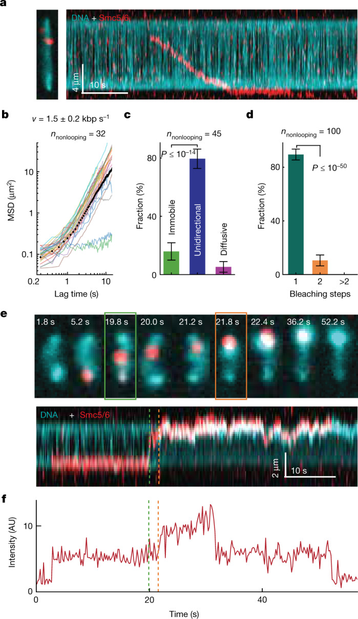
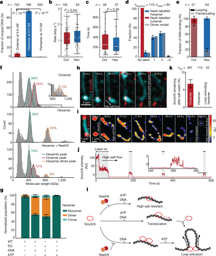
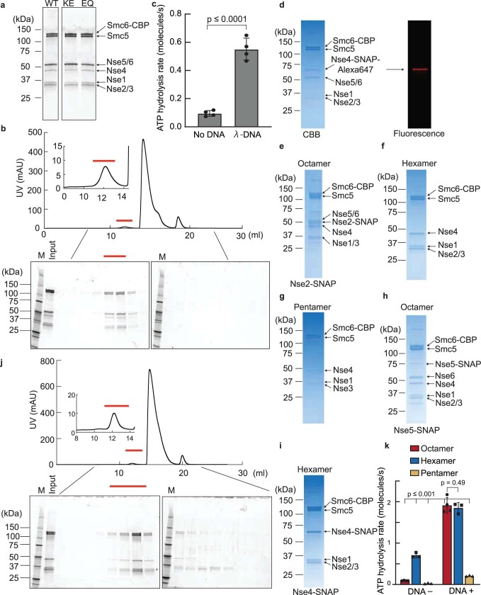
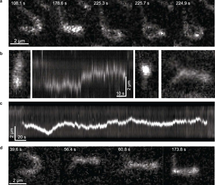
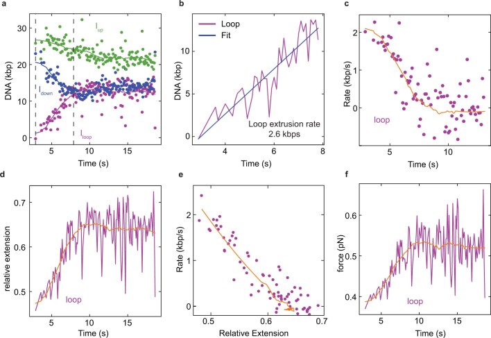
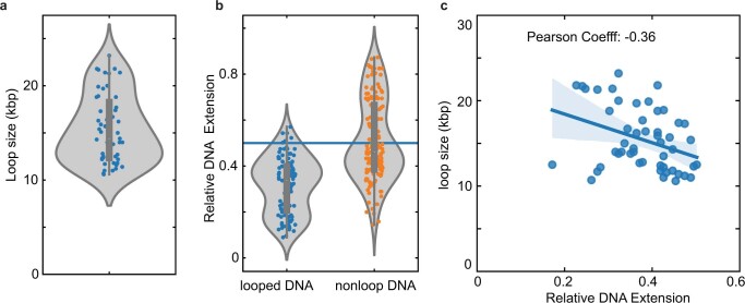
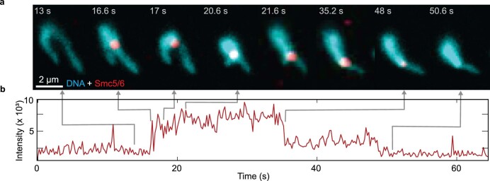
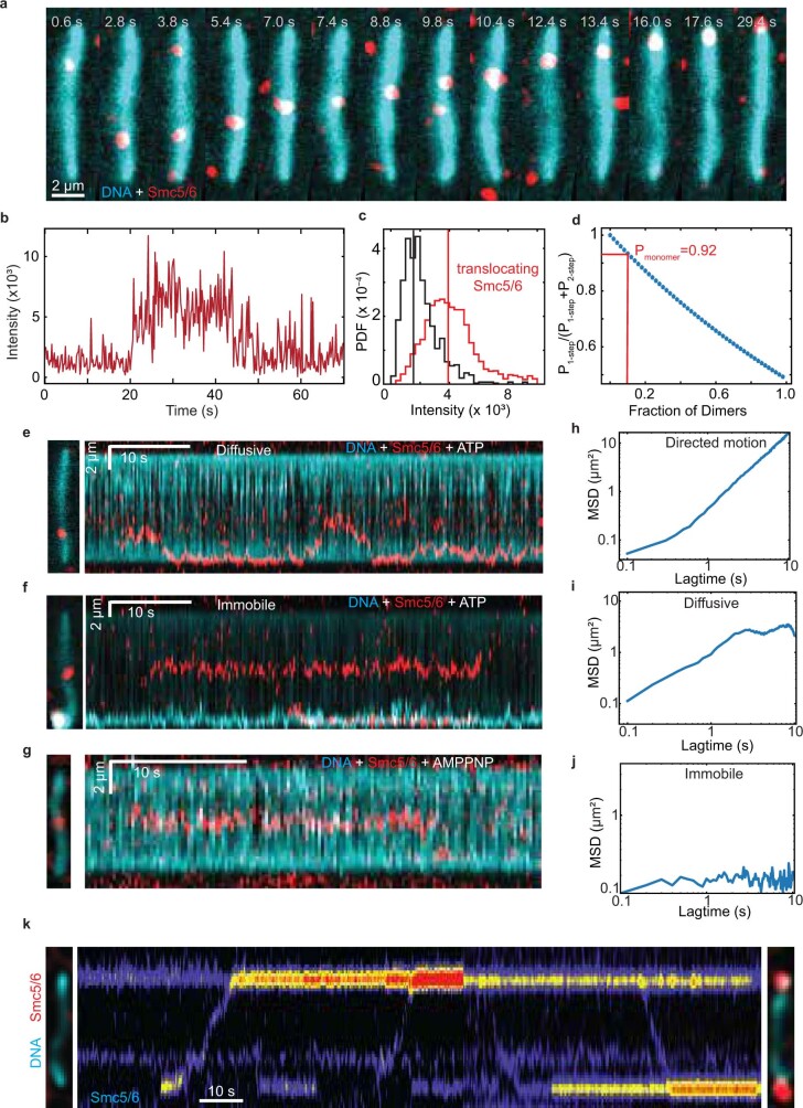
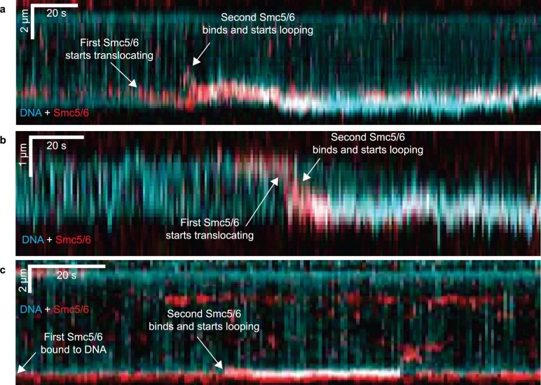
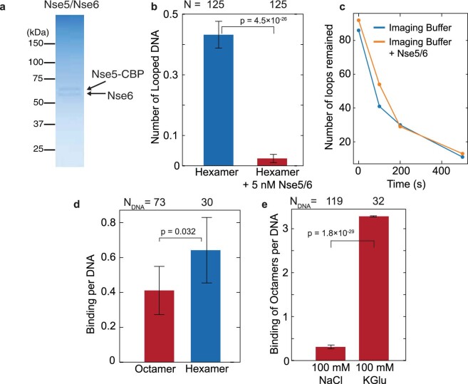
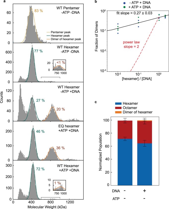
Structural maintenance of chromosomes (SMC) protein complexes are essential for the spatial organization of chromosomes1. Whereas cohesin and condensin organize chromosomes by extrusion of DNA loops, the molecular functions of the third eukaryotic SMC complex, Smc5/6, remain largely unknown2. Using single-molecule imaging, we show that Smc5/6 forms DNA loops by extrusion. Upon ATP hydrolysis, Smc5/6 reels DNA symmetrically into loops at a force-dependent rate of one kilobase pair per second. Smc5/6 extrudes loops in the form of dimers, whereas monomeric Smc5/6 unidirectionally translocates along DNA. We also find that the subunits Nse5 and Nse6 (Nse5/6) act as negative regulators of loop extrusion. Nse5/6 inhibits loop-extrusion initiation by hindering Smc5/6 dimerization but has no influence on ongoing loop extrusion. Our findings reveal functions of Smc5/6 at the molecular level and establish DNA loop extrusion as a conserved mechanism among eukaryotic SMC complexes.


Conflict of interest statement

The authors declare no competing interests.

Figures

Fig. 1
Fig. 1. Real-time imaging of loop extrusion by Smc5/6.
a, Cartoon of the S. cerevisiae Smc5/6 octameric structure. b, ATPase activity of the WT Smc5/6 octameric complex with different concentrations of DNA. Experimental data were fitted to a stimulatory dose–response model by nonlinear regression; mean ± s.d. from four independent measurements. c, ATPase activity of WT, KE and EQ Smc5/6 complexes in the absence or presence of 30 nM plasmid DNA; mean ± s.d. from three independent measurements. d, Schematic of DNA loop-extrusion assay. e, Series of images showing DNA loop-extrusion intermediates induced by Smc5/6 complex under constant buffer flow. f,g, Images (f) and fluorescence intensity kymograph (g) of a DNA molecule showing DNA loop extrusion in the absence of buffer flow. h, DNA lengths calculated from the kymograph in g for regions outside the loop (Iup and Idown) and the loop region itself (Iloop). i,j, Kymograph (i) and calculated DNA lengths (j) for a loop-extrusion event followed by loop release via gradual shrinkage. Dashed lines in i,j indicate the start of loop shrinkage. Data in ej represent typical events observed more than ten times in three independent experiments. k, DNA loop-forming fractions (mean ± s.d.) in the presence of ATP and 2 nM Smc5/6 WT, ATPase mutant complexes as in c and WT in the absence of ATP or presence of AMP-PNP. ntot = 233, 121, 93, 84 and 106, respectively. l,m, Box-and-whisker plots of Smc5/6 loop extrusion showing rates (l) and stalling force (m). ntot = 102 molecules, median ± 1.5× interquartile rate (IQR). n, Fraction of loop-extrusion events exhibiting two- or one-sided DNA reeling, as determined by observation of DNA length decrease in nonloop regions (Iup and Idown in h,j). Data in kn are from three independent experiments. Source data
Fig. 2
Fig. 2. Dimers of Smc5/6 complexes extrude DNA loops.
a,b,e, Snapshots of image overlays showing SxO-stained DNA (cyan) and Alexa 647-labelled Smc5/6 (red) during loop extrusion in the presence (a) and absence of buffer flow (b,e), and exhibiting one (b) or two photobleaching events (e). Arrows in a indicate the direction of Smc5/6 movement. c,f, Kymographs of the loop-extrusion events in b (c) and e (f) depicting overlays of DNA and Smc5/6 (top) and Smc5/6 (bottom). d,g, Time traces of DNA length (top) and Smc5/6 fluorescence intensity (bottom) determined from c (d) and f (g), with bleaching events indicated by dashed vertical lines. h, Probability density function (PDF) of fluorescence intensity for loop-extrusion events exhibiting either no (ntot = 8) Alexa 647 signal or one-(ntot = 11) or two-step (ntot = 21) bleaching. i, Fraction of loop-extruding Smc5/6 events that showed either none, one, two or more bleaching steps. Dashed bars denote the calculated probabilities for finding none, one or two labels assuming that all loop-extruding complexes are dimers with labelling efficiency of 68%. j, Fraction of the number of bleaching steps for Nse2-labelled Smc5/6 during loop extrusion with labelling efficiency of 70%. Data in ej represent five or more independent experiments. k, Histograms showing the number of bleaching steps observed during loop-extrusion events at indicated Smc5/6 concentrations. Data in ik indicate respective fractions of total looping events (nloop) with 95% confidence interval from at least three independent experiments. l, Langmuir–Hill plot showing the fraction of DNA substrates that formed loops as a function of Smc5/6 concentration (solid squares); mean ± s.d. from three independent experiments. The respective fit (solid line) indicates cooperative behaviour with Hill coefficient (nH) = 1.84, deviating from the Hill–Langmuir function expected for exclusively monomeric loop extrusion (nH = 1, dotted line). Experiments were performed using the WT octameric complex and at 1,000 s duration. AU, arbitrary units.
Fig. 3
Fig. 3. Single Smc5/6 complexes unidirectionally translocate along DNA.
a, Overlaid snapshots (left) and kymograph (right) showing an example of a labelled Smc5/6 complex translocating on a DNA molecule. Representative of five independent experiments. b, MSD plots determined from kymograph trajectories of individual Smc5/6 nonlooping events (solid lines). c, Fractions of nonlooping Smc5/6 complexes that remained immobile or translocated directional or diffusive on DNA before dissociating from the DNA. d, Fractions of nonlooping Smc5/6 complexes that exhibited either one (green), two (orange) or more bleaching steps. Error bars in c,d indicate 95% confidence interval. Data in bd were obtained from the respective nnonlooping events over five independent measurements. e,f Snapshots (top) and kymographs (bottom) of Smc5/6 and DNA (e) and the corresponding time trace of Smc5/6 label intensity (f), showing an event where a translocating Smc5/6 started to extrude a DNA loop upon forming a dimer with another Smc5/6 from the solution. The dashed lines in e,f indicate the start of the Smc5/6 translocation (green) and the binding of the additional Smc5/6 (orange). Representative of three independent measurements.
Fig. 4
Fig. 4. Nse5/6 downregulates loop extrusion by inhibiting dimerization of Smc5/6.
a, Fractions of DNA molecules that formed loops following the addition of octameric (+Nse5/6), hexameric (–Nse5/6) or pentameric (–Nse2/5/6) complexes. b,c, Loop-extrusion rates (b) and loop dwell times (c) for octameric and hexameric complexes; median ± 1.5× IQR. d, Fraction of loop-extruding complexes with no label or one, two or more than two bleaching steps for Nse4-labelled hexamers and Nse5-labelled octamers. e, Fractions of translocating and looping events per DNA binding for octamers and hexamers. P ≤ 10–28, two-sided binomial test. a,d,e, Error bars indicate 95% confidence interval. Data in ae are from indicated (n) events collected from more than three independent measurements. f, Histograms of mass distribution for octamer (top), hexamer (middle) and hexamer with additional Nse5/6 in a 1:1 ratio (bottom), measured in the presence of DNA without ATP. Peaks correspond to the molecular weights of hexamer (green), octamer (red) and dimer of hexamer (orange). Inset: zoomed-in peak centred at around 880 kDa. g, Fraction of monomers, dimers and trimers of WT and EQ mutant Smc5/6 observed under the indicated conditions, obtained from mass photometry; mean ± s.d. from three independent experiments. h, Snapshots of a loop (cyan) extruded by hexamers (red) on high-salt buffer flow showing an example of high-salt-induced loop disruption and subsequent protein dissociation. i, Snapshots of nonlooping Smc5/6 under high-salt buffer flow, showing that previously DNA-end accumulated complexes (16s) became redistributed along the DNA and subsequently dissociated (17s–353.6s), while a few molecules remained bound (562.6s). j, Time trace of fluorescence intensities from Nse2-labelled octamers during high-salt wash in i. Inset: zoomed-in trace towards the end of the high-salt wash. The laser was irradiated for short intervals (shaded area) to minimize photobleaching. Data in hj representative of three independent experiments. k, Fractions of labelled octamers and hexamers remaining bound on DNA after high-salt wash; mean ± s.d. from three independent experiments. ntot, number of Smc5/6 before salt wash. l, Model of Smc5/6-mediated loop extrusion. Source data
Extended Data Fig. 1
Extended Data Fig. 1. Purifications and ATPase activity of octameric and hexameric Smc5/6 complexes.
(a) Oriole stained SDS-PAGE gel for purified Smc5/6 and mutants. (b) Size-exclusion chromatography (SEC) of Smc5/6 octamers. The chromatogram and Oriole-stained SDS-PAGE for each fraction are shown. Peak fractions are indicated with red bar. (c) ATP hydrolysis rate for wild type Smc5/6 with/without λ-DNA. Mean ± S.D. obtained from 4 independent measurements. (d) Fluorescently-labeled Smc5/6 with a SNAP tag on Nse4. CBB staining and fluorescence detection of SDS-PAGE are shown. (e,f,g,h,i) CBB stained SDS-PAGE for the Smc5/6 complex with a SNAP tag on Nse2 (e), Hexameric Smc5/6 complex lacking Nse5/6 (f), Pentameric Smc5/6 complex lacking Nse2/5/6 (g), Octameric Smc5/6 complex with a SNAP tag on Nse5 (h), Hexameric Smc5/6 complex lacking Nse5/6 with a SNAP tag on Nse4 (i). (j) SEC of the Smc5/6 hexamer. The chromatogram and Oriole-stained SDS-PAGE for each fraction are shown. Peak fractions are indicated with red bar. (k) ATP hydrolysis rates of octamer, hexamer, and pentamer in the absence and presence of 30 nM plasmid DNA. Mean ± S.D. obtained from 3 or 4 independent measurements. P values are shown; two-tailed Student’s t-test. Source data
Extended Data Fig. 2
Extended Data Fig. 2. Characteristics of the Smc5/6-mediated DNA loop extrusion.
(a) Snapshots showing the process of loop extrusion on a sticky surface under side flow revealing the O-shape topology of the DNA loop. (b) Snapshots (first and third panel from the left) from the first and the last frames of the kymograph (second panel) showing a loop extrusion event in the absence of flow. The fourth panel shows the same loop upon the application of side-flow confirming that the observed DNA punctum is a loop. (c) A kymograph of a DNA molecule showing a Smc5/6-mediated loop that exhibits diffusion along the DNA. (d) Snapshots showing diffusion of an extruded loop. The side-flow reveals that the stem of the loop moves along the DNA. (b,c,d) are representative of five independent experiments.
Extended Data Fig. 3
Extended Data Fig. 3. Quantification of Smc5/6-mediated loop extrusion kinetics extracted from a single looping event.
(a) Kinetics of loop extrusion showing changes in DNA sizes from different sections (Idown, Iloop, Iup) over time. The linear region within the two dashed lines was used to obtain the initial loop extrusion rate via linear fit as shown in (b). (c) The change of loop extrusion rate during the loop growth, which was calculated using the change in loop sizes in a moving time window of 2 s. (d) The simultaneous change in relative DNA extension as a function of time. (c,d) shows that the decrease of extrusion rate is correlated with the increase in DNA tension. (e) Scatter plot of loop extrusion rate with the relative extension, taken from (c,d), showing that above extension value of 0.6 the rate is close to zero. (f) Change in tension on the DNA with time. The force values were extracted from the values of relative DNA extension in (d) and converted to the known force–extension relation. (a–f) correspond to the single DNA loop extrusion event shown in Fig. 1f–h, and solid lines in a, c–f show running averages over 50 points. (a,b,c,d,e,f) are representative of five independent experiments.
Extended Data Fig. 4
Extended Data Fig. 4. Statistics of the characteristics of Smc5/6-extruded DNA loops.
(a) Loop size distribution (violin plot) from DNA looping events (Nloop = 100 molecules). (b) Violin plot distributions of the initial relative extension of DNA which formed loops (blue, Ntot = 100 molecules) or not (orange, Ntot = 140 molecules). Extension values were collected before the loop extrusion events started. Only 6% of the loop extrusion events (N = 100) were observed on DNA molecules which were stretched beyond an extension value of 0.6 (indicated by the horizontal line). (c) Scatter plot of loop sizes versus the relative DNA extension showing a negative correlation with Pearson coefficient of −0.36, and linear regression fit with 95 % confidence interval indicated by the shaded area. (a–c) are obtained over five independent experiments.
Extended Data Fig. 5
Extended Data Fig. 5. Additional examples of photobleaching events from Alexa-647 labeled Smc5/6 during DNA loop extrusion.
Kymograph (top of each section) of DNA (cyan) and Smc5/6 (red). Time traces of DNA (middle of each section) and Smc5/6 fluorescence intensity (bottom of each section) determined from the corresponding kymographs showing two bleaching steps (a,b), one bleaching step (c), and no Smc5/6 signal at the loop initiation position (d). The corresponding bleaching times Δτ21 and Δτ22 in (a, bottom), and Δτ11 in (b, bottom) are indicated with arrows. Intensity values displayed in Smc5/6 time-traces were extracted on the same pixel positions as the DNA puncta intensities. (e) The bleaching time distributions extracted from two bleaching step traces. (f) The bleaching time distributions Δτ11 in a one-step bleaching traces (blue). When the two bleaching times Δτ21, Δτ22 from the two-step bleaching trace are put together in a histogram, they resemble the single-step distribution. Note that the average time of one-step bleaching traces (68 s) is similar to the average of the times (Δτ21, Δτ22) of the two-step bleaching traces (90 s). (g, left panel) Fraction of loop extruding Smc5/6 events that displayed either no, one, two or more bleaching steps as shown in Fig. 2i. Error bars with 95% confidence interval. Nloop = 168 from more than 3 independent experiments. (g, right panel) The probabilities for finding either 0 (gray shaded area), 1 (green shaded area) or 2 (orange shaded area) labels as a function of Smc5/6 dimer over monomer ratio estimated on basis of the labeling efficiency of 68 ± 10%. The error bars indicate 95% confidence interval estimated using binomial proportion. Images and time traces in (a–d) are representative of five independent measurements. (e,f) are extracted from three independent experiments.
Extended Data Fig. 6
Extended Data Fig. 6. Two bleaching steps observed from labelled Smc5/6 at the stem of the DNA loop during loop extrusion.
(a) Snapshots of DNA loop extrusion event showing labeled Smc5/6 at the stem of the extruded loop. (b) the corresponding time trace of Smc5/6 fluorescence intensity showing two-step bleaching. The arrows indicate the time points corresponding to the respective snapshots. (a,b) are the representative of five independent experiments.
Extended Data Fig. 7
Extended Data Fig. 7. DNA Translocation by a single Smc5/6 complex.
(a) Snapshots of labelled Smc5/6 (red) on DNA (cyan), showing a DNA translocation event, corresponding to Fig. 3a. (b) Intensity time trace of Smc5/6 depicted in (a) showing one-step bleaching. (c) Probability density functions (PDF) of fluorescence intensities for from Alexa647 signal on labeled Smc5/6 (red) and the background signal (black). (d) Calculated propability of observing single bleaching steps as a function of dimer fraction given that we experimentally only observe one-step and two-step bleaching events. Red line shows the experimentally observed value shown in Fig. 3d, indicating that 90% of events are likely come from single Smc5/6 complexes (Pmonomer) (e,f) Example kymographs of Smc5/6 (red) on DNA (cyan) in the presence of ATP showing diffusive and immobile characteristics. (g) Example kymograph of Smc5/6 remaining bound to DNA in the presence of AMP-PNP. (h,i,j) Mean square displacements (MSD) of Smc5/6 complexes displaying directed motion (h) corresponding to Fig. 3a, diffusive motion (i) extracted from the kymograph shown in (e), and immobile behaviour (j) corresponding to the kymograph in (f). (k) Example kymograph of Nse2-labeled Smc5/6 showing binding and translocation events leading to accumulation at the end of λ-DNA which is tethered on the surface. The snapshots of DNA (cyan) and Smc5/6 (red) before (left side of the kymograph) and after (right side of the kymograph) incubation of Smc5/6 for 1 h. Data in (a,b,c,e-g) are the representative of more than three independent experiments.
Extended Data Fig. 8
Extended Data Fig. 8. Dimerization of Smc5/6 on DNA results in initiation of loop extrusion.
Example kymographs showing different types of events where loop extrusion (cyan) starts upon dimerization of Smc5/6 (red). (a) Two translocating Smc5/6 merge together and start loop extrusion. (b) A single Smc5/6 bound on the end of DNA starts translocating. A second Smc5/6 from the solution binds the first one and loop extrusion is initiated. (c) A single Smc5/6 is bound to one end of the DNA at the start of the measurement. Loop extrusion is initiated when a second Smc5/6 from the solution associates. Data in (a,b,c) are the representative of more than three independent experiments.
Extended Data Fig. 9
Extended Data Fig. 9. The effect of Nse5/6 on DNA loading, loop initiation and loop disruption.
(a) CBB-stained SDS-PAGE for the Nse5/6 subcomplex. (b) Fraction of DNA that formed loops in the absence (blue) and the presence of purified Nse5/6 subcomplexes (5 nM, red) while the concentration of hexameric Smc5/6 complexes was held constant (0.5 nM). Note that Nse5/6 was mixed with hexamers shortly before introducing to the flow cell. (c) Number of loops remaining at different time points upon introducing buffer with or without a 10-fold excess of Nse5/6. t = 0 indicates the start of the buffer flow. The data shows that there are no additional loop disruption events induced by the presence of Nse5/6. (d) Number of DNA binding events of labeled octameric or hexameric Smc5/6 complexes per DNA molecule and per 1000 s. (e) Comparison between the numbers of DNA binding events obtained for octameric complexes in imaging buffers containing 100 mM NaCl or 100 mM Potassium Glutamate (KGlu). The error bars in b,d,e indicate 95% confidence interval estimated using binomial proportion.
Extended Data Fig. 10
Extended Data Fig. 10. Characterization of Smc5/6 dimerization.
(a) Mass distributions of pentameric (1st in row) and hexameric Smc5/6 complexes in different conditions, including no ligand (2nd in row), in the presence of DNA (3rd), ATP hydrolysis-deficient (EQ) mutant with ATP and DNA (4th), hexameric Smc5/6 with ATP and DNA (5th). The identified peaks exhibit mass distributions close to the molecular weight of pentamer (beige), single hexamer (green) and dimer of hexamer (orange). The inset panels show the zoom-in peak corresponding to the dimer of hexamers. The total number of counts for the fitted areas are displayed in percentage value compared to all detected events throughout the measurement period. (b) Fraction of dimers of hexamer EQ mutant in the absence (blue) or presence (green) of ATP with different protein:DNA ratios (2:1, 1:1, 1:10, and 1:100). The fraction of dimers is quantified as the total number of Smc5/6 that formed dimer normalized by the total number of Smc5/6. The red line with slope = 2 demonstrates that the expected outcome of a scenario where the dimer fraction observed reflects two monomeric Smc5/6 complexes binding to the same DNA without protein-protein interaction. Experimental data were fitted to a two-sided linear least-squares regression. Fitted slope of 0.27 signifies the theoretical power-law is not obeyed and dimer formed by Smc5/6 hexamer is due to additional interaction between the complexes, regardless of the presence of ATP. (c) Comparison of oligomeric states of octameric Smc5/6 complex in the absence (mean ± S.D. from 4 independent experiments) or presence of DNA (mean ± S.D. from 3 independent experiments) without ATP.

Comment in

Similar articles

Cited by

References

    1. Davidson IF, Peters JM. Genome folding through loop extrusion by SMC complexes. Nat. Rev. Mol. Cell Biol. 2021;22:445–464. doi: 10.1038/s41580-021-00349-7. - DOI - PubMed
    1. Aragón L. The Smc5/6 complex: new and old functions of the enigmatic long-distance relative. Annu. Rev. Genet. 2018;52:89–107. doi: 10.1146/annurev-genet-120417-031353. - DOI - PubMed
    1. Hassler M, Shaltiel IA, Haering CH. Towards a unified model of SMC complex function. Curr. Biol. 2018;28:R1266–R1281. doi: 10.1016/j.cub.2018.08.034. - DOI - PMC - PubMed
    1. Rao SSP, et al. Cohesin loss eliminates all loop domains. Cell. 2017;171:305–320. doi: 10.1016/j.cell.2017.09.026. - DOI - PMC - PubMed
    1. Dixon JR, et al. Topological domains in mammalian genomes identified by analysis of chromatin interactions. Nature. 2012;485:376–380. doi: 10.1038/nature11082. - DOI - PMC - PubMed

Publication types