

Acute hydrocephalus caused by a colloid cyst - a case report
- PMID: 37076791
- PMCID: PMC10114300
- DOI: 10.1186/s12245-023-00500-5
Acute hydrocephalus caused by a colloid cyst - a case report
Abstract
Background: Colloid cysts are rare benign, slowly growing intracranial tumors of endodermal origin. Most colloid cysts are found incidentally and are asymptomatic, but rarely, they can lead to sudden death.
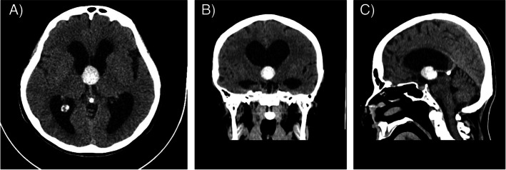
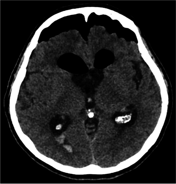
Case presentation: A 73-year-old female patient was admitted to our emergency department with complaints of dizziness, nausea, vomiting, fatigue, walking difficulties, and behavioral changes. CT imaging revealed acute obstructive hydrocephalus attributable to a third ventricular colloid cyst. The patient was immediately transferred to a tertiary center where she underwent successful neurosurgical resection of the mass. Pathology results of the lesion confirmed the diagnosis of colloid cyst.
Conclusion: The case we present emphasizes the critical importance of prompt identification of warning signs, complex thinking, and evaluation. Establishing the right diagnostic approach early on can facilitate accurate diagnosis.
Keywords: Acute hydrocephalus; Colloid cyst; Elevated intracranial pressure; Third ventricle.
© 2023. The Author(s).
Conflict of interest statement
The authors declare no competing interests.
Figures

Similar articles
-
Postexercise Death Due to Hemorrhagic Colloid Cyst of Third Ventricle: Case Report and Literature Review.World Neurosurg. 2019 Mar;123:351-356. doi: 10.1016/j.wneu.2018.12.057. Epub 2018 Dec 24. World Neurosurg. 2019. PMID: 30590211 Review.
-
Natural history of colloid cysts of the third ventricle.J Neurosurg. 2016 Dec;125(6):1420-1430. doi: 10.3171/2015.11.JNS151396. Epub 2016 Mar 11. J Neurosurg. 2016. PMID: 26967781
-
A 3rd ventricular colloid cyst causing acute hydrocephalus with stunned myocardium: A case report.Qatar Med J. 2020 Nov 12;2020(2):28. doi: 10.5339/qmj.2020.28. eCollection 2020. Qatar Med J. 2020. PMID: 33282712 Free PMC article.
-
Third Ventricle Cavernous Malformation and Obstructive Hydrocephalus Thought to Be a Colloid Cyst.World Neurosurg. 2021 Jan;145:315-319. doi: 10.1016/j.wneu.2020.09.136. Epub 2020 Oct 1. World Neurosurg. 2021. PMID: 33010503
-
Third-ventricle enterogenous cyst presentation mimicking a colloid cyst: uncommon presentation of a rare disease and literature review.Acta Neurochir (Wien). 2017 Mar;159(3):465-468. doi: 10.1007/s00701-016-3052-5. Epub 2016 Dec 16. Acta Neurochir (Wien). 2017. PMID: 27981391 Review.
Cited by
-
Acute hydrocephalus caused by colloid cyst of third ventricle: A case report.Radiol Case Rep. 2023 Aug 5;18(10):3662-3667. doi: 10.1016/j.radcr.2023.07.037. eCollection 2023 Oct. Radiol Case Rep. 2023. PMID: 37593333 Free PMC article.
-
Ominous Causes of Headache.Curr Pain Headache Rep. 2024 Mar;28(3):73-81. doi: 10.1007/s11916-023-01202-6. Epub 2023 Dec 13. Curr Pain Headache Rep. 2024. PMID: 38091239 Review.
-
Rapid Deterioration and Fatal Outcomes in Colloid Cyst-Induced Obstructive Hydrocephalus: A Case Report.Healthcare (Basel). 2024 Oct 29;12(21):2155. doi: 10.3390/healthcare12212155. Healthcare (Basel). 2024. PMID: 39517365 Free PMC article.
-
A quadrigeminal arachnoid cyst as a cause of neurological symptoms in an 11-month-old Brussels Griffon - A case study.Vet Med (Praha). 2023 Aug 31;68(8):343-348. doi: 10.17221/53/2023-VETMED. eCollection 2023 Aug. Vet Med (Praha). 2023. PMID: 37982125 Free PMC article.
References
-
- Schiff D, Hsu L, Wen P, Samuels M, Feske S. Office practice of neurology. 2003.
-
- Tenny S, Thorell W. StatPearls. Treasure Island (FL): StatPearls Publishing; 2022. Colloid brain cyst. - PubMed
LinkOut - more resources
Full Text Sources
