. 2023 Apr 19;20(1):12.
doi: 10.1186/s12989-023-00527-9.

Human dendritic cell maturation induced by amorphous silica nanoparticles is Syk-dependent and triggered by lipid raft aggregation

Affiliations

Human dendritic cell maturation induced by amorphous silica nanoparticles is Syk-dependent and triggered by lipid raft aggregation

Éléonore Guillet et al. Part Fibre Toxicol. .

Abstract

Background: Synthetic amorphous silica nanoparticles (SAS-NPs) are widely employed in pharmaceutics, cosmetics, food and concretes. Workers and the general population are exposed daily via diverse routes of exposure. SAS-NPs are generally recognized as safe (GRAS) by the Food and Drug Administration, but because of their nanoscale size and extensive uses, a better assessment of their immunotoxicity is required. In the presence of immune "danger signals", dendritic cells (DCs) undergo a maturation process resulting in their migration to regional lymph nodes where they activate naive T-cells. We have previously shown that fumed silica pyrogenic SAS-NPs promote the two first steps of the adaptative immune response by triggering DC maturation and T-lymphocyte response, suggesting that SAS-NPs could behave as immune "danger signals". The present work aims to identify the mechanism and the signalling pathways involved in DC phenotype modifications provoked by pyrogenic SAS-NPs. As a pivotal intracellular signalling molecule whose phosphorylation is associated with DC maturation, we hypothesized that Spleen tyrosine kinase (Syk) may play a central role in SAS-NPs-induced DC response.

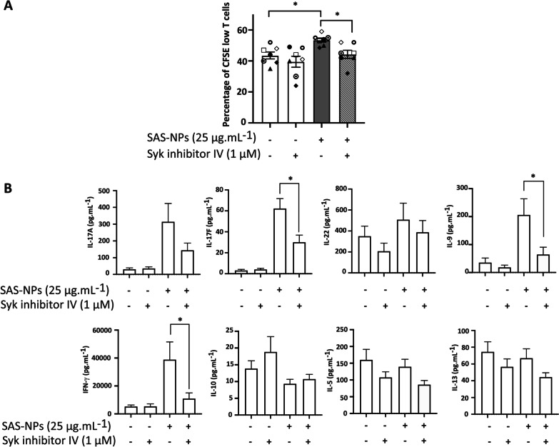
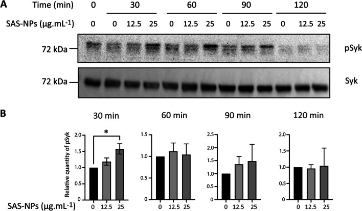
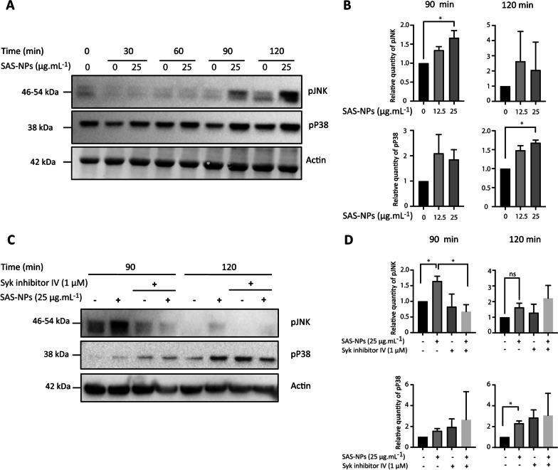
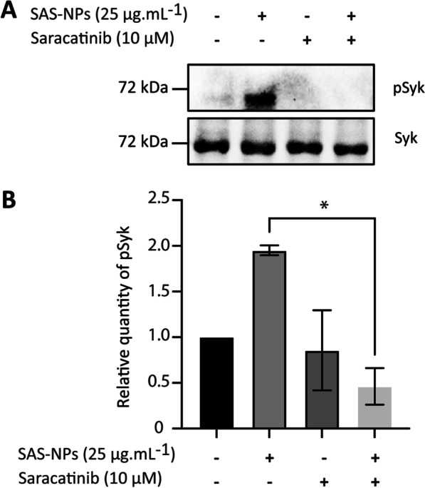
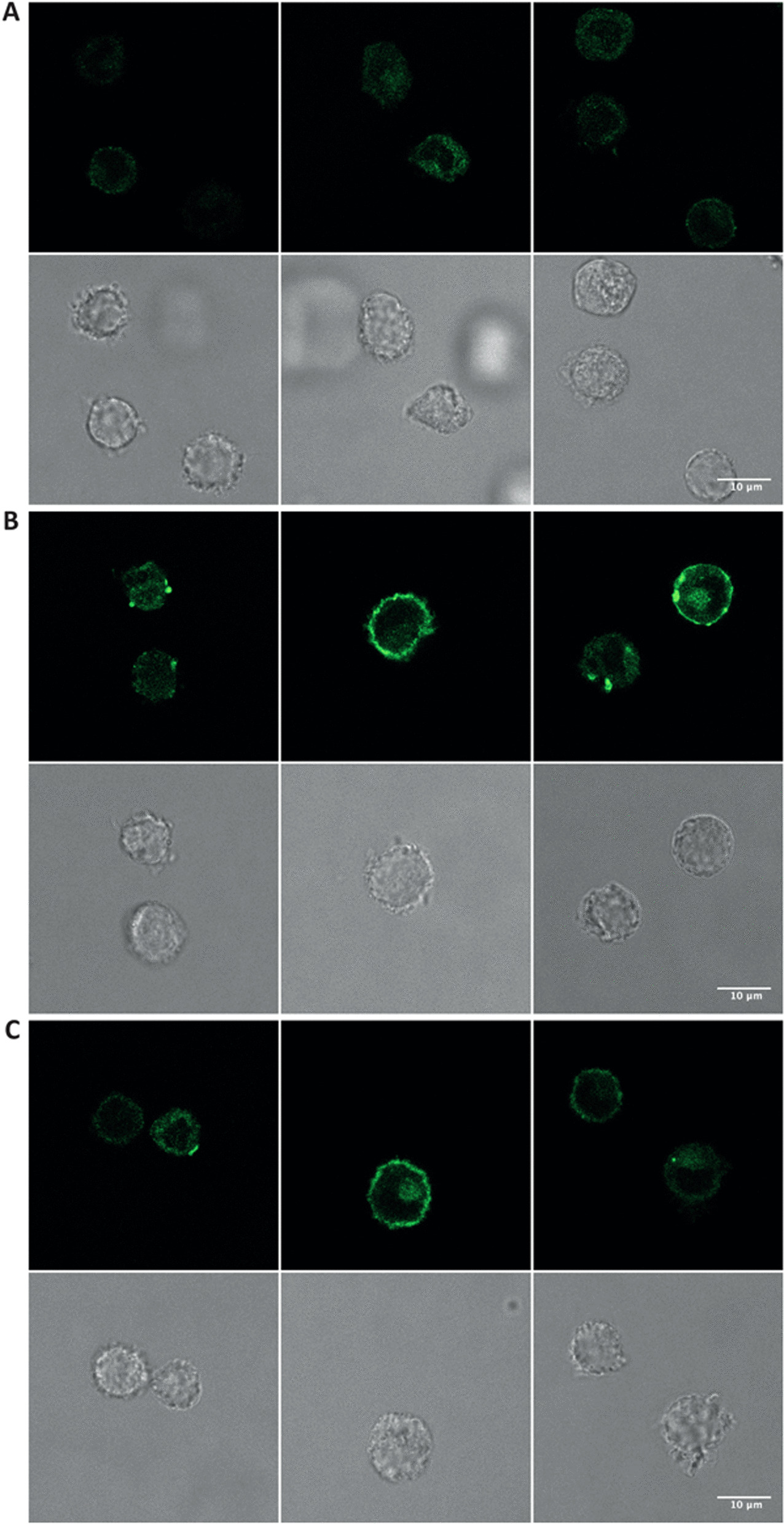
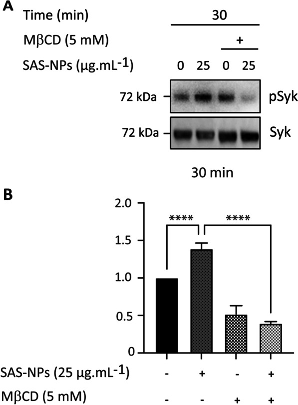
Results: In human monocyte-derived dendritic cells (moDCs) exposed to SAS-NPs, Syk inhibition prevented the induction of CD83 and CD86 marker expression. A significant decrease in T-cell proliferation and IFN-γ, IL-17F and IL-9 production was found in an allogeneic moDC:T-cell co-culture model. These results suggested that the activation of Syk was necessary for optimal co-stimulation of T-cells. Moreover, Syk phosphorylation, observed 30 min after SAS-NP exposure, occurred upstream of the c-Jun N-terminal kinase (JNK) Mitogen-activated protein kinases (MAPK) and was elicited by the Src family of protein tyrosine kinases. Our results also showed for the first time that SAS-NPs provoked aggregation of lipid rafts in moDCs and that MβCD-mediated raft destabilisation altered Syk activation.

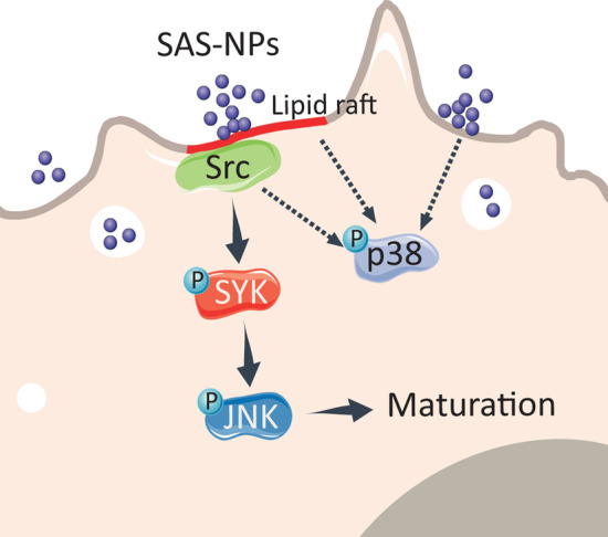
Conclusions: We showed that SAS-NPs could act as an immune danger signal in DCs through a Syk-dependent pathway. Our findings revealed an original mechanism whereby the interaction of SAS-NPs with DC membranes promoted aggregation of lipid rafts, leading to a Src kinase-initiated activation loop triggering Syk activation and functional DC maturation.

Keywords: Amorphous silica; Dendritic cells; Lipid rafts; Nanoparticles; Src kinases; Syk.


Conflict of interest statement

The authors declare that they have no competing interests.

Figures

Fig. 1
Fig. 1
Syk controls moDC phenotype in response to SAS-NPs. Immature moDCs were pre-treated for 1 h with Syk inhibitor IV (1 μM) and then treated for 16 h with SAS-NPs (12.5 or 25 μg.mL−1). Cells were then collected, washed, and analyzed by flow cytometry for surface marker expression. Results are expressed as relative fluorescence intensity (RFI) compared with non-stimulated cells and represented as the mean ± SEM of 3 independent experiments. *p < 0.05; **p < 0.01 One-way Anova, Kruskal–Wallis test
Fig. 2
Fig. 2
Syk inhibition alleviates allogeneic T-cell response to SAS-NP-treated moDCs. Immature moDCs were pre-treated for one hour with Syk inhibitor IV (1 μM) and then treated for 16 h with 25 µg.mL−1 of SAS-NPs. Treated moDCs were co-cultured with allogeneic CD4+ T-cells loaded with CFSE at a ratio of 1 moDC for 20 CD4+ T-cells. A Proliferation was measured after 5 days of co-culture as the percentage of CFSElow CD4+ T-cells. Untreated moDCs were used as control. B On day 5, cytokine levels were quantified in co-culture supernatants in duplicate, using an electroluminescent multiplex assay. Detection limits are indicated in the Methods section. Results for lymphocyte proliferation are expressed as percentage of CFSE low cells. Cytokine production is expressed in pg.mL−1. Both are represented as the mean ± SEM of 6 independent experiments. *p < 0.05 One-way Anova and Kruskal–Wallis test
Fig. 3
Fig. 3
Syk is phosphorylated in response to SAS-NPs. Immature moDCs were treated for 30, 60, 90 and 120 min with SAS-NPs (12.5 or 25 μg.mL−1). Immunoblotting of whole-cell extracts was used to quantify the phosphorylated form of Syk. A Representative experiment. B Results of 3 independent experiments are represented. Bands were quantified using the Image Lab software. Results are expressed as the fold induction, representing the ratio of the normalised intensity of specific bands of treated cells divided by the normalised intensity of bands of untreated cells (pSyk/Syk). *p < 0.05 One-way Anova, Kruskal–Wallis test
Fig. 4
Fig. 4
JNK and p38 MAPK are phosphorylated in response to SAS-NPs. A and B. Immature moDCs were treated for 30, 60, 90 and 120 min with SAS-NPs (12.5 or 25 μg.mL−1). A Representative experiment. B Immunoblotting of whole-cell extracts was used to quantify the phosphorylated forms of JNK and p38 kinases. C and D. Immature moDCs were pre-treated for 1 h with Syk inhibitor IV (1 μM) and then treated for 90 or 120 min with 25 µg.mL−1 of SAS-NPs. C Representative experiment. D Immunoblotting of whole-cell extracts was used to quantify the phosphorylated forms of JNK and p38. The results of 3 independent experiments are represented. Bands were quantified using the Image Lab software. Results are expressed as the fold induction, representing the ratio of the normalised intensity (pJNK/actin or pP38/actin) of specific bands of treated cells divided by the normalised intensity of bands of untreated cells, *p < 0.05 One-way Anova, Kruskal-Wallis test.
Fig. 5
Fig. 5
Src kinases are required to initiate Syk phosphorylation in response to SAS-NPs. Immature moDCs were pre-treated for 1 h with Saracatinib (10 µM) and then treated for 30 min with 25 µg.mL−1 of SAS-NPs. A Representative experiment. B Immunoblotting of whole-cell extracts was used to quantify the phosphorylated form of Syk. The results of 3 independent experiments are represented. Bands were quantified using the Image Lab software. Results are expressed as the fold induction, representing the ratio of the normalised intensity of specific bands of treated cells divided by the normalised intensity of bands of untreated cells, *p < 0.05 One-way Anova, Kruskal–Wallis test
Fig. 6
Fig. 6
SAS-NPs induce lipid raft aggregation in moDCs. Visualisation of lipid rafts by conventional fluorescence microscopy using cholera toxin subunit B conjugated with Alexa Fluor 488 which binds to the raft-associated monosialotetrahexosylganglioside (GM1). Immature moDCs were treated for 15 (B) or 30 min (C) with SAS-NPs (25 μg.mL−1) or left untreated (A) and then stained with cholera toxin subunit B conjugated with Alexa Fluor 488. The experiment was replicated twice. Left/middle/right panels: 3 different fields of the same experiment
Fig. 7
Fig. 7
Lipid rafts are necessary to activate Syk in response to SAS-NPs. Immature moDCs were pre-treated for 1 h with Methyl-ß-cyclodextrin (MßCD, 5 mM) and then treated for 30 min with 25 µg.mL−1 SAS-NPs. A Representative experiment. B Immunoblotting of whole-cell extracts was used to quantify the phosphorylated form of Syk. The results of 3 independent experiments are represented. Bands were quantified using the Image Lab software. Results are expressed as the fold induction, representing the ratio of the normalised intensity of specific bands of treated cells divided by the normalised intensity of bands of untreated cells, ****p < 0.0001 One-way ANOVA and Bonferroni’s multiple comparison test
Fig. 8
Fig. 8
Lipid raft aggregation following SAS-NP exposure leads to Src/Syk signalling pathway activation in human dendritic cells. Upon DC exposure to SAS-NPs, lipid raft aggregation leads to Src kinase recruitment required for the phosphorylation of the tyrosine kinase Syk. Syk phosphorylation is linked to JNK activation and DC maturation. p38 MAPK phosphorylation seems to be independent of Syk. Solid arrows: established links. Dashed arrows: hypotheses. The figure was partly generated using Servier Medical Art, provided by Servier, licensed under a Creative Commons Attribution 3.0 unported license (https://creativecommons.org/licenses/by/3.0/)

References

    1. Hochella MF, Mogk DW, Ranville J, Allen IC, Luther GW, Marr LC, et al. Natural, incidental, and engineered nanomaterials and their impacts on the Earth system. Science. 2019;363(6434):eaau8299. doi: 10.1126/science.aau8299. - DOI - PubMed
    1. Croissant JG, Butler KS, Zink JI, Brinker CJ. Synthetic amorphous silica nanoparticles: toxicity, biomedical and environmental implications. Nat Rev Mater. 2020;5(12):886–909. doi: 10.1038/s41578-020-0230-0. - DOI
    1. Zhang H, Dunphy DR, Jiang X, Meng H, Sun B, Tarn D, et al. Processing pathway dependence of amorphous silica nanoparticle toxicity: colloidal vs pyrolytic. J Am Chem Soc. 2012;134(38):15790–15804. doi: 10.1021/ja304907c. - DOI - PMC - PubMed
    1. Fruijtier-Pölloth C. The toxicological mode of action and the safety of synthetic amorphous silica-a nanostructured material. Toxicology. 2012;294(2–3):61–79. doi: 10.1016/j.tox.2012.02.001. - DOI - PubMed
    1. EFSA Panel on Food Additives and Nutrient Sources added to Food (ANS), Younes M, Aggett P, Aguilar F, Crebelli R, Dusemund B, et al. Re-evaluation of silicon dioxide (E 551) as a food additive. EFSA J. 2018;16(1):e05088. - PMC - PubMed

Publication types