. 2023 Apr 19;14(1):96.
doi: 10.1186/s13287-023-03318-3.

Serum- and xeno-free culture of human umbilical cord perivascular cells for pediatric heart valve tissue engineering

Affiliations

Serum- and xeno-free culture of human umbilical cord perivascular cells for pediatric heart valve tissue engineering

Shouka Parvin Nejad et al. Stem Cell Res Ther. .

Abstract

Background: Constructs currently used to repair or replace congenitally diseased pediatric heart valves lack a viable cell population capable of functional adaptation in situ, necessitating repeated surgical intervention. Heart valve tissue engineering (HVTE) can address these limitations by producing functional living tissue in vitro that holds the potential for somatic growth and remodelling upon implantation. However, clinical translation of HVTE strategies requires an appropriate source of autologous cells that can be non-invasively harvested from mesenchymal stem cell (MSC)-rich tissues and cultured under serum- and xeno-free conditions. To this end, we evaluated human umbilical cord perivascular cells (hUCPVCs) as a promising cell source for in vitro production of engineered heart valve tissue.

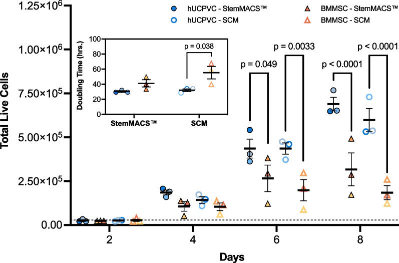
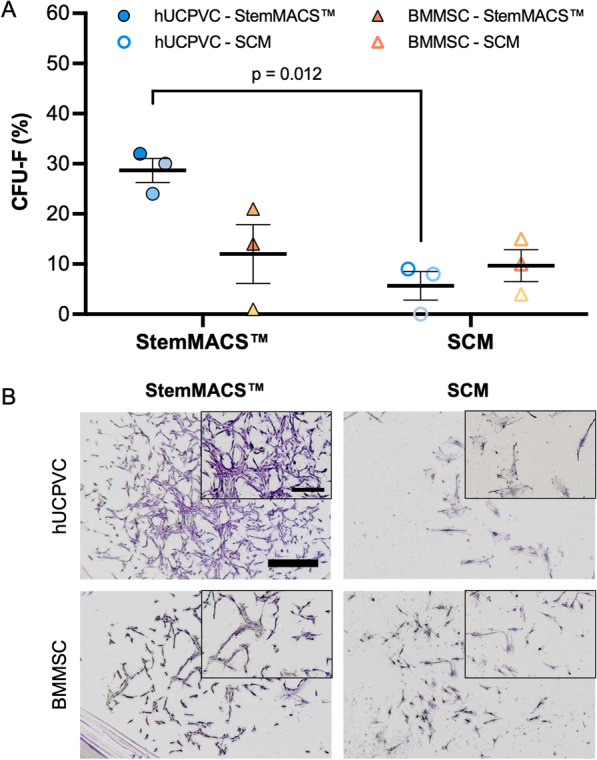
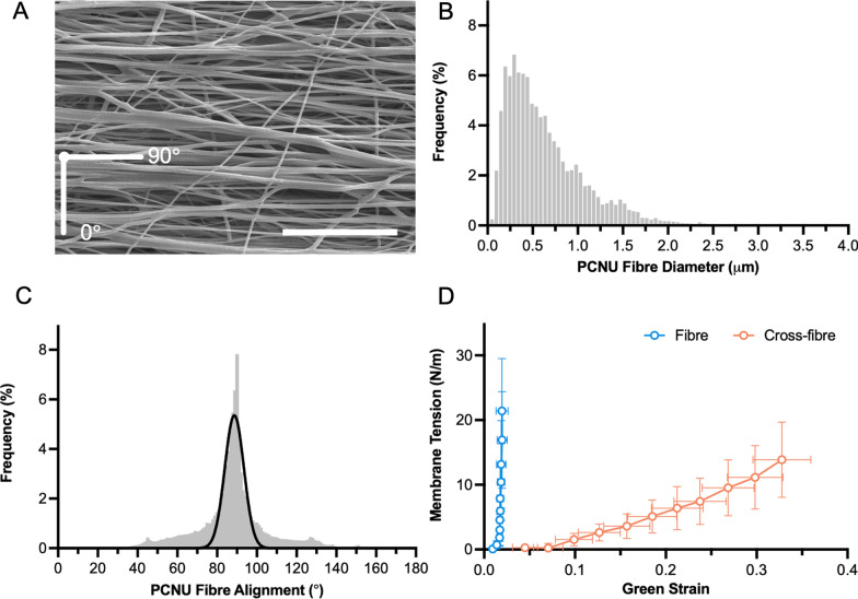
Methods: The proliferative, clonogenic, multilineage differentiation, and extracellular matrix (ECM) synthesis capacities of hUCPVCs were evaluated in a commercial serum- and xeno-free culture medium (StemMACS™) on tissue culture polystyrene and benchmarked to adult bone marrow-derived MSCs (BMMSCs). Additionally, the ECM synthesis potential of hUCPVCs was evaluated when cultured on polycarbonate polyurethane anisotropic electrospun scaffolds, a representative biomaterial for in vitro HVTE.

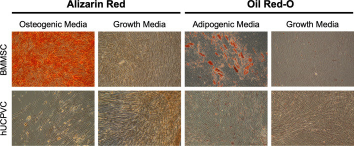
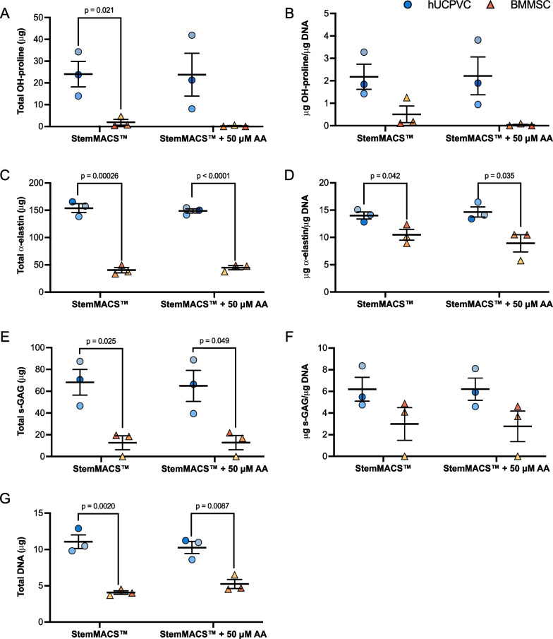
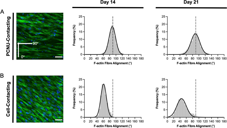
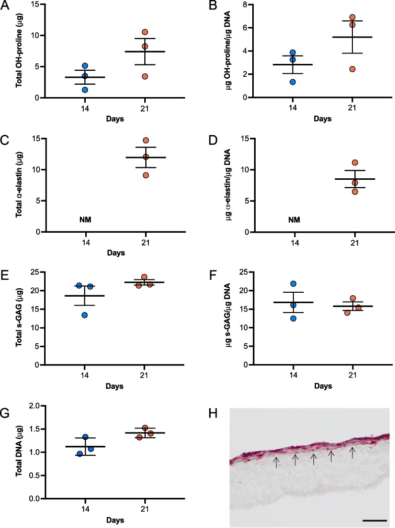
Results: hUCPVCs had greater proliferative and clonogenic potential than BMMSCs in StemMACS™ (p < 0.05), without differentiation to osteogenic and adipogenic phenotypes associated with valve pathology. Furthermore, hUCPVCs cultured with StemMACS™ on tissue culture plastic for 14 days synthesized significantly more total collagen, elastin, and sulphated glycosaminoglycans (p < 0.05), the ECM constituents of the native valve, than BMMSCs. Finally, hUCPVCs retained their ECM synthesizing capacity after 14 and 21 days in culture on anisotropic electrospun scaffolds.

Conclusion: Overall, our findings establish an in vitro culture platform that uses hUCPVCs as a readily-available and non-invasively sourced autologous cell population and a commercial serum- and xeno-free culture medium to increase the translational potential of future pediatric HVTE strategies. This study evaluated the proliferative, differentiation and extracellular matrix (ECM) synthesis capacities of human umbilical cord perivascular cells (hUCPVCs) when cultured in serum- and xeno-free media (SFM) against conventionally used bone marrow-derived MSCs (BMMSCs) and serum-containing media (SCM). Our findings support the use of hUCPVCs and SFM for in vitro heart valve tissue engineering (HVTE) of autologous pediatric valve tissue. Figure created with BioRender.com.

Keywords: Extracellular matrix; Heart valve tissue engineering; Human umbilical cord perivascular cells; Mesenchymal stromal cells; Serum- and xeno-free culture.


Conflict of interest statement

Dr. John E. Davies is the founding President and CEO of Tissue Regeneration Therapeutics, Inc. (Toronto, ON) who provided the human umbilical cord perivascular cells used in this study. The other authors declare no conflicts of interest.

Figures

Fig. 1
Fig. 1
Proliferation and population doubling time of hUCPVCs (N = 3 donors, n = 3 technical replicates per donor) and BMMSCs (N = 3 donors, n = 3 technical replicates per donor) in xeno-free StemMACS™ and SCM. StemMACS™ culture medium supported the proliferation of both hUCPVCs and BMMSCs similarly to conventionally used SCM in an 8-day proliferation assay. Proliferation of hUCPVCs in each media formulation was similar to that of BMMSCs after 2 and 4 days in culture. After 6 and 8 days in culture, there were significantly more hUCPVCs than BMMSCs in both StemMACS™ (Day 6: p = 0.049; Day 8: p < 0.0001) and SCM (Day 6: p = 0.0033; Day 8: p < 0.0001) media conditions, suggesting a superior proliferative capacity of hUCPVCs. Inset: The average population doubling time of hUCPVCs was lower than that of their BMMSC counterparts in both StemMACS™ and SCM (p = 0.038), though this difference was only statistically significant in the latter media condition. Reported values are mean ± SEM; statistical analysis by two-way ANOVA with post-hoc Tukey’s multiple comparisons test. Dashed grey line indicates initial cell seeding density in a 6-well plate. Each shade corresponds to a different donor of hUCPVCs (Donor 0917003; Donor 0917004; Donor 0317004 from dark to light shade of blue) and BMMSCs (Donor 8013L; Donor 36341; Donor 36550 from light to dark shade of yellow/orange)
Fig. 2
Fig. 2
A Frequency of CFU-F in hUCPVCs and BMMSCs (N = 3 donors each, n = 3 technical replicates per donor) cultured in StemMACS™ and SCM. Culture with StemMACS™ resulted in a similar frequency of CFU-F compared to culture with SCM for BMMSCs. hUCPVCs cultured in StemMACS™ had a significantly higher frequency of CFU-F than their counterparts cultured in SCM (p = 0.012). Reported values are mean ± SEM; statistical analysis by two-way ANOVA with post-hoc Tukey’s multiple comparisons test. Each shade corresponds to a different donor of hUCPVCs (Donor 0917003; Donor 0917004; Donor 0317004 from dark to light shade of blue) and BMMSCs (Donor 8013L; Donor 36341; Donor 36550 from light to dark shade of yellow/orange). B Crystal violet stained CFU-F colonies imaged under brightfield microscopy (scale bar = 1000 μm; inset scale bar = 500 μm). hUCPVCs cultured in StemMACS™ produced larger and more densely populated colonies than those cultured in SCM. BMMSCs consistently had smaller and less densely populated colonies than hUCPVCs in both media conditions
Fig. 3
Fig. 3
BMMSCs from all donors cultured in either osteogenic or adipogenic culture medium differentiated to osteogenic and adipogenic lineages as indicated by positive alizarin red and oil red-O staining, respectively. Conversely, none of the hUCPVC donors differentiated to either lineage. hUCPVC and BMMSC negative controls cultured in control SCM did not show differentiation to osteogenic or adipogenic lineages. Representative image from one biological donor shown for each cell type
Fig. 4
Fig. 4
Quantification of total and DNA-normalized hydroxyproline (OH-proline) (A, B), elastin (C, D), and sulphated glycosaminoglycans (s-GAG) (E, F) synthesized by hUCPVCs and BMMSCs (N = 3 donors each, n = 3 technical replicates per donor) cultured in StemMACS™ and ascorbic acid (AA)-supplemented StemMACS™ culture media. hUCPVCs synthesized significantly more total A OH-proline (p = 0.021), C elastin (p = 0.00026) and E s-GAG (p = 0.025) than their BMMSC counterparts cultured in StemMACS™. Supplementation with 50 μM AA did not significantly alter ECM protein synthesis in either cell population. Similarly, DNA-normalized B OH-proline, D elastin, and F s-GAG content in hUCPVC cultures was higher than that of BMMSCs in both StemMACS™ and AA-supplemented StemMACS™ culture media, although this difference was only statistically significant for DNA-normalized elastin content (p = 0.042 and p = 0.035, respectively). G Total DNA content of hUCPVCs cultured in StemMACS™ and AA-supplemented StemMACS™ culture media was significantly greater than that of their BMMSC counterparts (p = 0.0020 and p = 0.0087). Reported values are mean ± SEM; statistical analysis by unpaired t-test. Each shade corresponds to a different donor of hUCPVCs (Donor 0917003; Donor 0917004; Donor 0317004 from dark to light shade of blue) and BMMSCs (Donor 8013L; Donor 36341; Donor 36550 from light to dark shade of yellow/orange)
Fig. 5
Fig. 5
Characterization of PCNU scaffolds by scanning electron microscopy and biaxial tensile testing. A Representative scanning electron microscopy image of PCNU fibres in an electrospun biomaterial sheet (scale bar = 20 μm). Three separate regions of the electrospun sheet were isolated for scanning electron microscopy imaging and subsequent quantification of fibre diameter and orientation, and biaxial mechanical properties (N = 3). B Electrospun PCNU fibres were 447 ± 56.7 nm (mean ± SD) in diameter and C followed a principal direction of alignment, that conferred D anisotropic tensile properties to the PCNU scaffolds with greater compliance in the cross-fibre direction than the fibre direction
Fig. 6
Fig. 6
F-actin (green) and nuclear (blue) staining of hUCPVCs cultured on electrospun PCNU for 14 and 21 days in StemMACS™. Confocal images and frequency distribution plot of F-actin alignment in hUCPVC-seeded PCNU scaffolds (N = 3) showed two principal angles of cell alignment: A hUCPVCs in direct contact with PCNU fibres aligned with the scaffold fibres (90°), whereas B hUCPVCs growing on a monolayer of cells away from the surface of the scaffold aligned away from the principal direction of the PCNU fibres. PCNU fibres oriented at 90° marked by a grey dashed line on frequency distribution plots. Confocal images of hUCPVC-seeded PCNU constructs are representative of N = 3 on day 14 (scale bar = 50 μm)
Fig. 7
Fig. 7
Quantification of total and DNA-normalized hydroxyproline (OH-proline) (AB), elastin (CD), and sulphated glycosaminoglycans (s-GAG) (EF) synthesized by hUCPVCs from a single donor cultured on electrospun PCNU (N = 3) for 14 and 21 days in StemMACS™ culture medium. Total A OH-proline, E s-GAG and G DNA content increased between 14 and 21 days in culture, although this change was not statistically significant. Elastin deposition after 14 days of culture was not measured (NM) and time-dependent changes in elastin content were not evaluated. DNA-normalized B OH-proline and D elastin content was similar to or approaching that on tissue culture polystyrene (Fig. 4B and D), while DNA-normalized s-GAG content was greater than in tissue culture polystyrene (Fig. 4F). H Movat’s pentachrome staining of hUCPVC-seeded PCNU constructs shows that the cells and deposited ECM grew in layers above the PCNU scaffold. Black arrows label boundary between hUCPVC and scaffold (scale bar = 50 μm). Reported values are mean ± SEM; statistical analysis by unpaired t-test

Similar articles

Cited by

References

    1. Bacha E. Valve-sparing or valve reconstruction options in tetralogy of fallot surgery. Semin Thorac Cardiovasc Surg Pediatr Cardiac Surg Ann. 2017;20:79–83. doi: 10.1053/j.pcsu.2016.09.001. - DOI - PubMed
    1. Patukale A, Daley M, Betts K, Justo R, Dhannapuneni R, Venugopal P, et al. Outcomes of pulmonary valve leaflet augmentation for transannular repair of tetralogy of Fallot. J Thorac Cardiovasc Surg. 2021;162(5):1313–1320. doi: 10.1016/j.jtcvs.2020.12.145. - DOI - PubMed
    1. Fioretta ES, Motta SE, Lintas V, Loerakker S, Parker KK, Baaijens FPT, et al. Next-generation tissue-engineered heart valves with repair, remodelling and regeneration capacity. Nat Rev Cardiol. 2020;18(2):92–116. doi: 10.1038/s41569-020-0422-8. - DOI - PubMed
    1. Mirani B, Parvin Nejad S, Simmons CA. Recent progress toward clinical translation of tissue-engineered heart valves. Can J Cardiol. 2021;37(7):1064–1077. doi: 10.1016/j.cjca.2021.03.022. - DOI - PubMed
    1. Liu AC, Joag VR, Gotlieb AI. The emerging role of valve interstitial cell phenotypes in regulating heart valve pathobiology. Am J Pathol. 2007;171(5):1407–1418. doi: 10.2353/ajpath.2007.070251. - DOI - PMC - PubMed

Publication types

LinkOut - more resources