. 2023 Jun;14(3):1349-1364.
doi: 10.1002/jcsm.13228. Epub 2023 Apr 19.

Ginkgolide B facilitates muscle regeneration via rejuvenating osteocalcin-mediated bone-to-muscle modulation in aged mice

Affiliations

Ginkgolide B facilitates muscle regeneration via rejuvenating osteocalcin-mediated bone-to-muscle modulation in aged mice

Belle Yu-Hsuan Wang et al. J Cachexia Sarcopenia Muscle. 2023 Jun.

Abstract

Background: The progressive deterioration of tissue-tissue crosstalk with aging causes a striking impairment of tissue homeostasis and functionality, particularly in the musculoskeletal system. Rejuvenation of the systemic and local milieu via interventions such as heterochronic parabiosis and exercise has been reported to improve musculoskeletal homeostasis in aged organisms. We have shown that Ginkgolide B (GB), a small molecule from Ginkgo biloba, improves bone homeostasis in aged mice by restoring local and systemic communication, implying a potential for maintaining skeletal muscle homeostasis and enhancing regeneration. In this study, we investigated the therapeutic efficacy of GB on skeletal muscle regeneration in aged mice.

Methods: Muscle injury models were established by barium chloride induction into the hind limb of 20-month-old mice (aged mice) and into C2C12-derived myotubes. Therapeutic efficacy of daily administrated GB (12 mg/kg body weight) and osteocalcin (50 μg/kg body weight) on muscle regeneration was assessed by histochemical staining, gene expression, flow cytometry, ex vivo muscle function test and rotarod test. RNA sequencing was used to explore the mechanism of GB on muscle regeneration, with subsequent in vitro and in vivo experiments validating these findings.

Results: GB administration in aged mice improved muscle regeneration (muscle mass, P = 0.0374; myofiber number/field, P = 0.0001; centre nucleus, embryonic myosin heavy chain-positive myofiber area, P = 0.0144), facilitated the recovery of muscle contractile properties (tetanic force, P = 0.0002; twitch force, P = 0.0005) and exercise performance (rotarod performance, P = 0.002), and reduced muscular fibrosis (collagen deposition, P < 0.0001) and inflammation (macrophage infiltration, P = 0.03). GB reversed the aging-related decrease in the expression of osteocalcin (P < 0.0001), an osteoblast-specific hormone, to promote muscle regeneration. Exogenous osteocalcin supplementation was sufficient to improve muscle regeneration (muscle mass, P = 0.0029; myofiber number/field, P < 0.0001), functional recovery (tetanic force, P = 0.0059; twitch force, P = 0.07; rotarod performance, P < 0.0001) and fibrosis (collagen deposition, P = 0.0316) in aged mice, without an increased risk of heterotopic ossification.

Conclusions: GB treatment restored the bone-to-muscle endocrine axis to reverse aging-related declines in muscle regeneration and thus represents an innovative and practicable approach to managing muscle injuries. Our results revealed the critical and novel role of osteocalcin-GPRC6A-mediated bone-to-muscle communication in muscle regeneration, which provides a promising therapeutic avenue in functional muscle regeneration.

Keywords: Ginkgolide B; aging; osteocalcin; rejuvenation; skeletal muscle regeneration.


Conflict of interest statement

The authors declare that they have no competing interests.

Figures
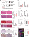
Figure 1
Figure 1
Ginkgolide B (GB) accelerates aged skeletal muscle repair and modulates muscle stem cells (MuSCs) in aged mice. (A) Experimental design. BaCl2 was intramuscularly injected into 20‐month‐old female mice to induce muscle injury. At 6 h after BaCl2 injection, the mice were treated with GB (12 mg/kg body weight) daily by oral gavage until they were sacrificed (n = 5). (B) Representative haematoxylin and eosin (HE) (×200), Masson's trichrome and F4/80 staining at 7 days post‐injury (DPI) (n = 5). Scale bar at 150 μm for HE and Masson's trichrome staining; scale bar at 150 μm for F4/80 staining. Quantification of myofiber numbers at the centre of the damage site under high magnification (×540). Myofibers are stained red, and collagen is stained blue in Masson's trichrome staining. F4/80 represents a pan‐macrophage marker. (C) Expression of myogenesis‐related genes at 7 DPI measured by real‐time PCR (n = 4). (D) Expression of inflammatory cytokines in muscle at 7 DPI (n = 4). (E) Quantification of MuSCs by flow cytometry analysis at 3 DPI. MuSCs, CD31, CD45, Sca1 and Vcam+ population (n = 3). (F) Representative embryonic myosin heavy chain (eMHC)/laminin/4′,6‐diamidino‐2‐phenylindole (DAPI) staining at 3 DPI. Quantitative analysis of the regenerating myofiber (centre nucleus, eMHC+) area versus injury area (n = 6 images from 2 animals in each group). Scale bar: 50 μm. ND, not detectable. Quantitative data are presented as the means ± SDs in the histogram with data points. For panels (A)–(D), statistical analyses were performed using two‐way analysis of variance (ANOVA) with Tukey's multiple comparison test; for panels (E) and (F), statistical analyses were performed using one‐way ANOVA with Tukey's multiple comparison test. Means not sharing any letters are significantly different (P < 0.05).
Figure 2
Figure 2
Ginkgolide B (GB) improves muscle functional recovery in injured aged mice. (A) Experimental design (n = 5 for the ex vivo muscle function test, n = 5 for the in vivo rotarod test for each group). BaCl2 was intramuscularly injected into the gastrocnemius (GA) muscle in 20‐month‐old female mice to induce muscle injury. At 6 h after BaCl2 injection, the mice were treated with GB (12 mg/kg body weight) daily by oral gavage until they were sacrificed. (B) GA muscle mass at Day 7 after BaCl2‐induced injury with GB treatment. (C–E) Ex vivo muscle contractility was assessed at 7 days post‐injury (DPI) and after GB treatment. Absolute and specific twitch force (C), absolute and specific tetanic force (D) and time to the peak twitch force and half relaxation time (E) were measured at 7 DPI. (F) Rotarod test of BaCl2‐treated mice at 14 DPI with GB treatment. Quantitative data are presented as the means ± SDs in the histogram with data points. Statistical analyses were performed using one‐way analysis of variance (ANOVA) with Tukey's multiple comparison test. Means not sharing any letters are significantly different (P < 0.05).
Figure 3
Figure 3
Ginkgolide B (GB) does not directly modulate proliferation or myogenic differentiation but enhances the migration of control and multiple population doubling (MPD) myocytes. (A, B) Cell viability (A) and expression of proliferation markers (B) in C2C12 cells after GB treatment for 48 h (n = 3). (C) Migration of C2C12 cells after GB treatment for 24 h. The results are normalized to the control C2C12 with vehicle treatment (n = 5). (D) The expression of myogenic‐specific genes in C2C12‐derived myotubes at myogenic differentiation Day 7 with or without GB treatment (n = 8 for control C2C12, n = 4 for MPD C2C12). (E) Representative images of myosin heavy chain (MyHC) staining in C2C12‐derived myotubes at myogenic differentiation Day 7 with or without GB treatment (n = 4). 4′,6‐Diamidino‐2‐phenylindole (DAPI) was used as a nuclear counterstain. Scale bar: 100 μm. (F) The percentage of MyHC‐positive area, relative fluorescence intensity, differentiation index and fusion index were quantified from (E) with ImageJ software (n = 5). Control C2C12 cells, Passages 2–5; MPD C2C12 cells, Passages 15–20. Quantitative data are presented as the means ± SDs in the histogram with data points. Statistical analyses were performed using two‐way analysis of variance (ANOVA) with Tukey's multiple comparison test. Means not sharing any letters are significantly different (P < 0.05).
Figure 4
Figure 4
Ginkgolide B (GB)‐primed osteoblast‐conditioned medium modulates myogenesis in multiple population doubling (MPD) myoblasts in vitro. (A) Cell viability in MPD myoblasts after OB CM or OB + GB treatment for 48 h (n = 6). The control C2C12 and MPD + control CM groups were treated with osteogenic induction medium (control CM) only. (B) Representative myosin heavy chain (MyHC) staining of aged myotubes treated with OB CM or OB + GB. Control C2C12‐derived myotubes served as a positive control for staining. 4′,6‐Diamidino‐2‐phenylindole (DAPI) was used as a nuclear counterstain. Scale bar: 50 μm. (C–E) The percentage of MyHC‐positive area (C), differentiation index (D) and fusion index (E) of MPD myotubes were determined with ImageJ software (n = 6). (F) Protein content (n = 4). Control C2C12 cells, Passages 2–5; MPD C2C12 cells, Passages 15–20; OB CM, conditioned medium from aged osteoblasts; OB + GB CM, conditioned medium from aged osteoblasts with GB stimulation. Quantitative data are presented as the means ± SDs in the histogram with data points. Statistical analyses were performed using one‐way analysis of variance (ANOVA) with Tukey's multiple comparison test. Means not sharing any letters are significantly different (P < 0.05).
Figure 5
Figure 5
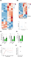
Ginkgolide B (GB) activates osteokines in aged osteoblasts and increases circulating osteocalcin levels in aged mice. (A) Heatmap of differentially expressed genes (DEGs) in aged osteoblasts with and without GB treatment (n = 3). Rows are centred; unit variance scaling is applied to rows. Both rows and columns were clustered using correlation distance and average linkage. (B) Secreted DEGs revealed by heatmap and volcano plot. Eleven downregulated secreted DEGs, which were undetectable in GB‐treated aged osteoblasts, were not shown in the volcano plot. (C) The expression of osteocalcin in aged OBs was measured by real‐time PCR (n = 6) and enzyme‐linked immunosorbent assay (ELISA) (n = 6). (D) The levels of serum osteocalcin in aged mice with or without GB treatment for 2 months were measured by ELISA (n = 5). (E, F) The protein (E) and transcript (F) levels of osteocalcin in muscle after BaCl2‐induced injury were measured by ELISA and real‐time PCR (n = 3). The Bglap primer can amplify all three transcripts and variants in mouse osteocalcin gene cluster, including NM_007541.3, NM_001032298.3, NM_001305448.1, NM_001305449.1, NM_031368.5 and NM_001305450.1. In the histogram, quantitative data are presented as the means ± SDs with data points, and the statistical analyses were performed using one‐way analysis of variance (ANOVA) with Tukey's multiple comparison test. Means not sharing any letters are significantly different (P < 0.05). In the line chart, quantitative data are presented as the means ± SDs, and statistical analyses were performed using two‐way ANOVA with a mixed‐effect model. *, P < 0.05; **, P < 0.01; ***, P < 0.001 at the indicated time points. Means not sharing any letters are significantly different in the same group (P < 0.05); ND, not detectable.
Figure 6
Figure 6
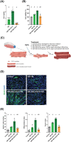
Osteocalcin mediates the effects of Ginkgolide B (GB) on muscle regeneration in an in vitro BaCl2‐induced muscle injury/regeneration model. (A) Enzyme‐linked immunosorbent assay (ELISA) revealed the expression levels of osteocalcin in OB CM after siOCN treatment (n = 6). (B) Cell viability of multiple population doubling (MPD) C2C12 cells after OB CM treatment (n = 4). The control group was treated with osteogenic induction medium. (C) Illustration of the in vitro BaCl2‐induced muscle injury/regeneration model in MPD C2C12‐derived myotubes. All groups received BaCl2 and were then treated with different CM. (D) Representative myosin heavy chain (MyHC) staining of MPD C2C12‐derived myotubes at myogenic differentiation Day 5 with GB‐primed OB CM treatment. 4′,6‐Diamidino‐2‐phenylindole (DAPI) was used as a nuclear counterstain (n = 4). Scale bar: 50 μm. (E) The percentage of MyHC‐positive area, differentiation index and fusion index of MPD C2C12‐derived myotubes subjected to various treatments were quantified with ImageJ software (n = 4). MPD C2C12 cells, Passages 15–20, represent replicatively aged myoblasts. OB CM, conditioned medium from aged osteoblasts; OB + GB CM + siCon, conditioned medium from aged osteoblasts with GB stimulation and scramble control siRNA; OB + GB + siOCN CM, conditioned medium from aged osteoblasts with GB stimulation and siOCN transfection; OCN, osteocalcin; siOCN, siRNA against osteocalcin. Quantitative data are presented as the means ± SDs in the histogram with data points. Statistical analyses were performed using one‐way analysis of variance (ANOVA) with Tukey's multiple comparison test. Means not sharing any letters are significantly different (P < 0.05).
Figure 7
Figure 7
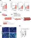
Osteocalcin is sufficient to promote myogenesis in an in vitro BaCl2‐induced muscle injury/regeneration model. (A) Viability of multiple population doubling (MPD) C2C12 cells after osteocalcin treatment under serum‐free conditions for 48 h (n = 5). Control group was treated with phosphate‐buffered saline (PBS). (B) Illustration of the in vitro BaCl2‐induced muscle injury/regeneration model. (C) Representative myosin heavy chain (MyHC) staining of MPD C2C12‐derived myotubes at myogenic differentiation Day 5 with osteocalcin treatment (n = 5). 4′,6‐Diamidino‐2‐phenylindole (DAPI) was used as a nuclear counterstain. Scale bar: 50 μm. (D) The percentage of MyHC‐positive area, differentiation index and fusion index of MPD C2C12‐derived myotubes subjected to osteocalcin treatment were quantified with ImageJ software (n = 5). (E) Comparison of the differentiation index (D‐index) and fusion index (F‐index) between control and MPD C2C12‐derived myotubes (n = 5–6). Control C2C12 cells, Passages 2–5; and MPD C2C12 cells, Passages 15–20 represent replicatively aged myoblasts. OCN, osteocalcin. Quantitative data are presented as the means ± SDs in the histogram with data points. Statistical analyses were performed using one‐way analysis of variance (ANOVA) with Tukey's multiple comparison test or multiple t‐tests (panel D). Means not sharing any letters are significantly different (P < 0.05).
Figure 8
Figure 8
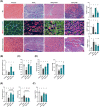
Exogenous osteocalcin promotes muscle regeneration and muscle functional recovery in aged mice. (A) Representative haematoxylin and eosin (HE), embryonic myosin heavy chain (eMHC) and Masson's trichrome staining and quantification of myofiber area, eMHC area and Masson's trichrome‐positive area at 4 days post‐injury (DPI) (n = 5). White arrows indicate mature regenerated myofibers (centre nucleus, eMHC‐negative myofibers). Myofibers are stained red, and collagen is stained blue. Scale bars: 100, 20 and 50 μm from top to bottom. (B) Flow cytometry analysis of muscle stem cells (MuSCs) (CD31, CD45, Sca1 and Vcam+ population) at 3 DPI (n = 3). (C) Gastrocnemius (GA) muscle mass at 7 DPI (n = 3–6). (D, E) Ex vivo muscle contractility was assessed at 7 DPI (n = 3–6). Twitch force and tetanic force (D) and half relaxation time and time to max (E) of GA muscle were measured at 7 DPI. (F) Rotarod test of BaCl2‐treated mice at 14 DPI (n = 5). Mesenchymal stem cell (MSC)‐derived osteoblasts served as a positive control. OCN, osteocalcin. Quantitative data are presented as the means ± SDs in the histogram with data points. Statistical analyses were performed using one‐way analysis of variance (ANOVA) with Tukey's multiple comparison test. Means not sharing any letters are significantly different (P < 0.05).

References

    1. Conboy IM, Conboy MJ, Wagers AJ, Girma ER, Weissman IL, Rando TA. Rejuvenation of aged progenitor cells by exposure to a young systemic environment. Nature 2005;433:760–764. - PubMed
    1. Chen WJ, Lin IH, Lee CW, Yoshioka K, Ono Y, Yan YT, et al. Ribonucleotide reductase M2B in the myofibers modulates stem cell fate in skeletal muscle. NPJ Regen Med 2022;7:37. - PMC - PubMed
    1. Blau HM, Cosgrove BD, Ho AT. The central role of muscle stem cells in regenerative failure with aging. Nat Med 2015;21:854–862. - PMC - PubMed
    1. Tagliaferri C, Wittrant Y, Davicco MJ, Walrand S, Coxam V. Muscle and bone, two interconnected tissues. Ageing Res Rev 2015;21:55–70. - PubMed
    1. Mera P, Laue K, Ferron M, Confavreux C, Wei J, Galan‐Diez M, et al. Osteocalcin signaling in myofibers is necessary and sufficient for optimum adaptation to exercise. Cell Metab 2016;23:1078–1092. - PMC - PubMed

Publication types