. 2023 Jan-Dec:32:9636897231166209.
doi: 10.1177/09636897231166209.

Mapping of the Human Amniotic Membrane: In Situ Detection of Microvesicles Secreted by Amniotic Epithelial Cells

Affiliations

Mapping of the Human Amniotic Membrane: In Situ Detection of Microvesicles Secreted by Amniotic Epithelial Cells

Mariangela Basile et al. Cell Transplant. 2023 Jan-Dec.

Abstract

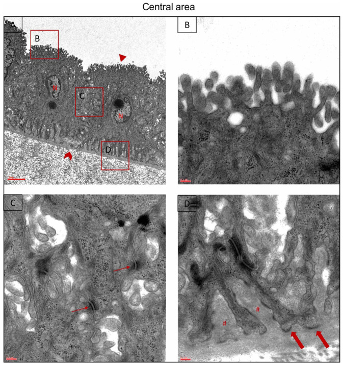
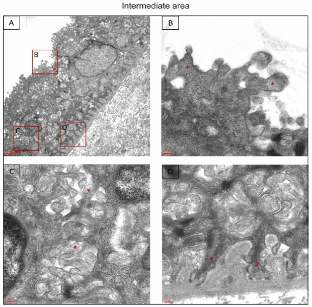
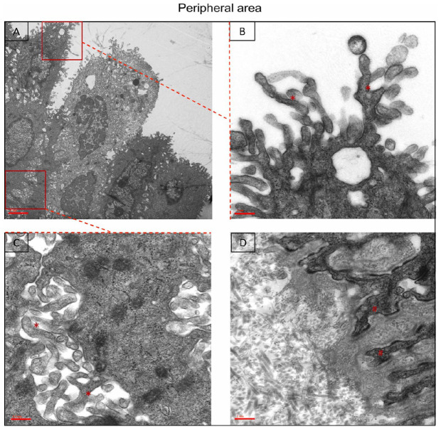
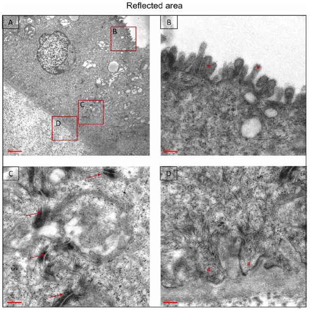
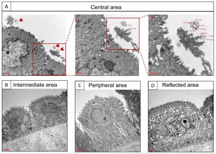
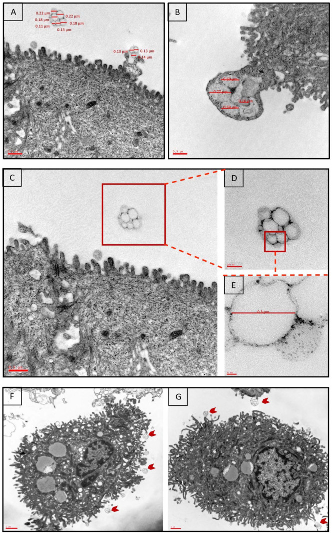
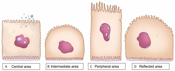
The potential clinical applications of human amniotic membrane (hAM) and human amniotic epithelial cells (hAECs) in the field of regenerative medicine have been known in literature since long. However, it has yet to be elucidated whether hAM contains different anatomical regions with different plasticity and differentiation potential. Recently, for the first time, we highlighted many differences in terms of morphology, marker expression, and differentiation capabilities among four distinct anatomical regions of hAM, demonstrating peculiar functional features in hAEC populations. The aim of this study was to investigate in situ the ultrastructure of the four different regions of hAM by means of transmission electron microscopy (TEM) to deeply understand their peculiar characteristics and to investigate the presence and localization of secretory products because to our knowledge, there are no similar studies in the literature. The results of this study confirm our previous observations of hAM heterogeneity and highlight for the first time that hAM can produce extracellular vesicles (EVs) in a heterogeneous manner. These findings should be considered to increase efficiency of hAM applications within a therapeutic context.

Keywords: amniotic epithelial cells; amniotic membrane; electron microscopy; microvesicles; stem cells.


Conflict of interest statement

The author(s) declared no potential conflicts of interest with respect to the research, authorship, and/or publication of this article.

Figures

Figure 1.
Figure 1.
Ultrastructure of the amniotic epithelium in the central area. (A) hAECs are in contact with the amniotic cavity (triangle) and the underlying avascular connective layer through a thick basal lamina (arrowhead). Nuclei are euchromatic (N) (3,000×; scale bar: 2 µm). (B, C) The (B) apical and (C) lateral portions are covered by microvilli. Numerous desmosomes are visible (thin arrows) (20,000×; scale bar: 0.2 µm). (D) Cytoplasmic extensions of hAECs enter the basal membrane (#). Emidesmosomes are present (thick arrows) (20,000×; scale bar: 0.2 µm). The most representative sample out of the five different samples is shown. hAECs: human amniotic epithelial cells.
Figure 2.
Figure 2.
Ultrastructure of the amniotic epithelium in the intermediate area. (A) hAECs present a characteristic dome shape (3,000×; scale bar: 2 µm). (B, C) The microvilli in the (B) apical region are thick and short (*) (20,000×; scale bar: 0.2 µm), while they appear more numerous in the (C) lateral region (*) (20,000×; scale bar: 0.2 µm). (D) The hAEC cytoplasmic extensions entering the basal membrane are short and spaced apart (#) (20,000×; scale bar: 0.2 µm). The most representative sample out of the five different samples is shown. hAECs: human amniotic epithelial cells.
Figure 3.
Figure 3.
Ultrastructure of the amniotic epithelium in the peripheral area. (A) hAECs possess a columnar shape and are organized on multiple layers (3,000×; scale bar: 2 µm). (B, C) The microvilli in the (B) apical and (C) lateral regions are long and thin (*) (20,000×; scale bar: 0.2 µm). (D) The hAEC cytoplasmic extensions appear thin (#) (20,000×; scale bar: 0.2 µm). The most representative sample out of the five different samples is shown. hAECs: human amniotic epithelial cells.
Figure 4.
Figure 4.
Ultrastructure of the amniotic epithelium in the reflected area. (A) hAECs possess a cuboidal shape and a smoother surface (3,000×; scale bar: 2 µm). (B, C) The microvilli in the (B) apical region are short, while they are few in the (C) lateral area. Numerous desmosomes are also visible (thin arrows) (20,000×; scale bar: 0.2 µm). (D) The hAEC cytoplasmic extensions are short and thick (#) (20,000×; scale bar: 0.2 µm). The most representative out of the five different samples is shown. hAECs: human amniotic epithelial cells.
Figure 5.
Figure 5.
Ultrastructural detection of extracellular vesicles (EVs) secreted by the central area of hAM. (A) The figure shows a detail from the central area at different magnifications, highlighting the presence of EVs near the apical surface of the epithelial cell (A: 7,000×, scale bar: 1 µm; A1: 12,000×, scale bar: 0.5 µm; A2: 20,000×, scale bar: 0.5 µm). (B–D) The presence of these structures was not detected in the other regions of hAM (7,000×, scale bars: 2 µm). The most representative sample out of the five different samples is shown. hAM: human amniotic membrane.
Figure 6.
Figure 6.
Ultrastructural detection of extracellular vesicles (EVs) secreted by the central area of hAM. (A–B) The figure shows a group of microvesicles identified by their range of size between 100 and 1,000 nm. They seem to bud from the apical surface of the cytoplasm (12,000×, scale bar: 0.5 µm). (C–E) A group of EVs secreted by the central area of hAM at different magnifications is shown (12,000×; 20,000×; 50,000×; scale bars: 0.5 µm, 200 nm, 50 nm). (F–G) The figure shows several multivesicular bodies around the apical surface of hAECs after their enzymatic separation from the basal lamina (arrowheads) (3,000×, scale bars: 1 µm). The most representative samples out of the 10 different samples are shown. hAM: human amniotic membrane; hAECs: human amniotic epithelial cells.
Figure 7.
Figure 7.
Schematic cartoon of hAEC structural features from the different regions of hAM. (A) Central area: cells are mainly cuboidal, characterized by short and thick microvilli distributed at the apical, lateral, and basal surface; round aggregates of microvesicles are visible near the apical surface. (B) Intermediate area: cells show a dome shape, numerous microvilli at the basal surface compared with the central area. (C) Peripheral area: cells present columnar shape, long and thin microvilli. (D) Reflected area: cells show a dome shape with squat microvilli. hAECs: human amniotic epithelial cells; hAM: human amniotic membrane.

Similar articles

Cited by

References

    1. Caruso M, Evangelista M, Parolini O. Human term placental cells: phenotype, properties and new avenues in regenerative medicine. Int J Mol Cell Med. 2012;1(2):64–74. - PMC - PubMed
    1. Parolini O, Alviano F, Bagnara GP, Bilic G, Bühring HJ, Evangelista M, Hennerbichler S, Liu B, Magatti M, Mao N, Miki T, et al.. Concise review: isolation and characterization of cells from human term placenta: outcome of the first international workshop on placenta derived stem cells. Stem Cells. 2008;26(2):300–11. - PubMed
    1. Silini AR, Di Pietro R, Lang-Olip I, Alviano F, Banerjee A, Basile M, Borutinskaite V, Eissner G, Gellhaus A, Giebel B, Huang YC, et al.. Perinatal derivatives: where do we stand? A roadmap of the human placenta and consensus for tissue and cell nomenclature. Front Bioeng Biotechnol. 2020;8:610544. - PMC - PubMed
    1. Davis JS. Skin grafting at the Johns Hopkins hospital. Ann Surg. 1909;50(3):542–49. - PMC - PubMed
    1. Dua HS, Gomes JA, King AJ, Maharajan V. The amniotic membrane in ophthalmology. Surv Ophthalmol. 2004;49:51–77. - PubMed

Publication types

LinkOut - more resources