. 2023 May 16;122(10):1846-1857.
doi: 10.1016/j.bpj.2023.04.016. Epub 2023 Apr 18.

Role of surfactants in electron cryo-microscopy film preparation

Affiliations

Role of surfactants in electron cryo-microscopy film preparation

Baptiste Michon et al. Biophys J. .

Abstract

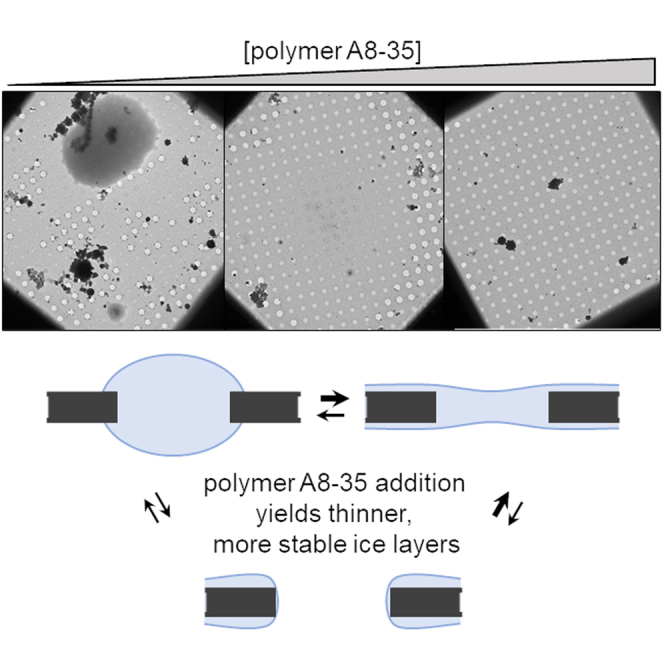
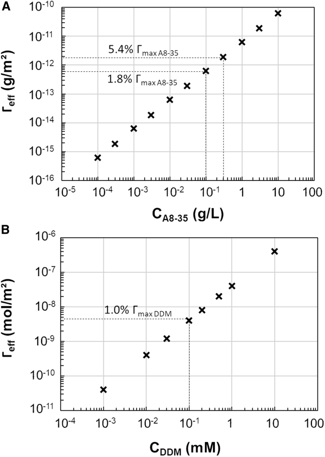
Single-particle electron cryo-microscopy (cryo-EM) has become an effective and straightforward approach to determine the structure of membrane proteins. However, obtaining cryo-EM grids of sufficient quality for high-resolution structural analysis remains a major bottleneck. One of the difficulties arises from the presence of detergents, which often leads to a lack of control of the ice thickness. Amphipathic polymers such as amphipols (APols) are detergent substitutes, which have proven to be valuable tools for cryo-EM studies. In this work, we investigate the physico-chemical behavior of APol- and detergent-containing solutions and show a correlation with the properties of vitreous thin films in cryo-EM grids. This study provides new insight on the potential of APols, allowing a better control of ice thickness while limiting protein adsorption at the air-water interface, as shown with the full-length mouse serotonin 5-HT3A receptor whose structure has been solved in APol. These findings may speed up the process of grid optimization to obtain high-resolution structures of membrane proteins.

Keywords: air-water interface; amphipol; detergent; ice thickness; membrane proteins; particle orientation.


Conflict of interest statement

Declaration of interests The authors declare no competing interests.

Figures

None
Graphical abstract
Figure 1
Figure 1
Illustration of the thickness measurement approach. (a) Low-magnification (500×) image of a grid prepared with a solution containing A8-35 at 0.003 g/L, with highlights on one of the holes used to acquire pairs of tilted images (scale bar: 10 μm). (b and c) Zooms on a pair of high-magnification (30,000×) tilted images, acquired at −15° and +15°, respectively (scale bar: 200 nm). (d and e) Zooms on the highlighted regions from (b) and (c), respectively (scale bar: 50 nm). Apparent distances between two impurities adsorbed on each side of the ice film (here, L1 and L2) are measured after projection onto a plane perpendicular to the rotation axis (dashed lines). (fh) Cartoon-illustrated calculation of the ice thickness. Each cartoon represents the section of an ice film, with impurities adsorbed on both sides. (f) No tilt. (g) Tilt at angle –α. (h) Tilt at angle +α. (For symbols and calculation description, see main text).
Figure 2
Figure 2
Calibration curve for ice thickness estimation. The hole intensity (mean gray value measured within a hole at low magnification, 500×) is correlated with the hole ice thickness, calculated from tilted pairs acquired at high magnification (30,000×). This conversion curve was used to draw all thickness distributions reported in Figs. 4 and 5.
Figure 3
Figure 3
Properties of solutions containing increasing concentrations of surfactants. Characterization of solutions containing APol A8-35 (a and b) and DDM (c and d). (a) and (c) show evolution of liquid film stability measured with the film pulling method for APol and detergent solutions, respectively (centimetric films, 100% humidity, error bars are standard deviations calculated with 300 measures per data point). Note that films generated at 3 g/L A8-35 and above 0.5 mM DDM were too stable to allow measurements. (b) and (d) show short-time surface tension variation at the air-water interface for APol and detergent solutions, respectively. To see this figure in color, go online.
Figure 4
Figure 4
Impact of A8-35 concentration on ice distribution and thickness on cryo-EM grids. (a) Low-magnification (LM, 500×) picture of a grid square (scale bar: 10 μm). The deposited sample was A8-35 at 0.003 g/L in Tris/NaCl buffer. Bright holes with sharp edges (highlighted by yellow circles) were empty, and those covered by thick ice patches (yellow stars) were categorized in the "ice patch" population. All remaining holes (except those recovered by ice crystal contaminations; red stars) were used for ice thickness estimation. (b) Proportion of each hole population in cryo-EM grid (error bars for each population are standard deviations calculated from proportions of holes on grids at each APol concentration). (ch) Distributions of ice thickness with A8-35 concentrations ranging from 10−4 g/L to 3 g/L. The ice thickness measurements were performed using a calibration curve (see Fig. 2) plotting the ice thickness obtained with stereo pairs of high-magnification images of holes at known angles (+15°/–15°) and the intensity of the same hole at LM (for more details, see materials and methods). The calibration curve allowed us to estimate the ice thickness of a large number of holes based on intensity measurements performed on sets of LM pictures (30–40 images/grid, 11,000 ± 4000 holes/grid). For each concentration, three to four grids were analyzed, leading to as many distributions, normalized on the number of exploitable holes with ice films. To see this figure in color, go online.
Figure 5
Figure 5
Variation of cryo-EM ice properties with DDM concentrations. (a) Proportion of each hole population in cryo-EM grid (error bars for each population are standard deviations calculated from proportions of holes on grids at each DDM concentration). (b and c) Distributions of ice thickness with DDM concentrations. The experimental procedure for ice thickness determination was as described in the legend for Fig. 4. To see this figure in color, go online.
Figure 6
Figure 6
Cryo-EM data set of the homomeric mouse 5-HT3A receptor reconstituted in APol A8-35, supplemented with 0.3 g/L of A8-35. (a) Representative micrograph of the analyzed data set (scale bar: 100 nm). (b) Representative 2D class averages separated into two main groups: top views (upper panels) and tilted views (lower panels) with indicated counts for each group. (c) Unsharpened cryo-EM map (3D reconstruction) colored by estimated local resolution, where the scale indicates the corresponding values in Angstroms (accession EMDB code: EMD-15471). The extracellular domain (ECD) and transmembrane domain (TMD) are also indicated. (d) Projections of the particles over azimuth and elevation angles calculated for the 3D reconstruction depicted in (c). To see this figure in color, go online.
Figure 7
Figure 7
Estimation of the effective surfactant coverage of the cryo-film interfaces, depending on the initial sample surfactant concentration. Those estimations were performed for APol A8-35 (a) and DDM (b) by modeling the films as cylinders with a diameter of 2 μm and a thickness of 0.04 μm. This allowed for a calculation of the total amount of surfactant within a film, leading to a surface concentration when assuming that all surfactants reach and adsorb to the air-water interface.

References

    1. Kühlbrandt W. The resolution revolution. Science. 2014;343:1443–1444. - PubMed
    1. Vinothkumar K.R., Henderson R. Single particle electron cryomicroscopy: trends, issues and future perspective. Q. Rev. Biophys. 2016;49:e13. - PubMed
    1. Nwanochie E., Uversky V.N. Structure determination by single-particle cryo-electron microscopy: only the sky (and intrinsic disorder) is the limit. Int. J. Mol. Sci. 2019;20:4186. - PMC - PubMed
    1. Liao M., Cao E., et al. Cheng Y. Structure of the TRPV1 ion channel determined by electron cryo-microscopy. Nature. 2013;504:107–112. - PMC - PubMed
    1. Popot J.-L., Althoff T., et al. Zoonens M. Amphipols from A to Z. Annu. Rev. Biophys. 2011;40:379–408. - PubMed

Publication types

LinkOut - more resources