

Cardiomyocyte-fibroblast crosstalk in the postnatal heart
- PMID: 37077417
- PMCID: PMC10106698
- DOI: 10.3389/fcell.2023.1163331
Cardiomyocyte-fibroblast crosstalk in the postnatal heart
Abstract
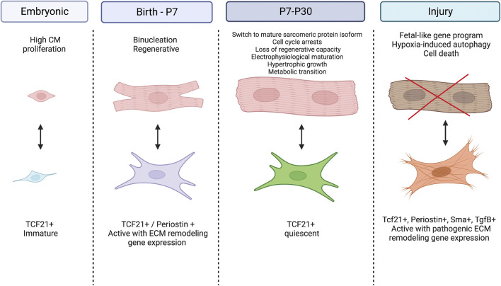
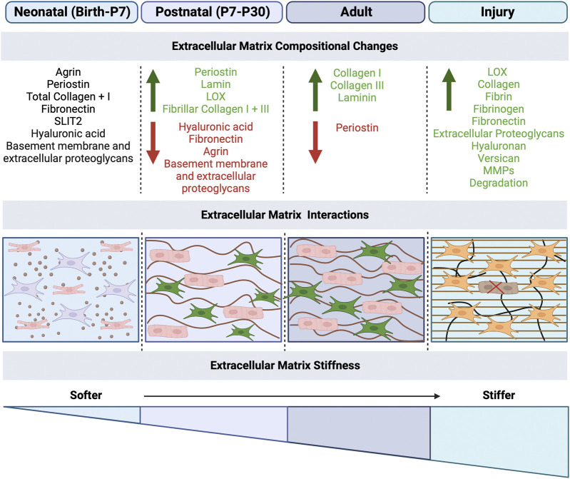
During the postnatal period in mammals, the heart undergoes significant remodeling in response to increased circulatory demands. In the days after birth, cardiac cells, including cardiomyocytes and fibroblasts, progressively lose embryonic characteristics concomitant with the loss of the heart's ability to regenerate. Moreover, postnatal cardiomyocytes undergo binucleation and cell cycle arrest with induction of hypertrophic growth, while cardiac fibroblasts proliferate and produce extracellular matrix (ECM) that transitions from components that support cellular maturation to production of the mature fibrous skeleton of the heart. Recent studies have implicated interactions of cardiac fibroblasts and cardiomyocytes within the maturing ECM environment to promote heart maturation in the postnatal period. Here, we review the relationships of different cardiac cell types and the ECM as the heart undergoes both structural and functional changes during development. Recent advances in the field, particularly in several recently published transcriptomic datasets, have highlighted specific signaling mechanisms that underlie cellular maturation and demonstrated the biomechanical interdependence of cardiac fibroblast and cardiomyocyte maturation. There is increasing evidence that postnatal heart development in mammals is dependent on particular ECM components and that resulting changes in biomechanics influence cell maturation. These advances, in definition of cardiac fibroblast heterogeneity and function in relation to cardiomyocyte maturation and the extracellular environment provide, support for complex cell crosstalk in the postnatal heart with implications for heart regeneration and disease mechanisms.
Keywords: cardiac fibroblast; cardiomyocyte; cell signaling; extracellular matrix; heart regeneration; postnatal heart.
Copyright © 2023 Uscategui Calderon, Gonzalez and Yutzey.
Conflict of interest statement
The authors declare that the research was conducted in the absence of any commercial or financial relationships that could be construed as a potential conflict of interest.
Figures
Similar articles
-
GDF10 promotes rodent cardiomyocyte maturation during the postnatal period.J Mol Cell Cardiol. 2025 Apr;201:16-31. doi: 10.1016/j.yjmcc.2025.01.010. Epub 2025 Feb 3. J Mol Cell Cardiol. 2025. PMID: 39909309
-
A specialized population of Periostin-expressing cardiac fibroblasts contributes to postnatal cardiomyocyte maturation and innervation.Proc Natl Acad Sci U S A. 2020 Sep 1;117(35):21469-21479. doi: 10.1073/pnas.2009119117. Epub 2020 Aug 17. Proc Natl Acad Sci U S A. 2020. PMID: 32817558 Free PMC article.
-
Modulation of Mammalian Cardiomyocyte Cytokinesis by the Extracellular Matrix.Circ Res. 2020 Sep 11;127(7):896-907. doi: 10.1161/CIRCRESAHA.119.316303. Epub 2020 Jun 21. Circ Res. 2020. PMID: 32564729
-
Cardiac Fibroblasts and the Extracellular Matrix in Regenerative and Nonregenerative Hearts.J Cardiovasc Dev Dis. 2019 Aug 20;6(3):29. doi: 10.3390/jcdd6030029. J Cardiovasc Dev Dis. 2019. PMID: 31434209 Free PMC article. Review.
-
Cardiomyocyte Proliferation and Maturation: Two Sides of the Same Coin for Heart Regeneration.Front Cell Dev Biol. 2020 Oct 15;8:594226. doi: 10.3389/fcell.2020.594226. eCollection 2020. Front Cell Dev Biol. 2020. PMID: 33178704 Free PMC article. Review.
Cited by
-
GDF10 promotes rodent cardiomyocyte maturation during the postnatal period.J Mol Cell Cardiol. 2025 Apr;201:16-31. doi: 10.1016/j.yjmcc.2025.01.010. Epub 2025 Feb 3. J Mol Cell Cardiol. 2025. PMID: 39909309
-
Histone H2A.Z Deacetylation and Dedifferentiation in Infarcted/Tip60-depleted Cardiomyocytes.bioRxiv [Preprint]. 2025 Jan 16:2024.01.11.575312. doi: 10.1101/2024.01.11.575312. bioRxiv. 2025. PMID: 38260622 Free PMC article. Preprint.
-
Metabolic Culture Medium Enhances Maturation of Human iPSC-Derived Cardiomyocytes via Cardiac Troponin I Isoform Induction.Int J Mol Sci. 2025 Jul 26;26(15):7248. doi: 10.3390/ijms26157248. Int J Mol Sci. 2025. PMID: 40806381 Free PMC article.
-
The epigenetic regulation of crosstalk between cardiac fibroblasts and other cardiac cell types during stress.Front Cardiovasc Med. 2025 Apr 8;12:1539826. doi: 10.3389/fcvm.2025.1539826. eCollection 2025. Front Cardiovasc Med. 2025. PMID: 40264508 Free PMC article. Review.
-
Matrix Architecture and Mechanics Regulate Myofibril Organization, Costamere Assembly, and Contractility in Engineered Myocardial Microtissues.Adv Sci (Weinh). 2024 Dec;11(47):e2309740. doi: 10.1002/advs.202309740. Epub 2024 Nov 18. Adv Sci (Weinh). 2024. PMID: 39558513 Free PMC article.
References
Publication types
LinkOut - more resources
Full Text Sources