

Treatment of corneal dermoid with lenticules from small incision lenticule extraction surgery: a surgery assisted by fibrin glue
- PMID: 37077475
- PMCID: PMC10089904
- DOI: 10.18240/ijo.2023.04.08
Treatment of corneal dermoid with lenticules from small incision lenticule extraction surgery: a surgery assisted by fibrin glue
Abstract
Aim: To observe the clinical efficacy of the combined use of small incision lenticule extraction (SMILE)-derived lenticule patches in corneal dermoid excision, with fixation of the lenticule patches assisted by fibrin glue.
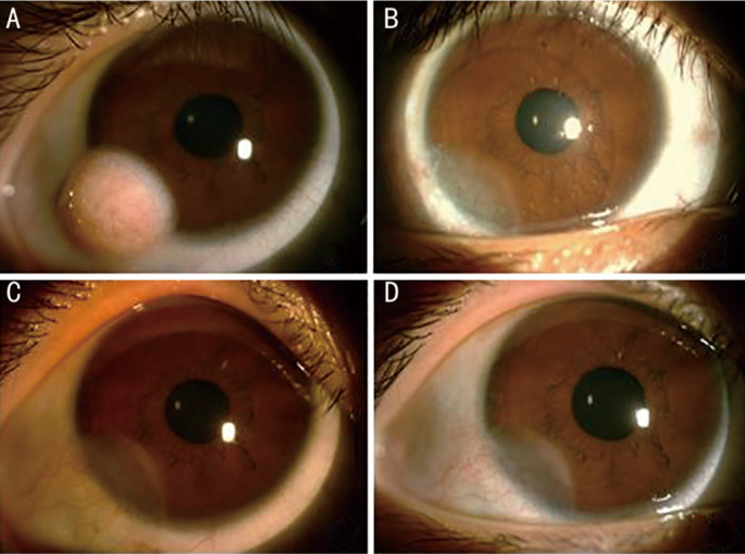
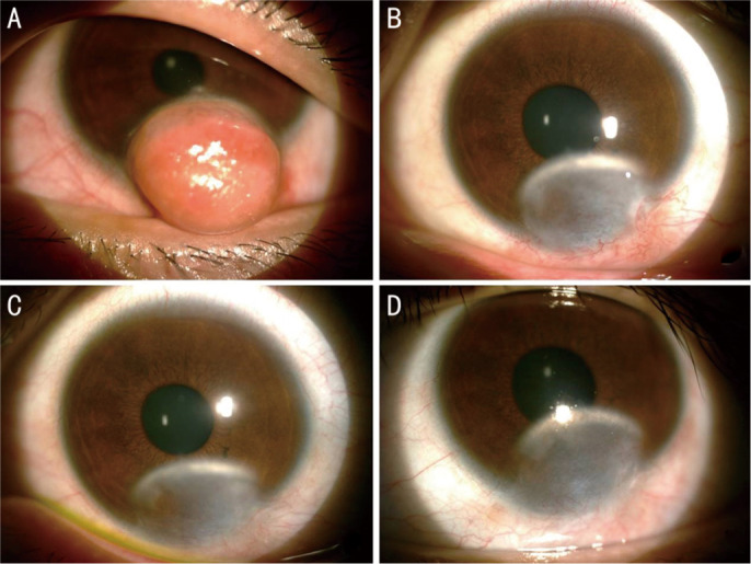
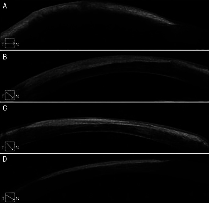
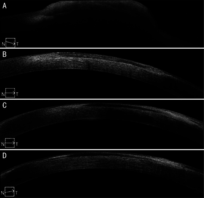
Methods: Seventeen eyes of 17 patients with corneal dermoid were treated with dermoid removal combined with SMILE-derived lenticule transplantation. All lenticule patches were fixed by fibrin glue. Ocular changes were assessed using slit lamp microscopy and anterior-segmental optical coherence tomography. The best-corrected visual acuity (BCVA) and ocular dioptric variations were examined preoperatively and postoperatively. Intraocular pressure (IOP) was also monitored in all visited time.
Results: Totally, 18 lenticule patches were used on 17 eyes of 17 cornea dermoid patients. The mean follow-up time was 11.47±5.28mo. All lenticule patches were successfully glued, kept on its location and maintained transparent during the follow-up time, with a consecutive epithelial cover for 1wk. Nine of the patients could coordinate visual and optometry exam well. Their preoperative BCVA is 0.60±0.35 in decimal, significantly improved to 0.80±0.26 in decimal at 6mo postoperatively (Z=-2.392, P=0.017), but the changes of their corneal astigmatism diopters showed no significance, with 2.22±1.91 D preoperatively, and 2.28±1.31 D at 6mo postoperatively (Z=-0.135, P=0.893). Limbal pannus formation occurred in 4 (23.52%) cases and decreased with the application of tacrolimus eyedrops. IOP increased in 2 (11.76%) cases, but well decreased by timolol maleate eyedrops. All the adult patients or guardians of minor patients were satisfied with the cosmetic improvement.
Conclusion: Dermoid excision combined with transplantation of SMILE-derived lenticule patches using fibrin glue is a safe and effective novel tectonic keratoplasty procedure for corneal dermoid.
Keywords: corneal dermoid; fibrin glue; small incision lenticule extraction.
International Journal of Ophthalmology Press.
Figures

Similar articles
-
Treatment of Corneal Dermoid with Fibrin Glue Boned Multi-Layer Lenticules from Small Incision Lenticules Extraction Surgery: A Preliminary Study of Five Patients.Curr Eye Res. 2025 Feb;50(2):132-138. doi: 10.1080/02713683.2024.2398121. Epub 2024 Sep 4. Curr Eye Res. 2025. PMID: 39229665
-
Surgical treatment of corneal dermoid by using intrastromal lenticule obtained from small-incision lenticule extraction.Int Ophthalmol. 2020 Jan;40(1):43-49. doi: 10.1007/s10792-019-01201-w. Epub 2019 Nov 17. Int Ophthalmol. 2020. PMID: 31735992
-
Combined interface tattooing and fibrin glue-assisted sutureless corneal resurfacing with donor lenticule obtained from small-incision lenticule extraction for limbal dermoid.J Cataract Refract Surg. 2017 Nov;43(11):1371-1375. doi: 10.1016/j.jcrs.2017.09.021. J Cataract Refract Surg. 2017. PMID: 29223224
-
Lamellar keratoplasty using femtosecond laser intrastromal lenticule for limbal dermoid: case report and literature review.J Int Med Res. 2018 Nov;46(11):4753-4759. doi: 10.1177/0300060518790874. Epub 2018 Aug 8. J Int Med Res. 2018. PMID: 30088427 Free PMC article. Review.
-
A novel case using femtosecond laser-acquired lenticule for recurrent pterygium: case report and literature review.J Int Med Res. 2018 Jun;46(6):2474-2480. doi: 10.1177/0300060518765303. Epub 2018 Apr 16. J Int Med Res. 2018. PMID: 29658366 Free PMC article. Review.
Cited by
-
Treatment of superficial corneal opacities with corneal stromal lenticule obtained through SMILE surgery.Int J Ophthalmol. 2024 Dec 18;17(12):2221-2228. doi: 10.18240/ijo.2024.12.09. eCollection 2024. Int J Ophthalmol. 2024. PMID: 39697877 Free PMC article.
-
Combined Multilayered Amniotic Membrane Graft and Fibrin Glue as a Surgical Management of Limbal Dermoid Cyst.J Clin Med. 2025 Jan 18;14(2):607. doi: 10.3390/jcm14020607. J Clin Med. 2025. PMID: 39860612 Free PMC article.
-
A Retrospective Analysis of Corneal Dermoid.Cureus. 2024 Jul 18;16(7):e64840. doi: 10.7759/cureus.64840. eCollection 2024 Jul. Cureus. 2024. PMID: 39156291 Free PMC article.
-
A novel sandwich technique of minimally invasive surgery for corneal perforation.Sci Rep. 2024 Nov 12;14(1):27675. doi: 10.1038/s41598-024-79376-1. Sci Rep. 2024. PMID: 39533004 Free PMC article.
References
-
- Jacob S, Narasimhan S, Agarwal A, Agarwal A, Ai S. Combined interface tattooing and fibrin glue-assisted sutureless corneal resurfacing with donor lenticule obtained from small-incision lenticule extraction for limbal dermoid. J Cataract Refract Surg. 2017;43(11):1371–1375. - PubMed
LinkOut - more resources
Full Text Sources