. 2023 Apr 18;16(4):514-522.
doi: 10.18240/ijo.2023.04.04. eCollection 2023.

Role of apigenin in high glucose-induced retinal microvascular endothelial cell dysfunction via regulating NOX4/p38 MAPK pathway in vitro

Affiliations

Role of apigenin in high glucose-induced retinal microvascular endothelial cell dysfunction via regulating NOX4/p38 MAPK pathway in vitro

Li-Li Liu et al. Int J Ophthalmol. .

Abstract

Aim: To investigate the retinoprotective role of Apigenin (Api) against high glucose (HG)-induced human retinal microvascular endothelial cells (HRMECs), and to explore its regulatory mechanism.

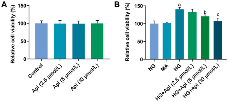
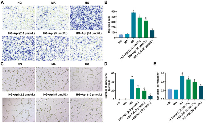
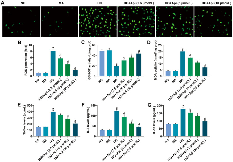
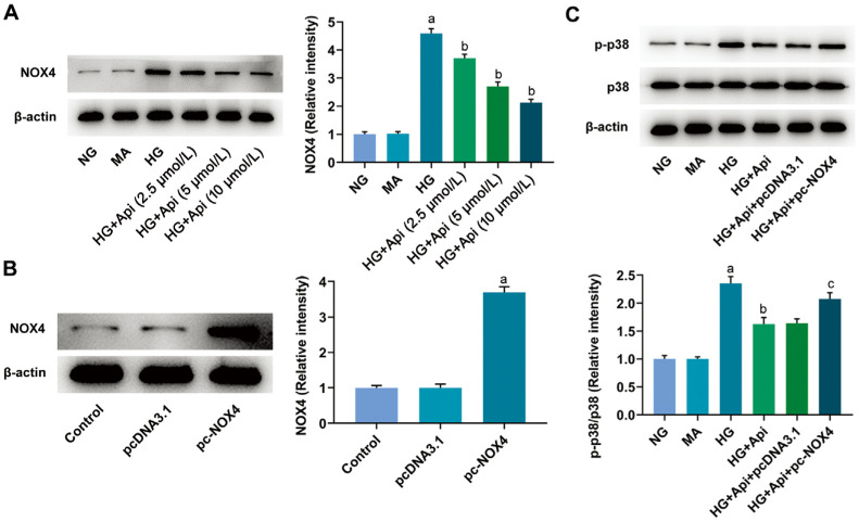
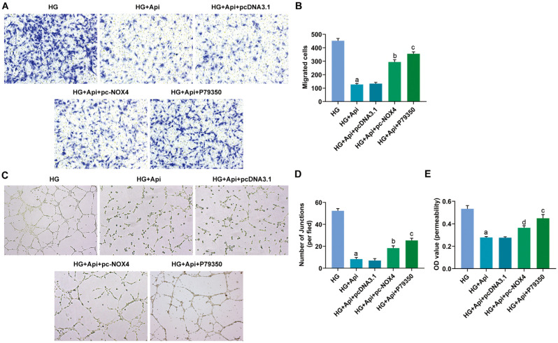
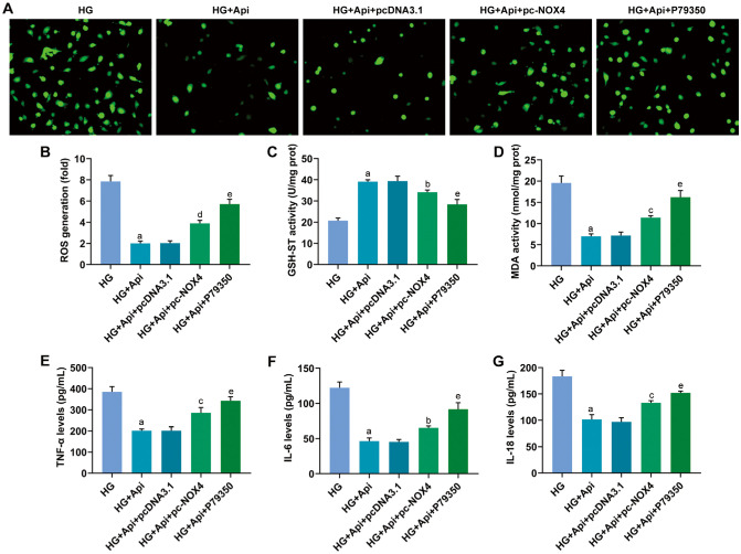
Methods: HRMECs were stimulated by HG for 48h to establish the in vitro cell model. Different concentrations of Api (2.5, 5, and 10 µmol/L) were applied for treatment. Cell counting kit-8 (CCK-8), Transwell, and tube formation assays were performed to examine the effects of Api on the viability, migration, and angiogenesis in HG-induced HRMECs. Vascular permeability was evaluated by Evans blue dye. The inflammatory cytokines and oxidative stress-related factors were measured using their commercial kits. Protein expression of nicotinamide adenine dinucleotide phosphate (NADPH) oxidase 4 (NOX4) and p38 mitogen-activated protein kinase (MAPK) was measured by Western blot.

Results: Api prevented HG-induced HRMECs viability, migration, angiogenesis, and vascular permeability in a concentration-dependent manner. Meanwhile, Api also concentration-dependently inhibited inflammation and oxidative stress in HRMECs exposed to HG. In addition, HG caused an elevated expression of NOX4, which was retarded by Api treatment. HG stimulation facilitated the activation of p38 MAPK signaling in HRMECs, and Api could weaken this activation partly via downregulating NOX4 expression. Furthermore, overexpression of NOX4 or activation of p38 MAPK signaling greatly weakened the protective role of Api against HG-stimulated HRMECs.

Conclusion: Api might exert a beneficial role in HG-stimulated HRMECs through regulating NOX4/p38 MAPK pathway.

Keywords: NOX4; apigenin; glucose; p38 MAPK; retinal microvascular endothelial cell.


Figures

Figure 1
Figure 1. Api inhibits cell viability of HRMECs under the HG environment
A: HRMECs were treated with increasing concentrations of Api (0, 2.5, 5, and 10 µmol/L) for 48h. Cell viability was detected using CCK-8 assay. B: HRMECs were stimulated by HG for 48h to establish the in vitro DR cell model, with or without treatment of Api. Cell viability was detected using CCK-8 assay. aP<0.001 vs NG; bP<0.05, cP<0.001 vs HG. Api: Apigenin; HRMECs: Human retinal microvascular endothelial cells; CCK-8: Cell counting kit-8; HG: High glucose; DR: Diabetic retinopathy; NG: Normal glucose; MA: Mannitol.
Figure 2
Figure 2. Api prevents HG-induced HRMECs migration, angiogenesis, and vascular permeability
A, B: HRMECs were stimulated by HG for 48h, with or without treatment of Api. HRMECs migration was examined using Transwell assay. C, D: The tube formation assay was carried out to evaluate the effect of Api on angiogenesis of HRMECs in HG environment. E: The vascular permeability was elevated by Evens blue dye. aP<0.001 vs NG; bP<0.01, cP<0.001 vs HG. Api: Apigenin; HRMECs: Human retinal microvascular endothelial cells; HG: High glucose; NG: Normal glucose; MA: Mannitol.
Figure 3
Figure 3. Api reduces HG-induced oxidative stress and inflammation in HRMECs
A, B: HRMECs were stimulated by HG for 48h, with or without treatment of Api. ROS generation of each group was determined by DCF-DA method. C, D: The activity of GSH-ST and MDA was measured using their corresponding commercial kits, respectively. E–G: The production of inflammatory cytokines, including TNF-α, IL-6, and IL-18, was examined by ELISA kits. aP<0.001 vs NG; bP<0.05, cP<0.01, dP<0.001 vs HG. Api: Apigenin; HRMECs: Human retinal microvascular endothelial cells; HG: High glucose; NG: Normal glucose; MA: Mannitol; ROS: Reactive oxygen species; GSH-ST: Glutathione S-transferase; MDA: Malondialdehyde; DCF-DA: 2′,7′-Dichlorofluorescin diacetate; TNF-α: Tumor necrosis factor-α; IL-6: Interleukin-6.
Figure 4
Figure 4. Api inactivates HG-stimulated p38 MAPK signaling via targeting NOX4 in HRMECs
A, B: HRMECs were stimulated by HG for 48h, with or without treatment of Api. The protein expression of NOX4 was detected using Western blot. aP<0.001 vs NG; bP<0.001 vs HG. B: HRMECs were transfected with pcDNA3.1 or pc-NOX4, and the expression level of NOX4 was detected by Western blot. aP<0.001 vs pcDNA3.1. C: HRMECs were transfected with pc-NOX4 to overexpress NOX4, followed by Api treatment and HG induction. The expression level of p-p38 and p38 was examined using Western blot. aP<0.001 vs NG; bP<0.001 vs HG; cP<0.01 vs HG+Api+pcDNA3.1. MAPK: Mitogen-activated protein kinase; Api: Apigenin; HRMECs: Human retinal microvascular endothelial cells; HG: High glucose; NG: Normal glucose; MA: Mannitol; NOX4: NADPH oxidase 4; pc-NOX4: pcDNA3.1-based NOX4 overexpression vector.
Figure 5
Figure 5. NOX4 and p38 MAPK signaling are involved in the protective role of Api against HG-stimulated HRMECs
A, B: HRMECs were transfected with pc-NOX4 to overexpress NOX4 or pre-treated with the p38 MAPK signaling agonist P79350, followed by Api treatment and HG induction. HRMECs migration was examined using Transwell assay. C, D: The tube formation assay was carried out to evaluate the effect of Api on angiogenesis of HRMECs in HG environment. E: The vascular permeability was elevated by Evens blue dye. aP<0.001 vs HG; bP<0.001 vs HG+Api+pcDNA3.1; cP<0.001 vs HG+Api; dP<0.01 vs HG+Api+pcDNA3.1. MAPK: Mitogen-activated protein kinase; Api: Apigenin; HRMECs: Human retinal microvascular endothelial cells; HG: High glucose; NG: Normal glucose; NOX4: NADPH oxidase 4; pc-NOX4: pcDNA3.1-based NOX4 overexpression vector.
Figure 6
Figure 6. NOX4 and p38 MAPK signaling are involved in the protective role of Api against HG-stimulated HRMECs
A, B: HRMECs were transfected with pc-NOX4 to overexpress NOX4 or pre-treated with the p38 MAPK signaling agonist P79350, followed by Api treatment and HG induction. ROS generation of each group was determined by DCF-DA method. C, D: The activity of GSH-ST and MDA was measured using their corresponding commercial kits, respectively. E–G: The production of inflammatory cytokines, including TNF-α, IL-6 and IL-18, was examined by ELISA kits. aP<0.001 vs HG; bP<0.05, cP<0.01, dP<0.001 vs HG+Api+pcDNA3.1; eP<0.001 vs HG+Api. MAPK: Mitogen-activated protein kinase; Api: Apigenin; HRMECs: Human retinal microvascular endothelial cells; HG: High glucose; NOX4: NADPH oxidase 4; pc-NOX4: pcDNA3.1-based NOX4 overexpression vector; ROS: Reactive oxygen species; GSH-ST: Glutathione S-transferase; MDA: Malondialdehyde; DCF-DA: 2′,7′-Dichlorofluorescin diacetate; TNF-α: Tumor necrosis factor-α; IL-6: Interleukin-6.

Similar articles

References

    1. Wong TY, Cheung CMG, Larsen M, Sharma S, Simó R. Diabetic retinopathy. Nat Rev Dis Primers. 2016;2:16012. - PubMed
    1. Cho NH, Shaw JE, Karuranga S, Huang Y, da Rocha Fernandes JD, Ohlrogge AW, Malanda B. IDF Diabetes Atlas: global estimates of diabetes prevalence for 2017 and projections for 2045. Diabetes Res Clin Pract. 2018;138:271–281. - PubMed
    1. Kang Q, Yang C. Oxidative stress and diabetic retinopathy: Molecular mechanisms, pathogenetic role and therapeutic implications. Redox Biol. 2020;37:101799. - PMC - PubMed
    1. Savage SR, McCollum GW, Yang R, Penn JS. RNA-seq identifies a role for the PPARβ/δ inverse agonist GSK0660 in the regulation of TNFα-induced cytokine signaling in retinal endothelial cells. Mol Vis. 2015;21:568–576. - PMC - PubMed
    1. Pincelli Netto M, Lima VC, Pacheco MA, Unonius N, Gracitelli CP, Prata TS. Macular inner retinal layer thinning in diabetic patients without retinopathy measured by spectral domain optical coherence tomography. Med Hypothesis Discov Innov Ophthalmol. 2018;7(3):133–139. - PMC - PubMed

LinkOut - more resources