Case Reports
. 2023 Apr 13:14:101832.
doi: 10.1016/j.jaccas.2023.101832. eCollection 2023 May 17.

Sinus of Valsalva Pseudoaneurysm: An Unusual Cause of Complete Heart Block

Affiliations
Case Reports

Sinus of Valsalva Pseudoaneurysm: An Unusual Cause of Complete Heart Block

Marinella Centemero et al. JACC Case Rep. .

Abstract

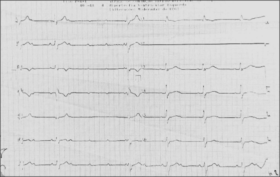
We present a case of a young man with complete atrioventricular block and aneurysm of the right sinus of Valsalva penetrating the interventricular septum and causing severe aortic regurgitation. Chest trauma and inflammatory or infectious diseases are potential causes. Bentall-de Bono surgical repair was performed. Anatomopathologic analysis demonstrated fibrosis, hyalinization, and extensive myxoid material. (Level of Difficulty: Beginner.).

Keywords: aneurysm; aorta; cardiac pacemaker; computed tomography; echocardiography; right ventricle.


Conflict of interest statement

The authors have reported that they have no relationships relevant to the contents of this paper to disclose.

Figures

None
Graphical abstract
Figure 1
Figure 1
Presentation 12-Lead Electrocardiogram Complete heart block with a broad complex ventricular escape rhythm of 42 beats/min.
Figure 2
Figure 2
Left Ventriculography and Aortography Aneurysmatic dilation at the level of the right coronary sinus observed during (A) left ventriculography in right anterior oblique 30° view and (B) aortography in left anterior oblique 45° view. Severe aortic regurgitation is observed.
Figure 3
Figure 3
Transthoracic Echocardiogram (A) Short axis at the aortic level and (B) parasternal longitudinal axis show the aneurysmatic dilation at the level of the right coronary sinus, measuring 24 mm in its largest axis. (C) Doppler evaluation revealed moderate to severe aortic regurgitation. (D) Dynamic obstruction of the right ventricle outflow tract was detected with a mid-ventricular gradient of 28 mm Hg.
Figure 4
Figure 4
Multidetector Cardiac Tomography (A and B) The aneurysm is located between the right aortic valve leaflet and the origin of the right coronary artery (RCA), promoting compression of both the left and the right ventricle outflow tracts (A). (C and D) 3-Dimensional reconstruction depicting the aneurysm relative to the aortic root. (E) 3-dimensional reconstruction showing the proximity of the aneurysm opening and the ostium of the RCA.
Figure 5
Figure 5
Intraoperative Findings (A) Left ventricle outflow tract (yellow star), right coronary valve leaflet (arrowhead), and edge of the aortic root at the level of the right coronary cusp pulled by the aortic retractor revealing the orifice of the wall discontinuity that forms the aortic-right ventricle fistula (blue arrow). (B) Interior aspect of the neocavity at the right side of the interventricular septum and the right ventricular infundibulum (asterisk).
Figure 6
Figure 6
Histologic Sections of Excised Fragments of the Aortic Valve Histologic sections of the excised fragments of the aortic valve showing (A) fibrosis, (B) hyalinization, and (C) deposits of myxoid material.

References

    1. Hanna M.F., Malguria N., Saboo S.S., et al. Cross-sectional imaging of sinus of Valsalva aneurysms: lessons learned. Diagn Interv Radiol. 2017;23:339–346. - PMC - PubMed
    1. Xu B., Kocyigit D., Betancor J., et al. Sinus of Valsalva aneurysms: a state-of-the-art imaging review. J Am Soc Echocardiogr. 2020;33:295–312. - PubMed
    1. Xu B., Kocyigit D., Godoy-Rivas C., et al. Outcomes of contemporary imaging-guided management of sinus of Valsalva aneurysms. Cardiovasc Diagn Ther. 2021;11:770–780. - PMC - PubMed
    1. Nguyen Q., Vervoort D., Phan K., Luc J.G.Y. Surgical management for unruptured sinus of Valsalva aneurysms: a narrative review of the literature. J Thorac Dis. 2021;13:1833–1850. - PMC - PubMed
    1. Yang K., Luo X., Tang Y., Hu H., Sun H. Comparison of clinical results between percutaneous closure and surgical repair of ruptured sinus of Valsalva aneurysm. Catheter Cardiovasc Interv. 2021;97:E354–E361. - PubMed

Publication types

LinkOut - more resources