. 2023 Mar 20;2(4):pgad088.
doi: 10.1093/pnasnexus/pgad088. eCollection 2023 Apr.

Commissural dentate granule cell projections and their rapid formation in the adult brain

Affiliations

Commissural dentate granule cell projections and their rapid formation in the adult brain

Matteo Egger et al. PNAS Nexus. .

Abstract

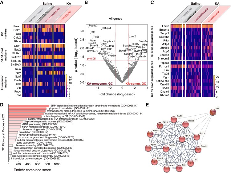
Dentate granule cells (GCs) have been characterized as unilaterally projecting neurons within each hippocampus. Here, we describe a unique class, the commissural GCs, which atypically project to the contralateral hippocampus in mice. Although commissural GCs are rare in the healthy brain, their number and contralateral axon density rapidly increase in a rodent model of temporal lobe epilepsies. In this model, commissural GC axon growth appears together with the well-studied hippocampal mossy fiber sprouting and may be important for the pathomechanisms of epilepsy. Our results augment the current view on hippocampal GC diversity and demonstrate powerful activation of a commissural wiring program in the adult brain.

Keywords: adult brain; circuit formation; commissural axon; contralateral projection; hippocampal granule cell; sprouting.


Figures

Fig. 1.
Fig. 1.
Commissural GC projections in the contralateral hippocampus. A) Tiled confocal image shows EGFP+ GCs at the ipsilateral injection site (left hippocampus), as well as EGFP+ axons entering the commissural/associational pathway (a), before entering the contralateral hippocampus (b), and in the contralateral hippocampal CA3 (c) and CA1 (d) areas. The bottom left inset shows the experimental design (also see main text). DG, dentate gyrus. B) Reconstruction of contralateral EGFP+ axons from saline- and KA-injected mice. Each image shows axons reconstructed from 20 to 26 consecutive, 50-µm-thick sections from one animal, labeled with sample ID and total axon length. C) Quantification of total contralateral EGFP+ axon length. Each data point represents the total axon length traced from one animal, as in B). Data represent mean ± SEM (Mann–Whitney test).
Fig. 2.
Fig. 2.
Retrograde labeling identifies commissural GCs. A) Experimental design (also see main text). B) Confocal images show retrogradely labeled, EGFP+ cells in GC layer (GCL) in the left hippocampus after ipsilateral saline or KA injection. Note the (i) presence of retrogradely labeled mossy cells (which also project to the contralateral hippocampus) and their axons in the inner molecular layer after saline injection and (ii) lack of mossy cells and axons and GCL dispersion (these are known effects of KA) after KA injection; retrogradely labeled CA3 pyramidal cells (which also project to the contralateral hippocampus) are visible in both images. C and D) Confocal image and morphological reconstruction of retrogradely labeled commissural GCs after saline (cell S1 is also shown in B, left) and KA injection (cell KA1–8 are also shown in B, right), respectively. E) Plot shows the number of retrogradely labeled commissural GCs in the ipsilateral (with respect to saline or KA injection) left hippocampus. Each data point represents the total number of commissural GCs detected in one animal. Data represent mean ± SEM (Mann–Whitney test). F) Plot shows the septo-temporal distribution of retrogradely labeled commissural GCs after ipsilateral saline and KA injection. For averaging, individual distributions were aligned to the ipsilateral (saline and KA) injection sites.
Fig. 3.
Fig. 3.
Trans-synaptic labeling identifies postsynaptic targets of commissural GCs. A) Experimental design (also see main text). B) Confocal images show mRuby+ cells in the contralateral hippocampus after ipsilateral saline or KA injection. The CA3 and CA1 areas are shown in higher magnification in both panels. SO, stratum oriens; SP, stratum pyramidale; SL, stratum lucidum; SR, stratum radiatum. C) Quantification of mRuby+ cells in contralateral hippocampal DG, CA3-CA2, and CA1-subiculum areas after ipsilateral saline or KA injection. Each point represents data from one animal. Data were normalized by subtracting mRuby+ cell counts obtained from wild-type animals [see main text; for nonnormalized data, see Fig. S4; two-way ANOVA, Farea(2, 16) = 56, P < 0.0001; Ftreatment(1, 8) = 38, P = 0.0003; Farea×treatment(2, 16) = 24, P < 0.0001; adjusted P-values (FDR) of post hoc analyses are indicated in the figure]. D) Confocal images show immunostaining for mRuby and DAPI as well as immunostaining for either GluR2/3 (left, in contralateral CA3), Wfs1 (middle, in contralateral CA1; note that Wfs1 immuno-positivity was mostly apparent in the superficial, but not deep, CA1 pyramidal cells), or PV (right, in contralateral CA3 and CA1) in 50-µm-thick sections after saline (top row) and KA injections (bottom row). Insets show magnification of the highlighted areas (the three staining methods are shown separately). E) Bar plots show the coexpression of mRuby with GluR2/3 (in CA3), or Wfs1 (in CA1), or PV (in CA3 and CA1). F) Reconstruction of trans-synaptically labeled mRuby+ neurons after saline and KA injections reveals pyramidal cell morphology. Dendrites and axons are shown in black and red or magenta, respectively. G) The upper left image shows reconstruction of anterogradely labeled EGFP+ axons in the contralateral hippocampus in a 50-µm-thick section after ipsilateral saline injection. The electron microscopy images show EGFP+ synapses (green and red arrowheads) in the CA3 (stratum oriens) and CA1 (stratum radium) area, as well as EGFP nonlabeled synapses (yellow and blue arrowheads), for comparison. In the lower right image, the asterisk labels a putative interneuron dendrite; synapses between interneuron dendrites and adjacent EGFP+ processes were not present.
Fig. 4.
Fig. 4.
Electrophysiological and morphological characterization of commissural GCs. A) Experimental design (also see main text). B) Example electrophysiological traces in response to 1.5-s-long depolarizing and hyperpolarizing current injections recorded from GCs after saline or KA. C) Quantification of resting membrane potential, input resistance, and capacitance of saline–noncommissural, saline–commissural, KA–noncommissural, and KA–commissural GCs [two-way ANOVA, resting membrane potential: Ftreatment(1, 189) = 75, P < 0.0001; Fcommissural(1, 189) = 2.1, P = 0.15; Ftreatment×commissural(1, 189) = 0.017, P = 0.90; input resistance: Ftreatment(1, 189) = 19, P < 0.0001; Fcommissural(1, 189) = 0.081, P = 0.78; Ftreatment×commissural(1, 189) = 0.71, P = 0.40; capacitance: Ftreatment(1, 189) = 23, P < 0.0001; Fcommissural(1, 189) = 0.25, P = 0.62; Ftreatment×commissural(1, 189) = 0.039, P = 0.84; post hoc analyses: for all noncommissural vs. commissural comparisons, the P-values were >0.05, indicated as nonsignificant or “ns” in figure; for all noncommissural vs. noncommissural and commissural vs. commissural comparisons between saline and KA, the P-values were <0.01; each circle represents a single cell; data represent mean ± SEM]. D) Quantification of action potential firing (AP count) in response to depolarizing current injections recorded from GCs after ipsilateral saline [left panel, two-way ANOVA, Ftreatment(1, 109) = 0.077, P = 0.78; Fcurrent(11, 1,177) = 65, P < 0.0001; Ftreatment×current(11, 1,177) = 0.29, P = 0.99] or KA injection [right panel, Ftreatment(1, 79) = 0.38, P = 0.54; Fcurrent(11, 854) = 180, P < 0.0001; Ftreatment×current(11, 854) = 1.7, P = 0.073]. E) Morphological reconstruction of retrogradely labeled EGFP+ commissural GCs, which were filled with biocytin during electrophysiological recordings. Arrowheads indicate ispilaterally sprouting axons.
Fig. 5.
Fig. 5.
Transcriptomic characterization of commissural GCs. A) Heat map shows expression GC, GABAergic versus glutamatergic, and interneuron marker genes. Scale shows log10-based gene expression level. B) Volcano plot shows differential gene expression between KA–noncommissural and KA–commissural GCs. C) Heat map shows expression of the top 10 up- and down-regulated genes in KA–commissural GCs compared to KA–noncommissural GCs. D) GO enrichment analysis based on genes, whose expression level was >2-fold higher in KA–commissural GCs compared to noncommissural GCs. Enriched biological processes (FDR < 0.005 for all; also see Fig. S7) are ranked by decreasing Enrichr combined score from bottom to top. The analysis of genes whose expression level was >2-fold lower in KA–commissural GCs compared to noncommissural GCs did not reveal significantly enriched biological processes (see Fig. S7). E) Plot shows the only enriched protein cluster identifiable by STRING cluster analysis based on the top 150 up-regulated genes in KA–commissural GCs compared to KA–noncommissural GCs (also see Fig. S8). Proteins in this cluster are related to ribosome function. STRING analysis of the 150 down-regulated genes did not reveal identifiable clusters (see Fig. S9).

Similar articles

Cited by

References

    1. Seng C, Luo W, Földy C. 2022. Circuit formation in the adult brain. Eur J Neurosci. 56(3):4187–4213. - PMC - PubMed
    1. Denoth-Lippuner A, Jessberger S. 2021. Formation and integration of new neurons in the adult hippocampus. Nat Rev Neurosci. 22(4):223–236. - PubMed
    1. 2012. Jasper's basic mechanisms of the epilepsies [internet]. 4th edition. Bethesda: (MD: ): National Center for Biotechnology Information (US). - PubMed
    1. Luo W, et al. . 2022. Pcdh11x controls target specification of mossy fiber sprouting. Front Neurosci. 16:888362. - PMC - PubMed
    1. Luo W, et al. . 2021. Recurrent rewiring of the adult hippocampal mossy fiber system by a single transcriptional regulator, Id2. Proc Natl Acad Sci U S A. 118(40):e2108239118. - PMC - PubMed