

Development and assessment of a novel gold immunochromatographic assay for the diagnosis of schistosomiasis japonica
- PMID: 37077910
- PMCID: PMC10106775
- DOI: 10.3389/fimmu.2023.1165480
Development and assessment of a novel gold immunochromatographic assay for the diagnosis of schistosomiasis japonica
Abstract
Background: The neglected zoonosis, schistosomiasis japonica, remains a major public health problem in the Philippines. The current study aims to develop a novel gold immunochromatographic assay (GICA) and evaluate its performance in the detection of Schistosoma japonicum infection.
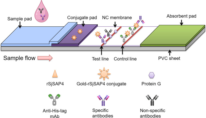
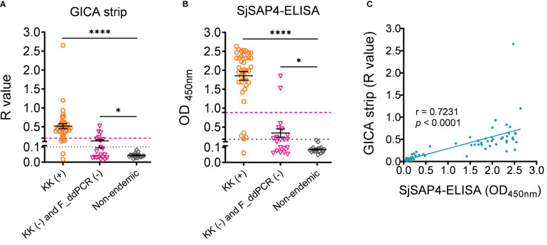
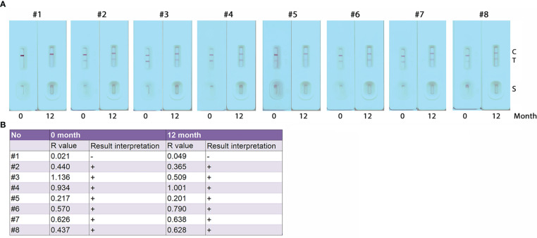
Methods: A GICA strip incorporating a S. japonicum saposin protein, SjSAP4 was developed. For each GICA strip test, diluted serum sample (50 µl) was loaded and strips were scanned after 10 min to convert the results into images. ImageJ was used to calculate an R value, which was defined as the signal intensity of the test line divided by the signal intensity of the control line within the cassette. After determination of optimal serum dilution and diluent, the GICA assay was evaluated with sera collected from non-endemic controls (n = 20) and individuals living in schistosomiasis-endemic areas of the Philippines (n = 60), including 40 Kato Katz (KK)-positive participants and 20 subjects confirmed as KK-negative and faecal droplet digital PCR assay (F_ddPCR)-negative at a dilution of 1:20. An ELISA assay evaluating IgG levels against SjSAP4 was also performed on the same panel of sera.
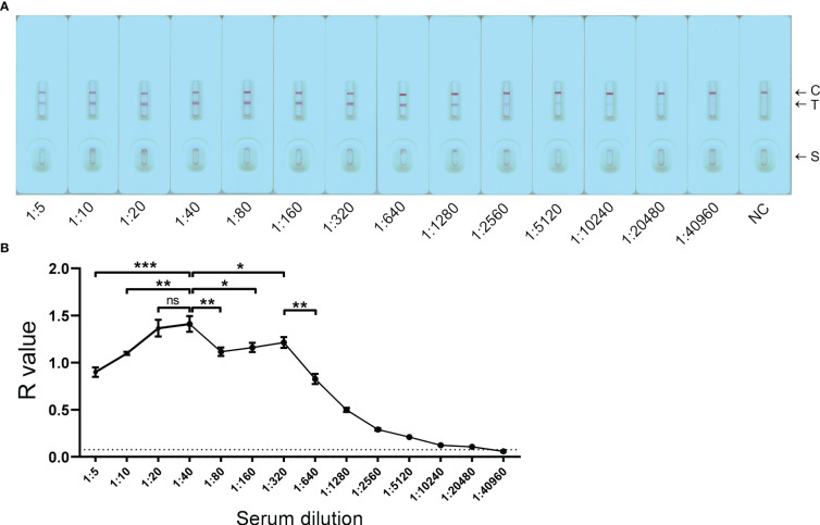
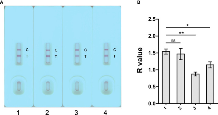
Results: Phosphate-buffered saline (PBS) and 0.9% NaCl were determined as optimal dilution buffer for the GICA assay. The strips tested with serial dilutions of a pooled serum sample from KK-positive individuals (n = 3) suggested that a relatively wide range of dilutions (from 1:10 to 1:320) can be applied for the test. Using the non-endemic donors as controls, the GICA strip showed a sensitivity of 95.0% and absolute specificity; while using the KK-negative and F_ddPCR-negative subjects as controls, the immunochromatographic assay had a sensitivity of 85.0% and a specificity of 80.0%. The SjSAP4-incorperated GICA displayed a high concordance with the SjSAP4-ELISA assay.
Conclusions: The developed GICA assay exhibited a similar diagnostic performance with that of the SjSAP4-ELISA assay, yet the former can be performed by local personnel with minimal training with no requirement for specialised equipment. The GICA assay established here represents a rapid, easy-to-use, accurate and field-friendly diagnostic tool for the on-site surveillance/screening of S. japonicum infection.
Keywords: ELISA; GICA strip; Schistosoma japonicum; lateral flow immunochromatographic test; point-of-care (POC); rapid diagnosis; schistosomiasis; surveillance.
Copyright © 2023 Mu, McManus, Gordon, You, Ross, Olveda and Cai.
Conflict of interest statement
The authors declare that the research was conducted in the absence of any commercial or financial relationships that could be construed as a potential conflict of interest.
Figures
Similar articles
-
Comparative assessment of the SjSAP4-incorporated gold immunochromatographic assay for the diagnosis of human schistosomiasis japonica.Front Public Health. 2023 Sep 1;11:1249637. doi: 10.3389/fpubh.2023.1249637. eCollection 2023. Front Public Health. 2023. PMID: 37736084 Free PMC article.
-
Development of a latex microsphere-based lateral flow immunoassay for the diagnosis of schistosomiasis japonica.PLoS Negl Trop Dis. 2024 Dec 16;18(12):e0012742. doi: 10.1371/journal.pntd.0012742. eCollection 2024 Dec. PLoS Negl Trop Dis. 2024. PMID: 39680611 Free PMC article.
-
Performance of the point-of-care circulating cathodic antigen test in the diagnosis of schistosomiasis japonica in a human cohort from Northern Samar, the Philippines.Infect Dis Poverty. 2021 Sep 23;10(1):121. doi: 10.1186/s40249-021-00905-5. Infect Dis Poverty. 2021. PMID: 34556183 Free PMC article.
-
Reviews and advances in diagnostic research on Schistosoma japonicum.Acta Trop. 2021 Jan;213:105743. doi: 10.1016/j.actatropica.2020.105743. Epub 2020 Nov 4. Acta Trop. 2021. PMID: 33159894 Review.
-
Unleashing the power of colloidal gold immunochromatographic assays for plant virus diagnostics.MethodsX. 2023 Nov 29;12:102498. doi: 10.1016/j.mex.2023.102498. eCollection 2024 Jun. MethodsX. 2023. PMID: 38089155 Free PMC article. Review.
Cited by
-
Schistosomiasis in the Philippines: A Comprehensive Review of Epidemiology and Current Control.Trop Med Infect Dis. 2025 Jan 21;10(2):29. doi: 10.3390/tropicalmed10020029. Trop Med Infect Dis. 2025. PMID: 39998033 Free PMC article. Review.
-
Comparative assessment of the SjSAP4-incorporated gold immunochromatographic assay for the diagnosis of human schistosomiasis japonica.Front Public Health. 2023 Sep 1;11:1249637. doi: 10.3389/fpubh.2023.1249637. eCollection 2023. Front Public Health. 2023. PMID: 37736084 Free PMC article.
-
Application of gold immunochromatographic assay strip combined with digital evaluation for early detection of Toxoplasma gondii infection in multiple species.Parasit Vectors. 2024 Feb 22;17(1):81. doi: 10.1186/s13071-024-06180-1. Parasit Vectors. 2024. PMID: 38389080 Free PMC article.
-
Development of a latex microsphere-based lateral flow immunoassay for the diagnosis of schistosomiasis japonica.PLoS Negl Trop Dis. 2024 Dec 16;18(12):e0012742. doi: 10.1371/journal.pntd.0012742. eCollection 2024 Dec. PLoS Negl Trop Dis. 2024. PMID: 39680611 Free PMC article.
-
Technologies for detecting biological risk factors in agricultural products and their applications.Curr Res Food Sci. 2025 May 6;10:101068. doi: 10.1016/j.crfs.2025.101068. eCollection 2025. Curr Res Food Sci. 2025. PMID: 40485901 Free PMC article. Review.
References
-
- WHO . Ending the neglect to attain the sustainable development goals: a road map for neglected tropical diseases 2021–2030: overview. Geneva: World Health Organization; (2020).
Publication types
MeSH terms
Substances
LinkOut - more resources
Full Text Sources