Review
. 2023 May 24;123(10):6839-6887.
doi: 10.1021/acs.chemrev.3c00016. Epub 2023 Apr 20.

Photocleavable Ortho-Nitrobenzyl-Protected DNA Architectures and Their Applications

Affiliations
Review

Photocleavable Ortho-Nitrobenzyl-Protected DNA Architectures and Their Applications

Michael P O'Hagan et al. Chem Rev. .

Abstract

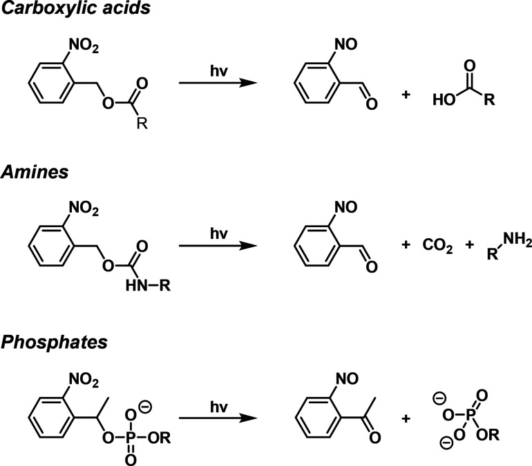
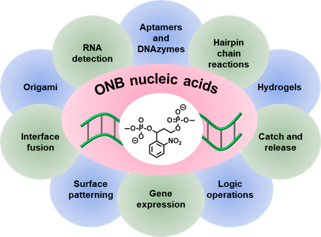
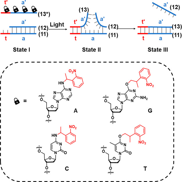
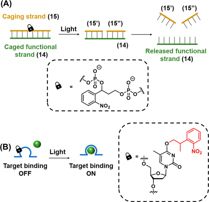
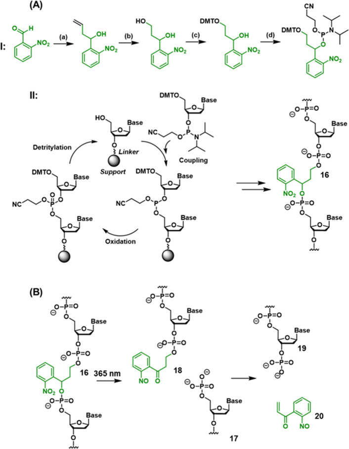
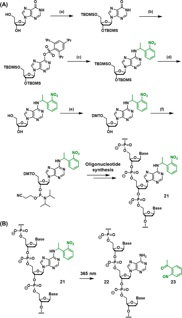
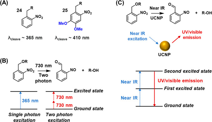
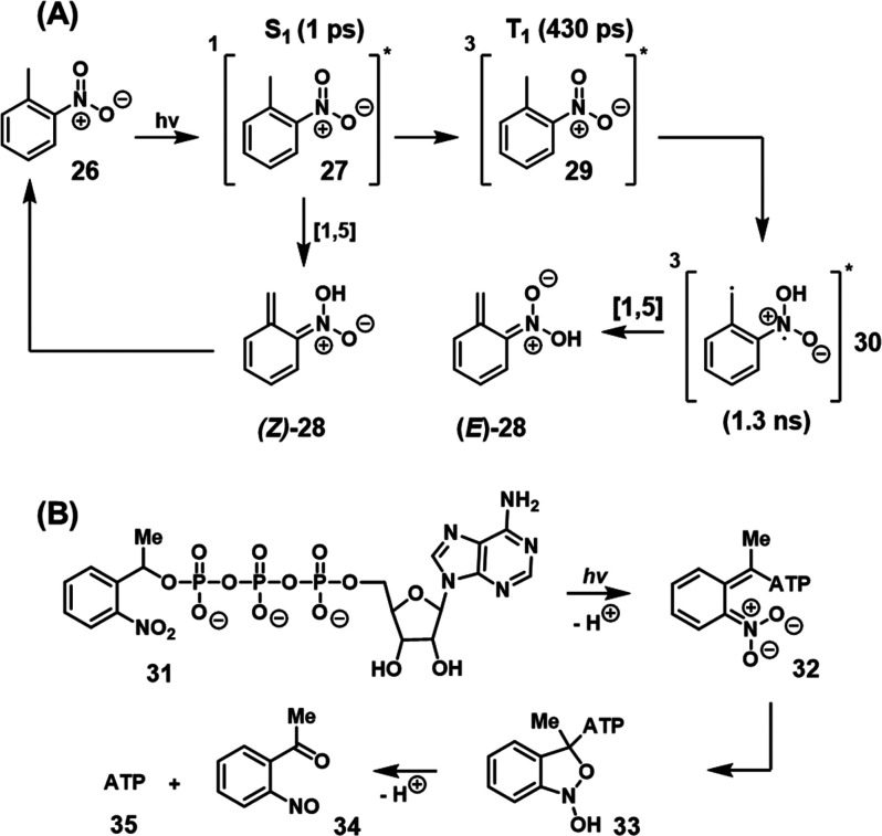
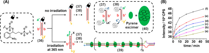
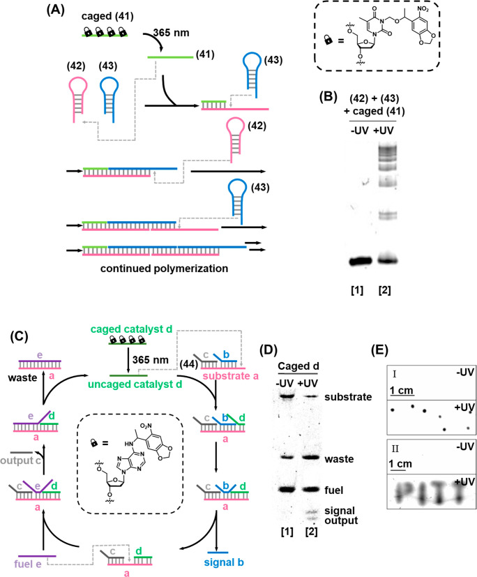
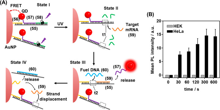
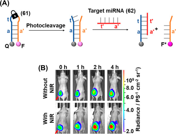
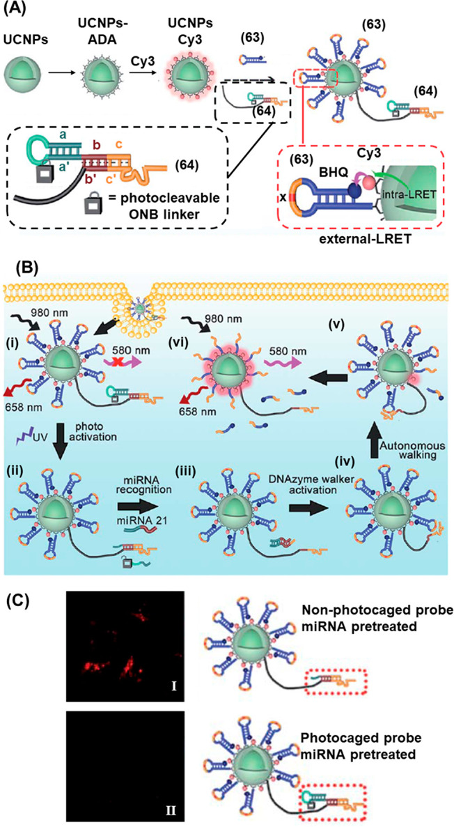
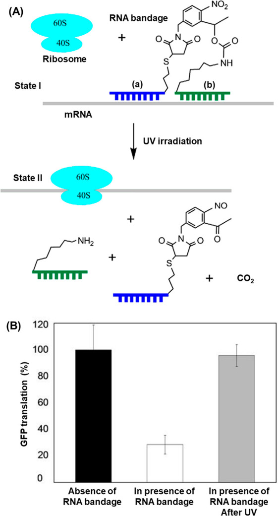
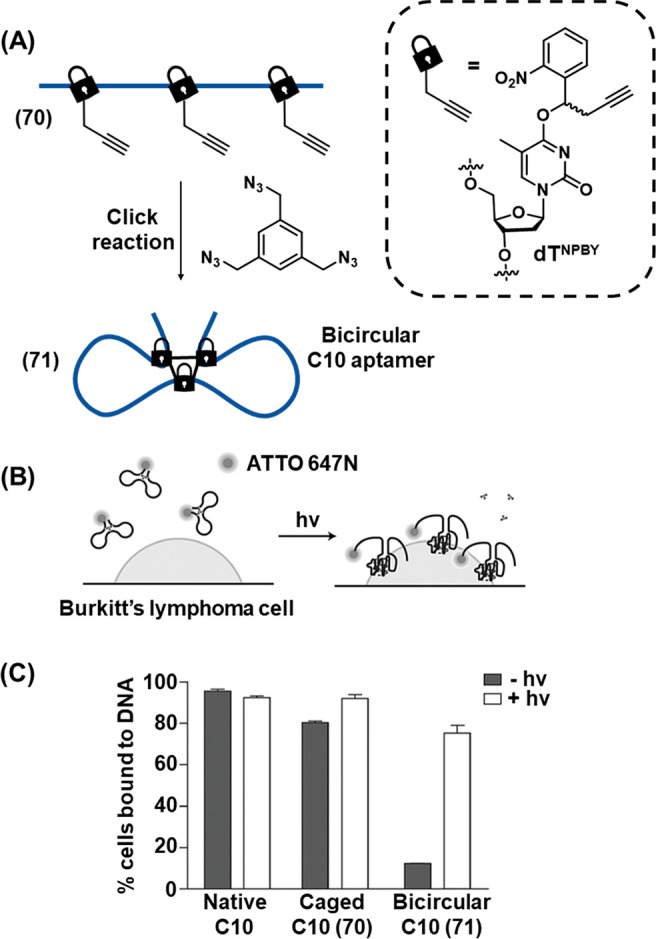
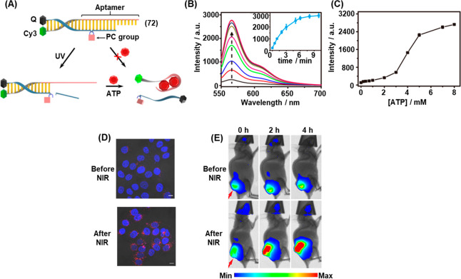
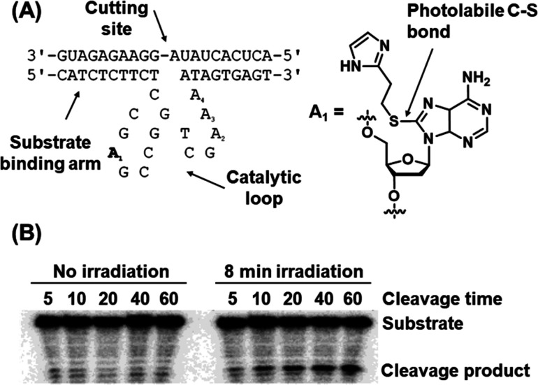
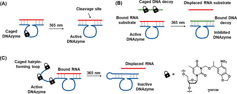
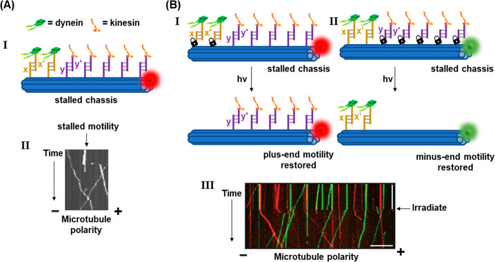
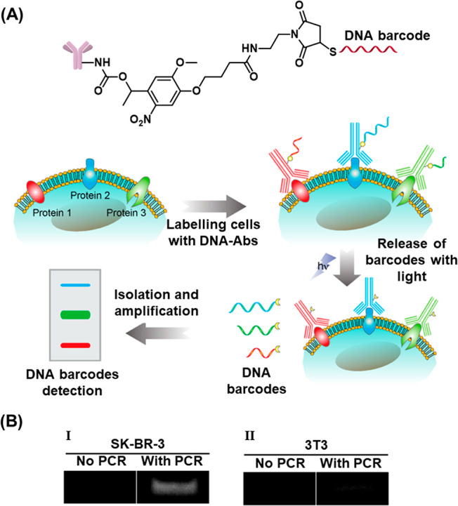
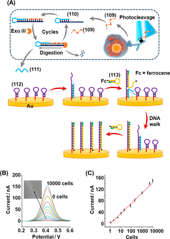
This review article introduces mechanistic aspects and applications of photochemically deprotected ortho-nitrobenzyl (ONB)-functionalized nucleic acids and their impact on diverse research fields including DNA nanotechnology and materials chemistry, biological chemistry, and systems chemistry. Specific topics addressed include the synthesis of the ONB-modified nucleic acids, the mechanisms involved in the photochemical deprotection of the ONB units, and the photophysical and chemical means to tune the irradiation wavelength required for the photodeprotection process. Principles to activate ONB-caged nanostructures, ONB-protected DNAzymes and aptamer frameworks are introduced. Specifically, the use of ONB-protected nucleic acids for the phototriggered spatiotemporal amplified sensing and imaging of intracellular mRNAs at the single-cell level are addressed, and control over transcription machineries, protein translation and spatiotemporal silencing of gene expression by ONB-deprotected nucleic acids are demonstrated. In addition, photodeprotection of ONB-modified nucleic acids finds important applications in controlling material properties and functions. These are introduced by the phototriggered fusion of ONB nucleic acid functionalized liposomes as models for cell-cell fusion, the light-stimulated fusion of ONB nucleic acid functionalized drug-loaded liposomes with cells for therapeutic applications, and the photolithographic patterning of ONB nucleic acid-modified interfaces. Particularly, the photolithographic control of the stiffness of membrane-like interfaces for the guided patterned growth of cells is realized. Moreover, ONB-functionalized microcapsules act as light-responsive carriers for the controlled release of drugs, and ONB-modified DNA origami frameworks act as mechanical devices or stimuli-responsive containments for the operation of DNA machineries such as the CRISPR-Cas9 system. The future challenges and potential applications of photoprotected DNA structures are discussed.


Conflict of interest statement

The authors declare no competing financial interest.

Figures

Figure 1
Figure 1
Examples of ortho-nitrobenzyl-protected chemical functionalities and light-induced cleavage to release the deprotected products.
Figure 2
Figure 2
Diverse applications of o-nitrobenzyl (ONB)-protected nucleic acids in DNA nanotechnology and DNA-based materials.
Figure 3
Figure 3
Displacement pathways of duplex nucleic acid structures. (A) Isoenergetic displacement of a duplex by a fuel strand. (B) Displacement of a toehold-functionalized duplex by a fuel strand. (C) Entropy-driven displacement of a duplex structure by a fuel strand.
Figure 4
Figure 4
(A) Photoinduced displacement of an ONB-locked duplex DNA in the presence of a fuel strand by the photochemical deprotection of the parent duplex and the toehold-mediated displacement of the cleaved product by the fuel strand. (B) Photoinduced cleavage of an ONB-protected hairpin structure containing a masked toehold in the loop region. Upon activating of the toehold, displacement by a fuel strand can proceed in the same manner as (A).
Figure 5
Figure 5
Schematic application of ONB-caged oligonucleotide bases for the light-induced toehold-mediated separation of a duplex nucleic acid. Photoprotected fuel strands are prohibited from initiating the toehold-mediated displacement process. Photodeprotection of the fuel strand releases the active fuel strand for the toehold-mediated displacement of the duplex structure.
Figure 6
Figure 6
Functional photoinduced reconfiguration of nucleic acid structures caged by an ONB protective group. (A) An ONB-caged duplex structure being cleaved by light into two subunits being separated from the duplex structure due to insufficient base stabilization of the resulting fragments. (B) Light-induced uncaging of an ONB-caged sequence in which target binding affinity is perturbed by the presence of the caging moiety. Cleavage of the ONB from the caged base activates binding of the DNA strand to the target.
Scheme 1
Scheme 1. Synthesis and Photocleavage of an ONB-Protected Nucleic Acid Containing the ONB in the DNA Backbone
(A) Panel I: Synthesis of the ONB-functionalized phosphoramite linker for incorporation into the oligonucleotide backbone by solid-phase synthesis. Synthetic pathway: (a) allylation, (b) ozonolysis/reduction, (c) DMT protection, (d) formation of phosphoramidite. For full synthetic details, see reference (206). Panel II: Solid-phase synthesis methodology for oligonucleotide synthesis. Each cycle incorporates an additional phosphoramidite-activated nucleotide (or linker) into the oligonucleotide chain. (B) Light-induced photocleavage of the ONB-functionalized strand.
Scheme 2
Scheme 2. (A) Synthetic Pathway Generating an ONB-Protected Nucleic Acid Containing a Caged Adenine Base; (B) Light-Induced Deprotection of the ONB-Protected Oligonucleotide Base
Synthetic pathway: (a) 3′/5′-OH TBDMS protection, (b) O6 activation, (c) C6 substitution, (d) 3′/5′ OH deprotection, (e) 5′-OH DMT protection, (f) formation of phosphoramidite. For full synthetic details, see reference (207).
Figure 7
Figure 7
Photophysical control of the photochemical deprotection of ONB-protecting groups by (A) red-shifting the excitation wavelength using electron donating substituents associated with the benzene ring, (B) two-photon excitation of the ONB chromophore and (C) application of up-conversion nanoparticles.
Scheme 3
Scheme 3. Mechanistic Issues Related to the Light-Induced Deprotection of the ONB Protecting Unit
(A) Light-induced phototautomerization of the ONB into the aci-nitro intermediate. (B) Mechanistic steps involving the light-induced phototautomeriztion of the ONB-protected ATP to an aci-nitro intermediate that undergoes reconfiguration and dark degradation into the o-nitrosobenzophenone product, releasing free ATP.
Figure 8
Figure 8
(A) Photochemical activation of an ONB-caged hairpin nucleic acid yielding a toehold-functionalized duplex to stimulate the hybridization chain reaction (HCR) between two pyrene-modified hairpins to yield oligomeric pyrene excimer structures. (B) Time-dependent exciplex emission intensities generated by the excimer oligomer formed upon the photochemical cleavage of the ONB-bridged activator for different time intervals: (a) 0 min, (b) 1 min, (c) 2 min, (d) 5 min, (e) 10 min, (f) 20 min. Figure adapted with permission from ref. (201). Copyright 2013, American Chemical Society.
Figure 9
Figure 9
(A) Photodeprotection of an ONB-caged strand that initiates the HCR process yielding the oligomerized duplex strand HCR product. (B) Electrophoretic separation of the resulting HCR products without (Lane 1) and with (Lane 2) light-induced activation of the HCR process. (C) Photochemically triggered activation of a fuel/catalyst driven toehold-mediated strand displacement cycle using the light-induced deprotection of an ONB-functionalized strand as the activator. (D) Imaging the light-induced fuel-driven strand displacement cycle by electrophoretic separation. (E) On-gel patterning of dots (Panel I) and text (Panel II). Figure adapted with permission from ref. (202). Copyright 2015, American Chemical Society.
Figure 10
Figure 10
(A) Fluorescence imaging of an mRNA analyte by the light-induced deprotection of an ONB-protected fluorophore/quencher labeled hairpin sensing probe. (B) Intracellular detection of the mRNA by the light-induced activation of the ONB-protected hairpins incorporated in the cell environment. Panel I: map of irradiation site (blue circle), probe-treated cell (red square) and untreated cell (green square). Panel II: intracellular beacon fluorescence prior to photoirradiation. Panel III: intracellular beacon fluorescence after photoirradiation. Figure adapted with permission from ref. (258). Copyright 2012, Royal Society of Chemistry.
Figure 11
Figure 11
(A) Schematic integration of an ONB-photoprotected fluorescent probe into cancer cells for the temporal activated sensing of the MnSOD mRNA. The light-induced cleavage of the probe yields a fluorescent quenched hairpin product in the absence of target. In the presence of the mRNA target the fluorescence of the remains switched on. (B) Intracellular activation of fluorescence imaging of intracellular mRNA upon light-induced activation of the probe. Figure adapted with permission from ref. (259). Copyright 2013, American Chemical Society.
Figure 12
Figure 12
(A) Light-triggered (365 nm) electrochemical sensing of MnSOD mRNA at the single-cell level using a methylene blue (MB) functionalized ONB-caged hairpin-modified nanoelectrode. (B) Intracellular voltammetric responses of the MB-functionalized ONB-caged hairpin-modified electrode: (a) before irradiation, (b) after irradiation and mRNA-induced displacement of the fragmented hairpin. (C) Intracellular detection of MnSOD mRNA using an ONB-caged FAM-modified hairpin hybridized to a dabcyl-quencher-modified strand. (D) Time-dependent fluorescence intensities upon sensing intracellular MnSOD mRNA by the optical probe: (a) without light-triggered cleavage of the hairpin, (b) after light-induced cleavage of the hairpin, (c) after upregulation of the MnSOD mRNA using LPS and light-induced cleavage of the probe. Figure adapted with permission from ref. (255). Copyright 2018, American Chemical Society.
Figure 13
Figure 13
(A) Intracellular amplified sensing of cMYC mRNA by UCNPs functionalized with an ONB-caged Cy3-modified hairpin. In the presence of an auxiliary Cy5-functionalized hairpin, the NIR-triggered UC-stimulated cleavage of the ONB-caged hairpin leads to a fragmented hairpin being opened by the c-MYC mRNA, a process initiating the HCR with the auxiliary hairpin, resulting in the Cy3/Cy5 FRET process providing the sensing readout signal. (B) Fluorescence output of the sensing module: (a) before irradiation and (b) after irradiation, resulting in the Cy5 FRET signal. Figure adapted with permission from ref. (260). Copyright 2019, John Wiley and Sons.
Figure 14
Figure 14
(A) Schematic amplified detection of miRNA in HeLa cells using an ONB-gated CdTe QD/AuNP conjugate. (B) Amplified luminescence signals observed upon sensing miRNA-21 with the Au nanoparticle/CdTe quantum dots in miRNA-containing HeLa cells and miRNA-negative HEK cells upon light-induced uncaging of the sensing probe for different time intervals. Figure adapted with permission from ref. (200). Copyright 2018, American Chemical Society.
Figure 15
Figure 15
(A) Photochemically triggered activation of an ONB-protected fluorophore/quencher caged hairpin miRNA sensing probe. (B) Application of the probe shown in (A) for the in vivo detection of miRNA-21 present in HeLa tumors in mice. Coupling of the probe UCNPs allowed NIR-triggered activation of the detection module. Images of probe fluorescence correspond to different time intervals in the absence of probe activation (upper) or following probe activation with NIR light (lower). Figure adapted with permission from ref. (257). Copyright 2019, American Chemical Society.
Figure 16
Figure 16
(A) Schematic synthesis of Cy3-modified UCNPs cofunctionalized with quencher-modified hairpin strands and ONB-caged DNAzyme units for the light induced deprotection of the DNAzyme structure that triggers the cleavage of the quencher-modified hairpin in the presence of the miRNA target, activating Cy3 fluorescence. (B) Intracellular application of the functionalized UCNPs for detection of miRNA-21 via the photochemical uncaging of the DNAzyme and miRNA-guided activation of the DNAzyme. (C) Confocal fluorescence imaging of miRNA-21 negative cells treated with (Panel I) uncaged UCNP probes pre-exposed to miRNA-21 to simulate extra-cellular activation, demonstrating false-positive intracellular fluorescence readout, resulting from extracellular miRNA-21 activation and (Panel II) photocaged UCNP probes pretreated with miRNA-21, demonstrating effective caging of the probes and elimination of false-positive intracellular fluorescence. Figure adapted with permission from ref. (229). Copyright 2020, Royal Society of Chemistry.
Figure 17
Figure 17
(A) Phototriggered unlocking of an ONB-protected mRNA template toward the ribosome translation of the protein on the deprotected template. (B) GFP yields in the presence of (I) the naked mRNA template, (II) the ONB-protected template in the absence of auxiliary light-induced deprotection, (III) the light-induced deprotected template. Figure adapted with permission from ref. (203). Copyright 2008, Elsevier.
Figure 18
Figure 18
(A) Schematic application of an ONB-caged antisense morpholino hairpin for the light-induced silencing of a target RNA. (B) In vivo application of the ONB-protected hairpin for the phototriggered silencing of ntl RNA in zebrafish. Panel I: zebrafish treated with ntl-targeting caged morpholino (ntl cMO) hairpin (65) in the absence of light (tail developed normally), Panel II: zebrafish treated with ntl cMO hairpin (65) and subjected to photoirradiation (tail development inhibited). (C) Spatially localized activation of the RNA-silencing hairpin in zebrafish chordamesoderm domains. Panel (I): in the absence of hairpin (65) normal development is observed in the photoirradiated (red) region. Panel (II): in the presence of hairpin (65) cell patterning defects are observed specifically in the photoirradiated (red) region. Figure adapted with permission from ref. (268). Copyright 2007, Springer Nature.
Figure 19
Figure 19
Light-induced switch-on/switch-off of gene expression using photocaged morpholino oligonucleotides. (A) Photodeprotection of the morpholino oligonucleotide that silences the target RNA and switches off gene expression. (B) Photodeprotection of sequestered target RNA activating gene expression. (C,D) Distribution of tail-development phenotypes of zebrafish embryos treated with (C) the photocaged inactive morpholino shown in (A) without exposure to light (80% normal tail development) and subjected to UV light (80% tail development perturbed), and (D) photocaged target RNA shown in (B) without exposure to light (∼100% abnormal tail development) and subjected to UV light (∼90% normal tail development). (E) Representative microscopy images of the different phenotypes in (C) and (D). (F) Spatially localized regulation of ntla gene expression in zebrafish tails. Panel I–III: treatment with the phototriggered switch-off construct in (A) results in normal tail development and observation of ntla antibody fluorescence throughout the tail in the dark (Panel I), global illumination disrupts tail development and ntla antibody fluorescence is not observed (Panel II) while irradiation at a specific site using a laser silences ntla expression in the illuminated cells (arrow shows absence of ntla antibody fluorescence in illuminated region). Panel IV–VI: treatment with the phototriggered switch-on module in (B) results in disrupted tail development and absence of ntla antibody fluorescence throughout the tail in the dark (Panel IV), global illumination restores tail development and ntla antibody fluorescence observed throughout the tail (Panel V) while irradiation at a specific site using a laser restores ntla expression specifically in the illuminated cells (asterisks show ntla antibody fluorescence in illuminated region). Figure adapted with permission from ref. (270). Copyright 2012, The Company of Biologists Ltd.
Figure 20
Figure 20
Phototriggered silencing of a target RNA by the photocleavage of a coumarin-labeled ONB-caged hairpin and displacement of the coumarin-labeled antisense strand to yield a silenced miRNA construct. (B) Schematic role of lys-6 in controlling the expression of GFP in the ASE neurons of C. elegans. (C) Temporal control of lys-6 expression in C. elegans treated with the caged hairpin (69) and illuminated at different stages of organism development. Phototriggered knockdown of lys-6 before the comma stage results in expression of GFP in both ASEL and ASER. Knockdown after the comma stage results in the wild-type phenotype. (D) Spatially localized activation of hairpin (69) in specific cells of four-cell C. elegans embryos. Panel I–II: specific irradiation of the ABa cell (ASEL precursor) leads to the knockdown phenotype. Panel III–IV: specific irradiation of the ABp cell (ASER precursor) results in no disruption of phenotype. The irradiation site is shown by the dotted circles and localized activation of coumarin fluorescence (corresponding to hairpin photocleavage) in the specific cells is demonstrated. Figure adapted with permission from ref. (267). Copyright 2011, American Chemical Society.
Figure 21
Figure 21
(A) An ONB-protected antithrombin aptamer generated by the photocaging of a thymine base in the thrombin binding site. (B) Concentration dependence of blood clotting times for (a) the native thrombin aptamer, (b) the inhibited ONB-caged thrombin aptamer, (c) the photodeprotected ONB-caged thrombin aptamer. Figure adapted with permission from ref. (204). Copyright 2005, American Chemical Society.
Figure 22
Figure 22
(A) Schematic bicircular caging of the ONB-acetylene functionalized C10 Burkitt’s lymphoma cell aptamer by the trimethyl azidophenyl bridging unit, applying click chemistry principles. (B) Photochemical uncaging of the ATTO 647M-labeled bicircular caged aptamer and binding of the uncaged aptamer to the lymphoma cells. (C) Binding of the native aptamer, the noncircularized ONB-modified aptamer and the bicircularized aptamer to the lymphoma cells before irradiation (black) and after irradiation (white). Figure adapted with permission from ref. (277). Copyright 2016, John Wiley and Sons.
Figure 23
Figure 23
(A) Schematic application of a fluorophore/quencher ONB-protected duplex DNA that includes a caged ATP-aptamer sequence as a functional module for optical detection of ATP. The photocleavage of the ONB units lead to an unstable duplex structure being a displaced by ATP to yield the fluorescent ATP-aptamer complex as optical readout. (B) Fluorescence intensities generated by the detection module shown in (A) upon sensing ATP, 5 mM, and photochemical uncaging the photoprotective unit by irradiation for different time intervals (from 0–9 min). Inset: derived intensity vs irradiation dose curve. (C) Fluorescence intensities generated by the module shown in (A) upon analyzing different concentrations of ATP after photodeprotection of the sensing module for a fixed time interval. (D) Intracellular imaging of ATP in cells by the introduction of the sensing module and UCNPs into the cells and near-IR (NIR) activation of the module. (E) In vivo imaging of ATP in HeLa tumors in mice using UCNPs and the sensing module shown upon NIR activation of the detection platform. Figure adapted with permission from ref. (278). Copyright 2017, American Chemical Society.
Figure 24
Figure 24
Schematic photocleavage of a thiolated adenonsine-protected Zn2+-ion-dependent DNAzyme/substrate complex leading to the activation of the DNAzyme activity. (B) Electrophoretic imaging of the DNAzyme activity before and after photoirradiation. Figure adapted with permission from ref. (293). Copyright 2004, American Chemical Society.
Figure 25
Figure 25
Schematic strategies to photochemically control DNAzyme activities of ONB-protected nucleic acid constructs. (A) Photodeprotection of an ONB-functionalized loop domain of a Mg2+-dependent DNAzyme leading to activation of catalytic activity. (B) Photodeprotection of an ONB-modified strand inducing the release of an inhibitor strand displacing the substrate strand associated with an active Mg2+-ion dependent DNAzyme/substrate complex. (C) An ONB-functionalized tether conjugated to an active Mg2+-ion dependent DNAzyme/substrate construct being deactivated by photodeprotection of the ONB-modified tether and the displacement of the substrate constituent by hairpin formation.
Figure 26
Figure 26
(A) Schematic application of an ONB-protected hairpin structure being photodeprotected to yield a duplex unit that is displaced by an RNA strand to yield a functional duplex for the guided cleavage of RNA by RNase. (B) Electrophoretic imaging of the cleavage of the target RNA by the photodeprotection of hairpin (73). Figure adapted with permission from ref. (294). Copyright 2006, John Wiley and Sons.
Figure 27
Figure 27
(A) Schematic reaction module executing an AND logic gate operation using two different wavelengths of light as inputs. (B) Truth table corresponding to the AND logic gate present in (A) immobilized in an agarose gel, with different regions illuminated with different combinations of I1 and I2. (C) Schematic reaction module using light and miRNA-21 as inputs for the intracellular sensing of miRNA-21 guided by the AND gate operation. (D) Application of the reaction module shown in (C) to trigger the logic gate optical transduction corresponding to the sensing of miRNA-21 in HeLa cells. Panel (A), (B) adapted with permission from ref. (302). Copyright 2012, American Chemical Society. Panel (C), (D) adapted with permission from ref. (303). Copyright 2013, American Chemical Society.
Figure 28
Figure 28
(A) Schematic probing of the c-Met and CD7 cell membrane receptor stimulated migration of cells using aptamer constructs controlling the receptor-induced migration functions by an AND gate logic operation. While the individual aptamer constructs do not affect the cell migration functionalities, the assembly of the receptors stimulated by uncaging the aptamer bridging units with simultaneous application of photochemical ONB-strand cleavage and auxiliary DNA strand inputs (Panel I) inhibits cell migration by preventing binding of HGF (Panel II). (B) Schematic activation of the aptamer assembly by the AND gate operation. By appropriate Cy3/Cy5 labeling of the aptamer constructs the dynamic bridging of the aptamers and inhibition of cell migration are probed by the resulting FRET signal output. (C) Fluorescence output signals demonstrating the AND gate induced bridging of the receptors (Panel I) and confocal microscopy imaging of cell migration demonstrating motility in the absence of both AND gate inputs (Panel II) and inhibited motility in the presence of both inputs (Panel III). Figure adapted with permission from ref. (304). Copyright 2019, John Wiley and Sons.
Figure 29
Figure 29
(A) Photochemical fusion of nucleic acid functionalized liposomes via the application of an ONB-hairpin-modified liposome loaded with UCNPsfor the NIR cleavage of the ONB-protected hairpins. The fusion leads to mixture of the loads in the liposomes. (B) Size increase of the fused liposomes induced by irradiation with light (curve a); no size changes of the liposomes occurs in the absence of light (curve b). (C) Time-dependent fluorescence changes corresponding to formation of the Tb3+/DPA complex generation upon the light-triggered fusion and the mixing of Tb3+ and DPA in the fused containment (curve a) triggered by NIR irradiation. No fluorescence changes are observed in the absence of light-induced fusion (curve b). (D) NIR-induced fusion of ONB-hairpin-functionalized, doxorubicin-loaded liposomes with MUC-1 aptamer functionalized HeLa cells. (E) Selective cytotoxicity toward HeLa and hESC cells demonstrated upon treatment of the cells with the UCNP/Dox-loaded ONB-protected liposomes and subsequent NIR fusion triggered by unblocking of the hairpins. Successful fusion proceeds only with the MUC1-aptamer-modified HeLa cells. Figure adapted with permission from ref. (308). Copyright 2019, Creative Commons CC-BY 3.0.
Figure 30
Figure 30
(A) Schematic photolithographic patterning of an ONB-protected hairpin monolayer-modified surface by selective UV light induced deprotection of the monolayer through a mask and the subsequent activation of the HCR in the presence of fluorescein-modified hairpins (87)/(88). The selective formation of fluorophore patterned domains of oligomeric DNA structures is imaged by fluorescence confocal microscopy and AFM. (B) Fluorescent image of the DNA-patterned surface generated upon photolithographic patterning of the monolayer using a grid-like mask. (C) AFM image of the square corner edges of the patterned DNA interface. (D) Cross-sectional profile of the patterned surface. (E) Schematic photopatterning of an ONB-protected monolayer-modified surface through a mask and selective deposition of nucleic acid-modified fluorophores or nanoparticles to the deprotected domains through complementary base-pair hybridization. Panel (A)–(D) adapted with permission from ref. (332). Copyright 2015, John Wiley and Sons.
Figure 31
Figure 31
(A) Photolithographic patterning of an ONB-functionalized DNA and the subsequent association of fluorophore-modified strands to the deprotected monolayer domains. (B) Three-color fluorescence pattern generated upon stepwise full-dose light deprotection of the monolayer through a hexagonal mask, Panel I, and partial dose light-induced deprotection of the monolayer, leading to mixed fluorescent colors in the photopatterned domains, Panel II. (C) Construction of a multicolor image by the sequential patterning of extracted red/green/blue colors leading to the reproduced multicolor image upon merging. (D) Photolithographic gene patterning on a target surface for guided gene expression. The yellow fluorescent protein (YFP) gene is photopatterned and imaged by a fluorescent label, Panel I. Subsequently the patterned gene is used to express YFP, Panel II. (E) Patterning of three different genes corresponding to yellow (YFP), red (RFP), and cyan (CFP) fluorescent proteins in different surface containments followed by the guided expression of the respective proteins in the patterned domains. Panel (B)–(D) adapted with permission from ref. (331). Copyright 2018, John Wiley and Sons.
Figure 32
Figure 32
(A) Photolithographic patterning of ONB-functionalized red-fluorophore-modified duplex monolayer coated microparticles using a scanning confocal microscope equipped with a laser source. The two-photon laser stimulated deprotection of the ONB groups leads to patterned nonfluorescent domains on the particles. (B) Square or triangle shaped patterns generated on the microparticles. (C) Schematic two-color patterning of microparticles; microparticles are coated with a monolayer mixture of ONB-functionalized red fluorophore-modified duplex and a ONB-functionalized hairpin-modified quencher hybridized with a green fluorescence strand, yielding a caged structure exhibiting quenched green fluorescence. The composite monolayer-modified microparticles yield continuous red fluorescence. Scanning confocal microscopy aided two-photon laser photopatterning of the microparticles leads to photodeprotection of the ONB units associated with the two fluorescent constituents associated with the patterned domain. This leads to the release of the red fluorescenct constituent from the coating and to the ungaging of the green fluorescent constituent yielding a green pattern on the red fluorescent background coating. (D) Circular green fluorescent doains patterned on the red fluorescent coated microparticles. Figure adapted with permission from ref. (334). Copyright 2019, American Chemical Society.
Figure 33
Figure 33
(A) Stepwise synthesis of ONB-modified nucleic acid-based tetramethyl rhodamine dextran (TMR-D) loaded microcapsules. (B) Light-induced degradation of the ODN-modified microcapsules resulting in the release of the load. (C) Fluorescence spectra of TMR-D released from the ONB-modified microcapsules irradiated for different time intervals: (a) 0 s to (g) 5 min. (D) Fluorescence spectra of doxorubicin-dextran (Dox-D) released from the ONB-modified microcapsules upon irradiation for different time intervals: (a) 0 s to (i) 120 s. (E) Cytotoxicity of the Dox-D-loaded microcapsules toward MDA-MB-231 breast cancer cells (red) and epithelial MCF-10A cells (blue) and measuring cell viability after 24 h: Panel I – without irradiation and deprotection of the microcapsules. Panel II – after irradiation for 5 min. Figure adapted with permission from ref. (370). Copyright 2016, American Chemical Society.
Figure 34
Figure 34
(A) Schematic photolithographic patterning of an ONB nucleic acid functionalized polyacrylamide hydrogel. (B) Fluorescent image of the photodeprotected patterned hydrogel. (C) Schematic photopatterning of an ONB nucleic acid functionalized cross-linked hydrogel matrix leading to photopatterned circular low stiffness quasi-liquid hydrogel domains with toehold functionalized duplex constituents. The subsequent association of the MUC-1 aptamer to the soft patterned domains guides the binding of HeLa cells to the patterned regions. (D) SEM image of the ONB-functionalized hydrogel before deprotection (Panel I) and after light-induced deprotection of the ONB units confirmed by the circular domains of the patterned hydrogel (Panel II). (E) HeLa cells caught in the patterned domains modified with the MUC-1 aptamer. Figure adapted with permission from ref. (369). Copyright 2021, the Authors.
Figure 35
Figure 35
(A) Schematic light-induced “mechanical” unlocking of a closed ONB-locked origami bundle tweezers-type structure leading upon deprotection to an open tweezers structure. (B) Schematic light-induced unlocking and opening of the tweezers structure, accompanied by a histogram corresponding to the populations of closed tweezer structures before illumination and the open tweezers after illumination. (C) Examples of the closed origami bundle tweezer before light-induced separation (left) and after light-induced deprotection of the ONB units and opening of the tweezers (right). Figure adapted with permission from ref. (387). Copyright 2018, John Wiley and Sons.
Figure 36
Figure 36
(A) Assembly of a photoresponsive ONB-protected origami tile/antifluorescein antibody hybrid system through the binding of the fluorescein (antigen)-ONB-protected nucleic acid (111) to the complementary origami tile edge-modified tether (112), and the light-induced separation of the hybrid by deprotection of the ONB linkage. (B) AFM images of the hybrid origami tile fluorescein conjugate before deprotection, Panel I, and after light-stimulated deprotection, Panel II. Figure adapted with permission from ref. (388). Copyright 2015, Royal Society of Chemistry.
Figure 37
Figure 37
(A) Schematic photochemical activation of the CRISPR-Cas9 machinery caged within an origami DNA ring toward the guided cleavage of a target site in a DNA duplex. The single guide RNA (sgRNA) Cas9 complex is inactivated by trapping in the ring via an ONB-protected strand linked to an anchoring strand protruding in the cavity of the origami ring. (B) The light induced deprotection of the Cas9-sgRNA machinery leads to the site-specific sgRNA-guided cleavage of the target duplex substrate. (C) AFM images corresponding to Panel I – the Cas9-sgRNA caged machinery in the DNA origami ring. Panel II – the vacant origami ring after deprotection of the ONB caging unit. (D) Electrophoretic analysis of the Cas9-sgRNA-guided site-specific cleavage of the duplex shown in (B). The cleavage band is only observed after light-induced release of the Cas9-sgRNA machinery from the cavity of the nanoring. Figure adapted with permission from ref. (389). Copyright 2021, Royal Society of Chemistry.
Figure 38
Figure 38
(A) Schematic assembly of a photoresponsive ONB-protected origami sphere consisting of two interlinked origami hemispheres bridged into the sphere structure by complementary ONB-functionalized tethers protruding from the hemisphere edges. (B) Photodeprotection of the hemisphere bridging duplex leads to the separation of the sphere into two tethered hemispheres. (C) AFM images corresponding to Panel I – the intact sphere structure composed of the ONB duplex bridged edges of the hemispheres. Panel II – the separation of the sphere into two tethered hemispheres unpon the light-induced deprotection of the crossover ONB-functionalized bridging units. Figure adapted with permission from ref. (390). Copyright 2015, Royal Society of Chemistry.
Figure 39
Figure 39
(A) Panel I – Assembly of two motor proteins, dynein (D) and kinesin (K), on a fluorescent DNA origami bundle acting as a chassis for motility along microtubules. The functionalization of D with nucleic acid x′ and modification of K with y′ allowed their linkage, through hybridization, to protruding nucleic acid anchors linked to the origami chassis. At a D:K ratio corresponding to 2:5, stalled motility of the chassis is observed, Panel II. (B) Functionalization of the DNA origami chassis with photocleavable x (Panel I) or photocleavable y (Panel II) yielded photoresponsive bundles modified by the motor proteins. Upon the selective light-induced removal of D (Panel I), the resulting K-rich origami restores, in the presence of ATP, motility toward the plus-end of the microtubule. In turn, photorelease of the K motor protein yields the D-rich origami chassis that restores, in the presence of ATP, motility toward the minus-end of the microtubule (Panel II). By labeling the two chassis with red or green fluorophores, respectively, the resulting restoration of motility of the stalled chassis upon light-induced deprotection is observed (Panel III). Figure adapted with permission from ref. (391). Copyright 2012, The American Association for the Advancement of Science.
Figure 40
Figure 40
(A) Schematic probing of proteins associated with cell membranes using specific antibody-barcode nucleic acid conjugates bridged by photocleavable ONB bridges. The association of the antibody conjugate(s) to the specific protein followed by the UV-light deprotection of the cell-bound barcode(s) as labels and their PCR amplification provide the means to analyze the respective protein(s). The PCR-amplified barcode is analyzed by electrophoretic separation. (B) Electrophoretic analysis of: Panel I – SK-BR-3 cells, containing the HER2/neu receptor protein and Panel II – 3T3 cells lacking the HER2/neu receptor protein using the ONB-bridged barcode conjugated HER2/neu antibody using the light/PCR-amplified detection platform outlined in (A). Figure adapted with permission from ref. (405). Copyright 2012, American Chemical Society.
Figure 41
Figure 41
(A) Schematic application of an ONB-bridged antibody-nucleic acid-based conjugate for the detection of the K+-ion channel protein in HEK 293 cells using an auxiliary amplification machinery for the DNA label amplification and its electrochemical sensing. The photoresponsive ONB-antibody-nucleic acid barcode conjugate associated with the cells is photocleaved and the resulting cleaved barcode (109) is amplified by a probe (110)/Exo III machinery, recycling (109), and generating strand (111) as a “waste” product. The “waste” strand opens the hairpin-monolayer (112) functionalized electrode and the resulting toehold-modified monolayer is quantitatively analyzed by a ferrocene reporter unit (113). The intensity of the voltammetric response of the functionalized electrode directly relates to the number of cells containing the K+-ion channesl. (B) Panel I – Voltammetric responses of the sensing module upon analyzing different number of HEK-293 cells. Panel II – Derived calibration curve. Figure adapted with permission from ref. (406). Copyright 2019, Elsevier.

Similar articles

Cited by

References

    1. Merrifield R. B. Solid Phase Peptide Synthesis. I. The Synthesis of a Tetrapeptide. J. Am. Chem. Soc. 1963, 85, 2149–2154. 10.1021/ja00897a025. - DOI
    1. Hartrampf N.; Saebi A.; Poskus M.; Gates Z. P.; Callahan A. J.; Cowfer A. E.; Hanna S.; Antilla S.; Schissel C. K.; Quartararo A. J.; Ye X.; Mijalis A. J.; Simon M. D.; Loas A.; Liu S.; Jessen C.; Nielsen T. E.; Pentelute B. L. Synthesis of Proteins by Automated Flow Chemistry. Science 2020, 368, 980–987. 10.1126/science.abb2491. - DOI - PubMed
    1. Laps S.; Satish G.; Brik A. Harnessing the Power of Transition Metals in Solid-Phase Peptide Synthesis and Key Steps in the (Semi)Synthesis of Proteins. Chem. Soc. Rev. 2021, 50, 2367–2387. 10.1039/D0CS01156H. - DOI - PubMed
    1. Laps S.; Atamleh F.; Kamnesky G.; Sun H.; Brik A. General Synthetic Strategy for Regioselective Ultrafast Formation of Disulfide Bonds in Peptides and Proteins. Nat. Commun. 2021, 12, 870.10.1038/s41467-021-21209-0. - DOI - PMC - PubMed
    1. Isidro-Llobet A.; Álvarez M.; Albericio F. Amino Acid-Protecting Groups. Chem. Rev. 2009, 109, 2455–2504. 10.1021/cr800323s. - DOI - PubMed

Publication types