. 2023 Jun 15;11(3):e0475322.
doi: 10.1128/spectrum.04753-22. Epub 2023 Apr 20.

MARCO Inhibits Porcine Reproductive and Respiratory Syndrome Virus Infection through Intensifying Viral GP5-Induced Apoptosis

Affiliations

MARCO Inhibits Porcine Reproductive and Respiratory Syndrome Virus Infection through Intensifying Viral GP5-Induced Apoptosis

Xiaoxiao Zhang et al. Microbiol Spectr. .

Abstract

Studying viral glycoprotein-host membrane protein interactions contributes to the discovery of novel cell receptors or entry facilitators for viruses. Glycoprotein 5 (GP5), which is a major envelope protein of porcine reproductive and respiratory syndrome virus (PRRSV) virions, is a key target for the control of the virus. Here, the macrophage receptor with collagenous structure (MARCO), which is a member of the scavenger receptor family, was identified as one of the host interactors of GP5 through a DUALmembrane yeast two-hybrid screening. MARCO was specifically expressed on porcine alveolar macrophages (PAMs), and PRRSV infection downregulated MARCO expression both in vitro and in vivo. MARCO was not involved in viral adsorption and internalization processes, indicating that MARCO may not be a PRRSV-entry facilitator. Contrarily, MARCO served as a host restriction factor for PRRSV. The knockdown of MARCO in PAMs enhanced PRRSV proliferation, whereas overexpression suppressed viral proliferation. The N-terminal cytoplasmic region of MARCO was responsible for its inhibitory effect on PRRSV. Further, we found that MARCO was a proapoptotic factor in PRRSV-infected PAMs. MARCO knockdown weakened virus-induced apoptosis, whereas overexpression aggravated apoptosis. MARCO aggravated GP5-induced apoptosis, which may result in its proapoptotic function in PAMs. The interaction between MARCO and GP5 may contribute to the intensified apoptosis induced by GP5. Additionally, the inhibition of apoptosis during PRRSV infection weakened the antiviral function of MARCO, suggesting that MARCO inhibits PRRSV through the regulation of apoptosis. Taken together, the results of this study reveal a novel antiviral mechanism of MARCO and suggest a molecular basis for the potential development of therapeutics against PRRSV. IMPORTANCE Porcine reproductive and respiratory syndrome virus (PRRSV) has been one of the most serious threats to the global swine industry. Glycoprotein 5 (GP5) exposed on the surface of PRRSV virions is a major glycoprotein, and it is involved in viral entry into host cells. A macrophage receptor with collagenous structure (MARCO), which is a member of the scavenger receptor family, was identified to interact with PRRSV GP5 in a DUALmembrane yeast two-hybrid screening. Further investigation demonstrated that MARCO may not serve as a potential receptor to mediate PRRSV entry. Instead, MARCO was a host restriction factor for the virus, and the N-terminal cytoplasmic region of MARCO was responsible for its anti-PRRSV effect. Mechanistically, MARCO inhibited PRRSV infection through intensifying virus-induced apoptosis in PAMs. The interaction between MARCO and GP5 may contribute to GP5-induced apoptosis. Our work reveals a novel antiviral mechanism of MARCO and advances the development of control strategies for the virus.

Keywords: MARCO; apoptosis; glycoprotein 5; porcine reproductive and respiratory syndrome virus; yeast two-hybrid screening.


Conflict of interest statement

The authors declare no conflict of interest.

Figures
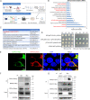
FIG 1
FIG 1
MARCO is identified as one of the host factors that interacts with PRRSV GP5. (A) Schematic of porcine cDNA library construction and yeast two-hybrid screening using PRRSV GP5 as a bait. The lung tissue samples from piglets challenged with or without PRRSV were pooled, and total RNA was subsequently extracted. Double-stranded cDNA was synthesized and cloned into each of three reading frames of the pDONR222 plasmid, and this was followed by recombination with the pPR3-N-DEST vector based on gateway DNA recombinant technology to obtain an expressed yeast porcine cDNA library (upper panel). A DUALmembrane Y2H screening was performed to identify potential host factors interacting with PRRSV GP5. PRRSV GP5 was cloned into the pBT3-SUC vector to generate the bait plasmid GP5-pBT3-SUC. The cDNA library was cloned into the pPR3-N-DEST vector to generate prey plasmids. All prey plasmids, together with the GP5-pBT3-SUC bait plasmid, were cotransformed into the yeast strain Y2HGold. The positive colonies were analyzed via DNA sequencing, and candidates interacting with PRRSV GP5 were identified via BLAST searches of the NCBI database (lower panel). (B) The gene ontology (GO) enrichment analysis of the candidate proteins that interact with PRRSV GP5. (C) The 10 selected interest proteins, including MARCO (marked red) from 49 candidates. (D) Confirmation of interaction between MARCO and PRRSV GP5, using Y2H for testing. The yeast strain NMY51 was cotransformed with the bait plasmid GP5-pBT3-SUC and the prey plasmid pPR3-N-MARCO. The cotransformation of pTSU2-APP/pNubG-Fe65 and GP5-pBT3-SUC/pPR3-N or pTSU2-APP/pPR3-N were used as the positive and negative controls, respectively. The transformants were grown on synthetic dropout (SD) medium. The interaction between GP5 and MARCO was identified by the growth of colonies on SD/-Leu/-Trp/-His/-Ade plates. SD/-Leu/-Trp/-His/-Ade, SD agar medium without leucine, tryptophan, histidine, and adenine; SD/-Leu/-Trp, SD agar medium without leucine and tryptophan. (E) The colocalization of the MARCO protein with PRRSV GP5 in 293T cells. The cells were cotransfected with Myc-tagged MARCO and mCherry-tagged GP5 plasmids for 24 h. The cells were fixed and immunostained with the anti-Myc antibody. The nuclei were stained with DAPI. Fluorescent images were acquired using laser-scanning fluorescent confocal microscopy. Bar, 5 μm. (F) Confirmation of interaction between MARCO and PRRSV GP5 using a co-IP assay. 293T cells were cotransfected with mCherry-tagged GP5 and Myc-tagged MARCO plasmids for 24 h. The cells were then lysed, and an IP analysis was performed using a mouse anti-Myc antibody. (G) The interaction between MARCO and PRRSV GP5 in the case of a PRRSV infection. Marc-145 cells were transiently transfected with the Myc-tagged MARCO plasmid for 24 h, prior to a PRRSV infection. At 24 hpi, the cells were harvested for an IP analysis using the anti-Myc antibody.
FIG 2
FIG 2
PRRSV infection downregulates MARCO expression in vivo and in vitro. (A) The mRNA level of MARCO and CD163 in PAMs was detected via RT-qPCR. (B) The mRNA level of MARCO and CD163 in various tissues. Different tissues were separated from 4- to 6-week-old PRRSV-negative, healthy piglets, and total RNA was extracted to perform a RT-qPCR analysis. (C) The transcriptional level change of MARCO in PAMs during PRRSV infection. The PAMs were mock-infected or were infected with PRRSV at a multiplicity of infection (MOI) of 0.5 for various times. At the indicated time points, the PAMs were harvested for RT-qPCR. (D) The transcriptional level change of MARCO in the lungs of PRRSV-infected pigs. PRRSV-negative piglets were challenged with PRRSV at 4 to 6 weeks of age. On days 3 and 7 postchallenge, three piglets from the infected and control group were euthanized, and their lung tissues were collected to perform the RT-qPCR analysis. HPRT, the hypoxanthine phosphoribosyltransferase of Sus scrofa, was used as an endogenous reference in RT-qPCR. The data are represented as the mean ± SE of the three biological replicates. Significant differences are indicated as: *, P < 0.05; **, P < 0.01; and ***, P < 0.001.
FIG 3
FIG 3
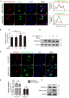
MARCO may not be involved in the entry of PRRSV into PAMs. (A) Route of MARCO internalization. PAMs were infected with recombinant adenovirus for 24 h to express MARCO-EGFP and were incubated with MARCO ligand Dxs (50 μg/mL, Sigma, D8787) at 37°C for 30 min. The cells were fixed for the immunostaining of clathrin heavy chain. The nuclei were stained with DAPI. Bar, 10 μm. Colocalization quantifications were done using the Plot Profile plugin in ImageJ. (B and C) Effects of MARCO ligand Poly(I) on PRRSV adsorption. PAMs were incubated with Poly(I) (Sigma, P4154) at 37°C for 30 min, and this was followed by PRRSV incubation at a MOI of 5 at 4°C for 2 h. After washing with cold PBS three times to remove the unbound virions, PAMs were collected for RT-qPCR and Western blotting analyses to detect the mRNA (panel B) and protein (panel C) levels of PRRSV N on the surfaces of the PAMs. (D–F) Effects of MARCO overexpression on PRRSV adsorption and internalization. PAMs infected with recombinant adenovirus to overexpress MARCO were incubated with PRRSV at a MOI of 5 at 4°C for 2 h and were washed with cold PBS to remove the unbound virions. Adsorbed PRRSV was detected using immunofluorescence staining (panel D), RT-qPCR (panel E), or Western blotting (panel F). Bar, 10 μm in the immunofluorescence analysis. For the internalized PRRSV, the PAMs were switched to 37°C for another 1 h after the removal of the unbound virus. RT-qPCR was performed to detect the mRNA level of PRRSV N in the PAMs (E). HPRT, the hypoxanthine phosphoribosyltransferase of Sus scrofa, was used as an endogenous reference in the RT-qPCR. The data are represented as the mean ± SE of three biological replicates. n.s. indicates no difference.
FIG 4
FIG 4
MARCO is a host restriction factor for PRRSV. (A–E) Effects of MARCO overexpression on PRRSV proliferation in PAMs. PAMs were infected with MARCO recombinant adenovirus for 24 h, and this was followed by infection with PRRSV at a MOI of 0.5. Cells were harvested at 12 or 24 hpi for RT-qPCR and Western blotting to examine the mRNA (A) and protein levels (B) of PRRSV N. (C) PAMs were infected with MARCO recombinant adenovirus for 24 h, and this was followed by infection with PRRSV at a MOI of 0.5. At 12 or 24 hpi, the cell supernatant was collected to determine the virus titers via a TCID50 assay. (D and E) PAMs were infected with different doses of MARCO recombinant adenovirus for 24 h and were then infected with PRRSV at a MOI of 0.5. At 24 hpi, the cells were lysed to detect the PRRSV N (panel D), and the corresponding supernatant was collected to examine the virus titers using a TCID50 assay (panel E). (F–I) Effects of MARCO knockdown on PRRSV proliferation in PAMs. (F) PAMs were transiently transfected with three independent small interfering RNAs (siRNA) targeting MARCO for 24 h and were harvested for RT-qPCR to examine the silencing efficiency of MARCO. (G–I) PAMs were transiently transfected with siRNA2 for 24 h, and this was followed by infection with PRRSV at a MOI of 0.5. At 12 or 24 hpi, cells were harvested to determine the PRRSV N mRNA (panel G) and protein (panel H) levels via RT-qPCR and Western blotting. The cell supernatant was collected to examine the virus titers via a TCID50 assay (panel I). HPRT, the hypoxanthine phosphoribosyltransferase of Sus scrofa, was used as an endogenous reference in the RT-qPCR. The data are represented as the mean ± SE of three biological replicates. Significant differences are indicated as: **, P < 0.01 and ***, P < 0.001.
FIG 5
FIG 5
The N-terminal intracellular domain of MARCO is required for its inhibitory effect on PRRSV. (A) Schematic representations of full-length or truncated MARCO constructs. Cyto, cytoplasmic domain; TM, transmembrane domain; Spa, spacer domain; collagen, collagenous domain; SRCR, scavenger receptor cysteine-rich domain. (B) PAMs were exogenously infected with recombinant adenovirus to express MARCO-WT or MARCO-D with N-terminal cytoplasmic domain deletion for 24 h, and they were infected with PRRSV at a MOI of 0.5 for 24 h. Cells were harvested for Western blotting to detect the PRRSV N level. (C–E) Marc-145 cells were transiently transfected with Myc-tagged MARCO-WT or MARCO-D plasmid for 24 h, and they were infected with PRRSV at a MOI of 0.5. At 24 hpi, immunofluorescence (C), RT-qPCR (D), and Western blotting (E) analyses were performed to examine the PRRSV N levels. Bar, 100 μm. (F) Marc-145 cells were infected with recombinant adenovirus to express MARCO-WT-EGFP or MARCO-D-EGFP heterogeneously for 24 h. Cells were fixed, and this was followed by nuclei staining with DAPI. Images of cells were acquired via laser-scanning fluorescent confocal microscopy. Bar, 10 μm. GAPDH, the glyceraldehyde-3-phosphate dehydrogenase of Chlorocebus sabaeus, was used as an endogenous reference in RT-qPCR. The data are represented as the mean ± SE of the three biological replicates. Significant differences are indicated as: *, P < 0.01 and **, P < 0.05.
FIG 6
FIG 6
MARCO is a proapoptotic factor in PRRSV-infected PAMs. (A) PAMs were mock-infected or infected with PRRSV at a MOI of 0.5 for various times, and cells were lysed for Western blotting to determine the levels of cleaved caspase3 and PRRSV N. (B) PAMs were mock-infected or infected with PRRSV at a MOI of 0.5 for 24 h, and cells were digested for an Annexin-V/PI double staining analysis. (C) PAMs were transiently transfected with siRNA2 for 24 h and were then mock-infected or infected with PRRSV at a MOI of 0.5. At 12 or 24 hpi, cells were harvested to determine the levels of cleaved caspase3 and PRRSV N. (D) PAMs were transiently transfected with siRNA2 for 24 h and were then infected with PRRSV at a MOI of 0.5. At 24 hpi, cells were digested for Annexin-V/PI double staining analysis. (E) PAMs were infected with recombinant adenovirus to express MARCO-WT or MARCO-D exogenously for 24 h and were then mock-infected or infected with PRRSV at a MOI of 0.5 for 24 h. The cells were harvested for Western blotting to detect the levels of cleaved caspase3 and PRRSV N.
FIG 7
FIG 7
The interaction between MARCO and PRRSV GP5 may aggravate PRRSV-induced apoptosis, which contributes to the inhibition function of MARCO on PRRSV. (A) PRRSV GP5 is an inducer of apoptosis, and MARCO overexpression further aggravates apoptosis. Marc-145 cells were transiently transfected with an empty vector, transfected with an GP5-expressing plasmid, or cotransfected with plasmids encoding GP5 and MARCO or an empty vector for 24 h. The cells were digested for a flow cytometry analysis of the apoptosis. The expression of Myc-tagged GP5 and Myc-tagged MARCO in the Marc-145 cells was examined via Western blotting. (B) PAMs were infected with PRRSV at a MOI of 0.5 for various times in the presence or absence of the caspase3 inhibitor Z-DEVD-FMK (30 μM, Abmole, 210344-95-9). The cells were lysed for Western blotting to determine the levels of cleaved caspase3 and PRRSV N. (C) The quantization of the PRRSV N protein abundance in panel B. (D) The PAMs were infected with recombinant adenovirus for 24 h to overexpress MARCO, and this was followed by infection with PRRSV at a MOI of 0.5 in the presence or absence of Z-DEVD-FMK (30 μM). At 24 hpi, the PAMs were lysed to examine the levels of PRRSV N and cleaved caspase3 via Western blotting.
FIG 8
FIG 8
The schematic model of MARCO regulation of PRRSV infection. PRRSV infection downregulates the expression of MARCO, which acts as a host restriction factor for PRRSV. MARCO interacts with PRRSV GP5 and aggravates GP5-induced apoptosis. The apoptotic cells are not conducive to PRRSV propagation. Thus, MARCO inhibits PRRSV infection through the regulation of apoptosis.

Similar articles

Cited by

References

    1. An T-Q, Li J-N, Su C-M, Yoo D. 2020. Molecular and cellular mechanisms for PRRSV pathogenesis and host response to infection. Virus Res 286:197980. doi:10.1016/j.virusres.2020.197980. - DOI - PMC - PubMed
    1. Lunney JK, Fang Y, Ladinig A, Chen N, Li Y, Rowland B, Renukaradhya GJ. 2016. Porcine reproductive and respiratory syndrome virus (PRRSV): pathogenesis and interaction with the immune system. Annu Rev Anim Biosci 4:129–154. doi:10.1146/annurev-animal-022114-111025. - DOI - PubMed
    1. Kappes MA, Faaberg KS. 2015. PRRSV structure, replication and recombination: origin of phenotype and genotype diversity. Virology 479-480:475–486. doi:10.1016/j.virol.2015.02.012. - DOI - PMC - PubMed
    1. Fang Y, Snijder EJ. 2010. The PRRSV replicase: exploring the multifunctionality of an intriguing set of nonstructural proteins. Virus Res 154:61–76. doi:10.1016/j.virusres.2010.07.030. - DOI - PMC - PubMed
    1. Huang C, Zhang Q, Feng W-h. 2015. Regulation and evasion of antiviral immune responses by porcine reproductive and respiratory syndrome virus. Virus Res 202:101–111. doi:10.1016/j.virusres.2014.12.014. - DOI - PMC - PubMed

Publication types

LinkOut - more resources