

Quantitative application of dual-phase 99mTc-sestamibi SPECT/CT imaging of parathyroid lesions: identification of optimal timing in secondary hyperparathyroidism
- PMID: 37079194
- PMCID: PMC10119365
- DOI: 10.1186/s40658-023-00548-5
Quantitative application of dual-phase 99mTc-sestamibi SPECT/CT imaging of parathyroid lesions: identification of optimal timing in secondary hyperparathyroidism
Abstract
Purpose: In this retrospective study, we compared the maximum standardized uptake values (SUVmax) of parathyroid lesions and the target-to-background ratio (TBR) of parathyroid lesions to thyroid tissue in early-phase single-photon emission computed tomography/computed tomography (SPECT/CT) versus delayed-phase SPECT/CT in patients with secondary hyperparathyroidism (SHPT) in order to determine the optimal timing of 99mTc- methoxyisobutylisonitrile (99mTc-MIBI) SPECT/CT imaging.
Methods: Seventeen patients with a history of chronic kidney failure stage 5 on hemodialysis, underwent pre-operative parathyroid scintigraphy for detection and localization of parathyroid lesions. Retrospective analysis was conducted for lesions with focal accumulation of 99mTc-MIBI. All patients underwent dual-phase 99mTc-MIBI parathyroid scintigraphy and dual-phase SPECT/CT. SUVmax of parathyroid lesions and thyroid tissues was measured.
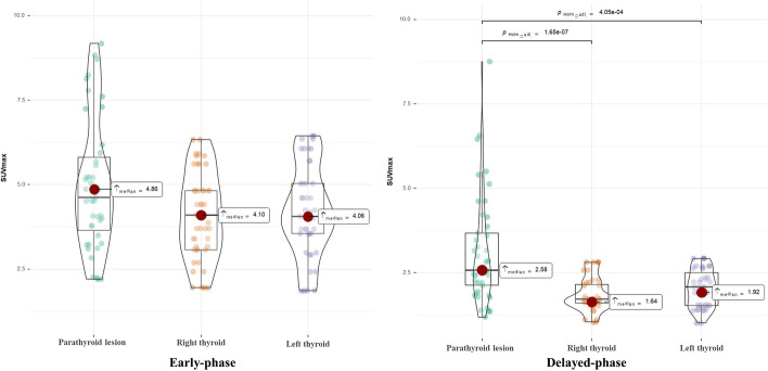
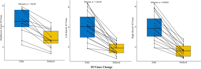
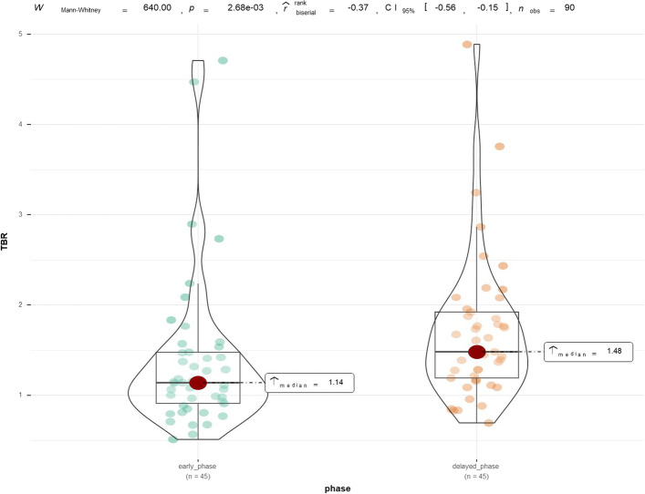
Results: Mean SUVmax of parathyroid lesions was 4.86 on early-phase and 2.58 on delayed-phase SPECT/CT, respectively. Mean TBR was 1.14 on early phase and 1.48 on delayed-phase SPECT/CT, respectively. Statistically significant differences in SUVmax and TBR between dual-phase SPECT/CT were observed (P < 0.001).
Conclusions: Delayed-phase SPECT/CT in SHPT is required because of the better image contrast.
Keywords: 99mTc-MIBI; Chronic kidney disease; Hyperparathyroidism; Maximum standardized uptake; SPECT/CT.
© 2023. The Author(s).
Conflict of interest statement
The authors have no relevant financial or non-financial interests to disclose.
Figures
Similar articles
-
Diagnostic performance of ultrasonography, dual-phase 99mTc-MIBI scintigraphy, early and delayed 99mTc-MIBI SPECT/CT in preoperative parathyroid gland localization in secondary hyperparathyroidism.BMC Med Imaging. 2020 Aug 3;20(1):91. doi: 10.1186/s12880-020-00490-3. BMC Med Imaging. 2020. PMID: 32746794 Free PMC article.
-
Comparison of biochemical markers and technetium 99m methoxyisobutylisonitrile imaging in primary and secondary hyperparathyroidism.Front Endocrinol (Lausanne). 2023 Mar 27;14:1094689. doi: 10.3389/fendo.2023.1094689. eCollection 2023. Front Endocrinol (Lausanne). 2023. PMID: 37051197 Free PMC article.
-
The diagnostic value of dual-phase SPECT/CT scintigraphy based on transport kinetics of 99mTc-sestamibi confirmed with histopathological findings in patients with secondary hyperparathyroidism - practical consideration.Nucl Med Rev Cent East Eur. 2020;23(2):71-77. doi: 10.5603/NMR.a2020.0017. Nucl Med Rev Cent East Eur. 2020. PMID: 33007093
-
Preoperative ⁹⁹mTc-sestamibi scintigraphy in patients with primary hyperparathyroidism and concomitant nodular goiter: comparison of SPECT-CT, SPECT, and planar imaging.Nucl Med Commun. 2012 Oct;33(10):1070-6. doi: 10.1097/MNM.0b013e32835710b6. Nucl Med Commun. 2012. PMID: 22825041 Clinical Trial.
-
Radionuclide imaging of the parathyroid glands.Semin Nucl Med. 2005 Oct;35(4):266-76. doi: 10.1053/j.semnuclmed.2005.06.001. Semin Nucl Med. 2005. PMID: 16150247 Review.
Cited by
-
Surgical Management of Mediastinal Ectopic Parathyroids.J Pers Med. 2025 Jun 30;15(7):276. doi: 10.3390/jpm15070276. J Pers Med. 2025. PMID: 40710393 Free PMC article. Review.
References
LinkOut - more resources
Full Text Sources
