

Oncogenic roles of GPR176 in breast cancer: a potential marker of aggressiveness and a potential target of gene therapy
- PMID: 37079213
- PMCID: PMC10462518
- DOI: 10.1007/s12094-023-03174-w
Oncogenic roles of GPR176 in breast cancer: a potential marker of aggressiveness and a potential target of gene therapy
Abstract
Background: Belonging to the G-protein coupled receptor 1 family, G protein-coupled receptor 176 (GPR176) is associated with the Gz/Gx G-protein subclass and is capable of decreasing cAMP production.
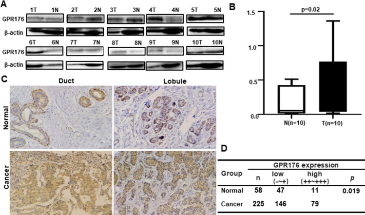
Methods: GPR176 expression was detected by qRT-PCR, bioinformatics analysis, Western blot and immunohistochemistry, and compared with clinicopathological characteristics of breast cancer. GPR176-related genes and pathways were subjected to bioinformatic analysis. We also explored the effects of GPR176 on the phenotypes of breast cancer cells.
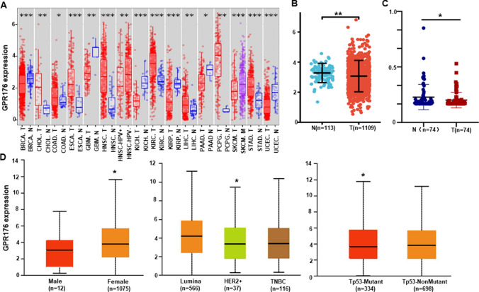
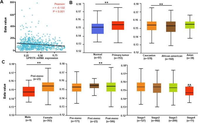
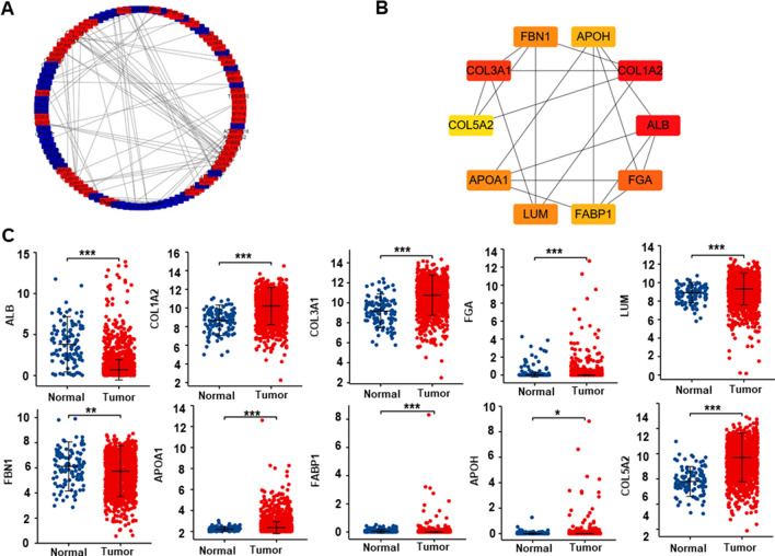
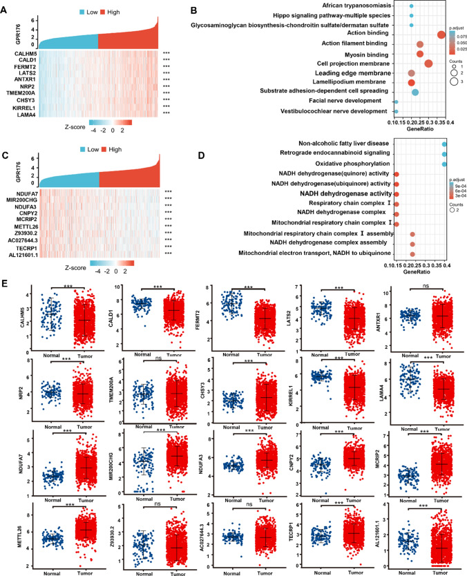
Results: Lower expression of GPR176 mRNA was seen in breast cancer than in normal tissues, but the opposite pattern was found for its protein (p < 0.05). GPR176 mRNA was associated with female sex, low T staging, non-Her-2+ subtypes, non-mutant p53 status in breast cancer (p < 0.05). GPR176 methylation was negatively correlated with its mRNA level and T staging in breast cancer, and was higher in breast cancer than normal tissues (p < 0.05). GPR176 protein expression was positively correlated with older age, small tumor size, and non-luminal-B subtype of breast cancers (p < 0.05). The differential genes of GPR176 were involved in receptor-ligand interaction, RNA maturation, and so forth (p < 0.05). GPR176-related genes were categorized into cell mobility, membrane structure, and so on (p < 0.05). GPR176 knockdown weakened the proliferation, glucose catabolism, anti-apoptosis, anti-pyroptosis, migration, invasion, and epithelial-mesenchymal transition of breast cancer cells.
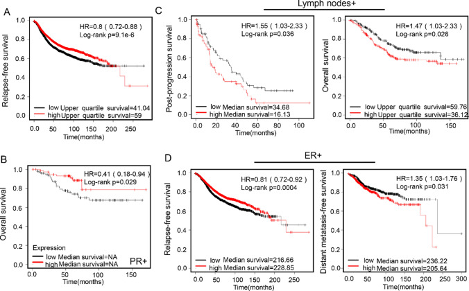
Conclusion: These results indicate that GPR176 might be involved in the tumorigenesis and subsequent progression of breast cancer by deteriorating aggressive phenotypes. It might be utilized as a potential biomarker to indicate the aggressive behaviors and poor prognosis of breast cancer and a potential target of genetic therapy.
Keywords: Aggressiveness; Breast cancer; GPR176; Prognosis; Target therapy.
© 2023. The Author(s).
Conflict of interest statement
The authors declare that they have no conflict of interest.
Figures


Similar articles
-
High expression of GPR176 predicts poor prognosis of gastric cancer patients and promotes the proliferation, migration, and invasion of gastric cancer cells.Sci Rep. 2023 Jun 8;13(1):9360. doi: 10.1038/s41598-023-36586-3. Sci Rep. 2023. PMID: 37291181 Free PMC article.
-
The promoting effects of GPR176 expression on proliferation, chemoresistance, lipogenesis and invasion of oesophageal cancer.J Cancer Res Clin Oncol. 2023 Nov;149(16):14641-14655. doi: 10.1007/s00432-023-05256-2. Epub 2023 Aug 16. J Cancer Res Clin Oncol. 2023. PMID: 37584712 Free PMC article.
-
The oncogenic roles of GPR176 in ovarian cancer: a molecular target for aggressiveness and gene therapy.J Obstet Gynaecol. 2024 Dec;44(1):2347430. doi: 10.1080/01443615.2024.2347430. Epub 2024 Jun 4. J Obstet Gynaecol. 2024. PMID: 38835234
-
Time-Restricted G-Protein Signaling Pathways via GPR176, Gz, and RGS16 Set the Pace of the Master Circadian Clock in the Suprachiasmatic Nucleus.Int J Mol Sci. 2020 Jul 17;21(14):5055. doi: 10.3390/ijms21145055. Int J Mol Sci. 2020. PMID: 32709014 Free PMC article. Review.
-
G-protein-coupled receptor signaling through Gpr176, Gz, and RGS16 tunes time in the center of the circadian clock [Review].Endocr J. 2017 Jun 29;64(6):571-579. doi: 10.1507/endocrj.EJ17-0130. Epub 2017 May 13. Endocr J. 2017. PMID: 28502923 Review.
Cited by
-
High expression of GPR176 predicts poor prognosis of gastric cancer patients and promotes the proliferation, migration, and invasion of gastric cancer cells.Sci Rep. 2023 Jun 8;13(1):9360. doi: 10.1038/s41598-023-36586-3. Sci Rep. 2023. PMID: 37291181 Free PMC article.
-
The promoting effects of GPR176 expression on proliferation, chemoresistance, lipogenesis and invasion of oesophageal cancer.J Cancer Res Clin Oncol. 2023 Nov;149(16):14641-14655. doi: 10.1007/s00432-023-05256-2. Epub 2023 Aug 16. J Cancer Res Clin Oncol. 2023. PMID: 37584712 Free PMC article.
References
MeSH terms
Substances
Grants and funding
LinkOut - more resources
Full Text Sources
Medical
Research Materials
Miscellaneous
