Incidence of Underlying Abnormal Findings on Routine Magnetic Resonance Imaging for Bell Palsy
- PMID: 37079301
- PMCID: PMC10119737
- DOI: 10.1001/jamanetworkopen.2023.9158
Incidence of Underlying Abnormal Findings on Routine Magnetic Resonance Imaging for Bell Palsy
Abstract
Importance: There is no consensus on the benefits of routine magnetic resonance imaging (MRI) of the facial nerve in patients with suspected idiopathic peripheral facial palsy (PFP) (ie, Bell palsy [BP]).
Objectives: To estimate the proportion of adult patients in whom MRI led to correction of an initial clinical diagnosis of BP; to determine the proportion of patients with confirmed BP who had MRI evidence of facial nerve neuritis without secondary lesions; and to identify factors associated with secondary (nonidiopathic) PFP at initial presentation and 1 month later.
Design, setting, and participants: This retrospective multicenter cohort study analyzed the clinical and radiological data of 120 patients initially diagnosed with suspected BP from January 1, 2018, to April 30, 2022, at the emergency department of 3 tertiary referral centers in France.
Interventions: All patients screened for clinically suspected BP underwent an MRI of the entire facial nerve with a double-blind reading of all images.
Main outcomes and measures: The proportion of patients in whom MRI led to a correction of the initial diagnosis of BP (any condition other than BP, including potentially life-threating conditions) and results of contrast enhancement of the facial nerve were described.
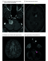
Results: Among the 120 patients initially diagnosed with suspected BP, 64 (53.3%) were men, and the mean (SD) age was 51 (18) years. Magnetic resonance imaging of the facial nerve led to a correction of the diagnosis in 8 patients (6.7%); among them, potentially life-threatening conditions that required changes in treatment were identified in 3 (37.5%). The MRI confirmed the diagnosis of BP in 112 patients (93.3%), among whom 106 (94.6%) showed evidence of facial nerve neuritis on the affected side (hypersignal on gadolinium-enhanced T1-weighted images). This was the only objective sign confirming the idiopathic nature of PFP.
Conclusions and relevance: These preliminary results suggest the added value of the routine use of facial nerve MRI in suspected cases of BP. Multicentered international prospective studies should be organized to confirm these results.
Conflict of interest statement
Figures


Comment in
-
Beyond the AJR: Routine MRI May Provide Utility in Identifying Secondary Causes in Adult Patients With Suspected Bell Palsy at Initial Presentation.AJR Am J Roentgenol. 2024 Feb;222(2):e2329811. doi: 10.2214/AJR.23.29811. Epub 2023 Jun 28. AJR Am J Roentgenol. 2024. PMID: 37377361 No abstract available.
Similar articles
-
Usefulness of High-Resolution 3D Multi-Sequences for Peripheral Facial Palsy: Differentiation Between Bell's Palsy and Ramsay Hunt Syndrome.Otol Neurotol. 2017 Dec;38(10):1523-1527. doi: 10.1097/MAO.0000000000001605. Otol Neurotol. 2017. PMID: 29135869
-
Diagnostic value of dynamic contrast-enhanced magnetic resonance imaging in Bell's palsy.Acta Radiol. 2021 Sep;62(9):1163-1169. doi: 10.1177/0284185120958414. Epub 2020 Sep 24. Acta Radiol. 2021. PMID: 32972214
-
Contrast-enhanced MRI findings of patients with acute Bell palsy within 7 days of symptom onset: A retrospective study.Medicine (Baltimore). 2023 Dec 1;102(48):e36337. doi: 10.1097/MD.0000000000036337. Medicine (Baltimore). 2023. PMID: 38050278 Free PMC article.
-
Facial nerve decompression for idiopathic Bell's palsy: report of 13 cases and literature review.J Laryngol Otol. 2010 Mar;124(3):272-8. doi: 10.1017/S0022215109991265. Epub 2009 Oct 2. J Laryngol Otol. 2010. PMID: 19796438 Review.
-
Facial Nerve Palsy: Clinical Practice and Cognitive Errors.Am J Med. 2020 Sep;133(9):1039-1044. doi: 10.1016/j.amjmed.2020.04.023. Epub 2020 May 20. Am J Med. 2020. PMID: 32445717 Review.
Cited by
-
3D T1-Weighted Black-Blood MRI in the Diagnosis and Follow-Up of Facial Neuritis: a Single-Center Prospective Study.Clin Neuroradiol. 2025 Jul 14. doi: 10.1007/s00062-025-01540-5. Online ahead of print. Clin Neuroradiol. 2025. PMID: 40659900
-
Serum levels of heavy metals in patients with Bell's palsy: a case-control study.Eur Arch Otorhinolaryngol. 2024 Feb;281(2):891-896. doi: 10.1007/s00405-023-08253-w. Epub 2023 Sep 28. Eur Arch Otorhinolaryngol. 2024. PMID: 37768371
References
Publication types
MeSH terms
LinkOut - more resources
Full Text Sources
Medical
Miscellaneous

