

Porcine Cornea Storage Ex Vivo Model as an Alternative to Human Donor Tissues for Investigations of Endothelial Layer Preservation
- PMID: 37079319
- PMCID: PMC10148656
- DOI: 10.1167/tvst.12.4.24
Porcine Cornea Storage Ex Vivo Model as an Alternative to Human Donor Tissues for Investigations of Endothelial Layer Preservation
Abstract
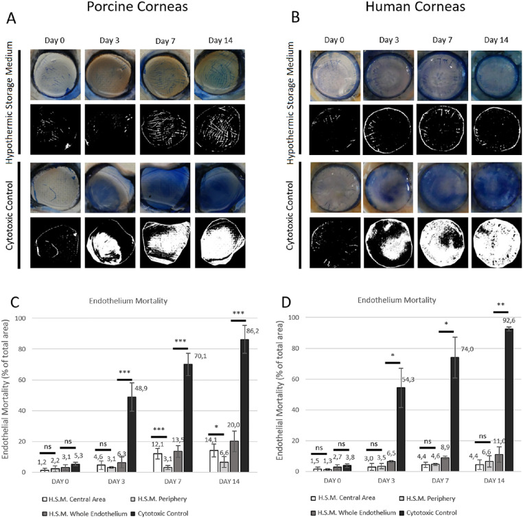
Purpose: Due to the growing shortage of human corneas for research, we developed a porcine cornea storage model with qualitative features comparable to human tissues.
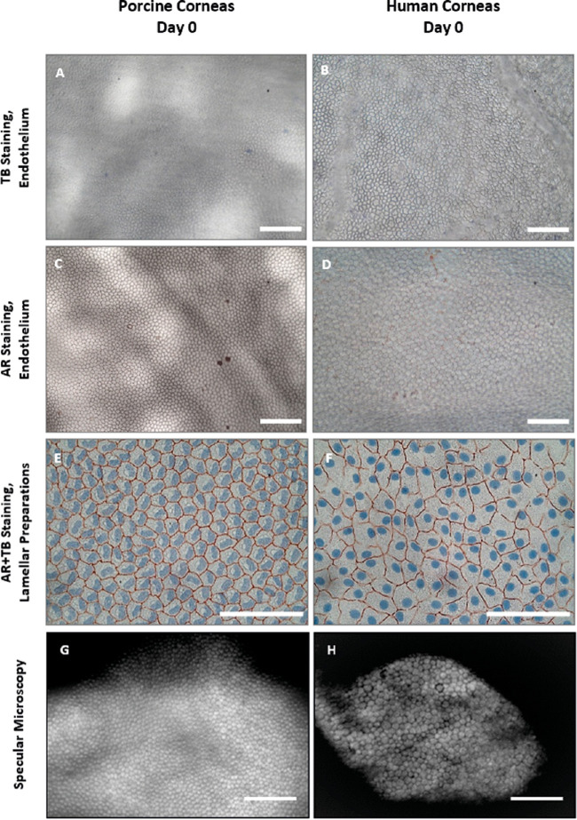
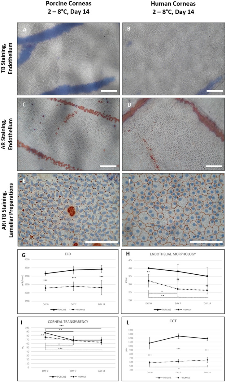
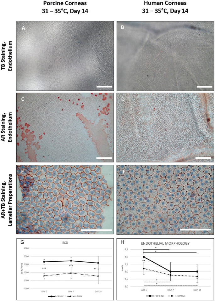
Methods: We established a decontamination procedure for porcine eye bulbs to ensure corneal storage at 31°C to 35°C for up to 28 days without contamination. We compared human and porcine corneas under hypothermic (2-8°C) or culture (31-35°C) conditions for central corneal thickness (CCT), corneal transparency, endothelial morphology, endothelial cell density (ECD), and a novel method to quantify whole endothelial mortality. We also examined portions of lamellar tissues consisting of Descemet's membrane and endothelial cells under the microscope after Alizarin red staining.
Results: Our decontamination procedure reduced corneal contamination from 94% (control corneas without decontamination) to 18% after 28 days of storage at 31°C to 35°C. ECD, CCT, transparency, and morphology were significantly higher in porcine corneas than in human corneas at day 0. Nevertheless, the qualitative parameters of porcine and human corneas showed comparable trends under both investigated storage conditions for up to 14 days.
Conclusions: The presented corneal storage model provides a reliable alternative to human tissues for preliminary corneal investigations.
Translational relevance: The porcine cornea storage model can be used to investigate the efficacy and safety of new media, substances, or storage conditions. Furthermore, the method developed to assess the percentage of endothelial mortality is tissue conservative and can be used in eye banks to monitor endothelial mortality during storage of tissues intended for transplantation.
Conflict of interest statement
Disclosure:
Figures
Similar articles
-
Assessment of performance and safety of Corneal Chamber hypothermic storage medium and PSS-L corneal rinsing solution in human and porcine corneas.BMJ Open Ophthalmol. 2024 Feb 22;9(1):e001453. doi: 10.1136/bmjophth-2023-001453. BMJ Open Ophthalmol. 2024. PMID: 38388003 Free PMC article.
-
18 A porcine cornea and lamellar tissue model to investigate effects of storage conditions on corneal preservation.BMJ Open Ophthalmol. 2022 Nov;7(Suppl 2):A8. doi: 10.1136/bmjophth-2022-EEBA.18. BMJ Open Ophthalmol. 2022. PMID: 37282691 Free PMC article.
-
A corneal and whole eye globe bovine ex vivo model to mimic human donor corneal storage conditions and eye surgeries.Cell Tissue Bank. 2025 Mar 4;26(2):14. doi: 10.1007/s10561-025-10162-7. Cell Tissue Bank. 2025. PMID: 40038115
-
[Transplantation of corneal endothelial cells].Nippon Ganka Gakkai Zasshi. 2002 Dec;106(12):805-35; discussion 836. Nippon Ganka Gakkai Zasshi. 2002. PMID: 12610838 Review. Japanese.
-
Ex vivo expansion and characterization of human corneal endothelium for transplantation: a review.Stem Cell Res Ther. 2021 Oct 30;12(1):554. doi: 10.1186/s13287-021-02611-3. Stem Cell Res Ther. 2021. PMID: 34717745 Free PMC article. Review.
Cited by
-
Chemical, Physical, and Biological Corneal Decellularization Methods: A Review of Literature.J Ophthalmol. 2024 Mar 25;2024:1191462. doi: 10.1155/2024/1191462. eCollection 2024. J Ophthalmol. 2024. PMID: 38567029 Free PMC article. Review.
-
Retrospective Clinical Outcomes of Keratoplasty Using Human Donor Corneas Preserved in Eusol-C Hypothermic Storage Medium.J Clin Med. 2024 Dec 13;13(24):7606. doi: 10.3390/jcm13247606. J Clin Med. 2024. PMID: 39768528 Free PMC article.
-
Assessment of performance and safety of Corneal Chamber hypothermic storage medium and PSS-L corneal rinsing solution in human and porcine corneas.BMJ Open Ophthalmol. 2024 Feb 22;9(1):e001453. doi: 10.1136/bmjophth-2023-001453. BMJ Open Ophthalmol. 2024. PMID: 38388003 Free PMC article.
-
Herpes simplex virus type-1 infection and spread in a novel porcine corneal explant model is restricted to the epithelium.PLoS Pathog. 2025 May 2;21(5):e1013162. doi: 10.1371/journal.ppat.1013162. eCollection 2025 May. PLoS Pathog. 2025. PMID: 40315239 Free PMC article.
References
-
- Guindolet DM, Crouzet E, He Z, et al. .. Storage of porcine cornea in an innovative bioreactor. Invest Ophthalmol Vis Sci . 2017; 58: 5905–5917. - PubMed
-
- Directive 2004/23/EC of the European Parliament and of the Council of 31 March 2004, https://eur-lex.europa.eu/legal-content/EN/TXT/PDF/?uri=CELEX:32004L0023. Accessed May 6, 2022.
-
- Commission Directive 2006/17/EC of 8 February 2006, https://eur-lex.europa.eu/legal-content/EN/TXT/PDF/?uri=CELEX:32006L0017.... Accessed May 6, 2022.
-
- Mistò R, D'Amato Tóthová J, Khan S, Limongelli L, Pateri F. A new approach to extend the storage of donor corneas after 28 days of corneal culture in an Italian eye bank. Cell Tissue Bank . 2020; 21: 47–55. - PubMed
-
- Gain P, Julienne R, He Z, et al. .. Global survey of corneal transplantation and eye banking. JAMA Ophthalmol. 2016; 134: 167–173. - PubMed
Publication types
MeSH terms
LinkOut - more resources
Full Text Sources
