. 1986 May 21;876(3):646-57.
doi: 10.1016/0005-2760(86)90054-8.

Metabolism of low-density lipoproteins by cultured hepatocytes from normal and homozygous familial hypercholesterolemic subjects

Metabolism of low-density lipoproteins by cultured hepatocytes from normal and homozygous familial hypercholesterolemic subjects

J M Hoeg et al. Biochim Biophys Acta. .

Abstract

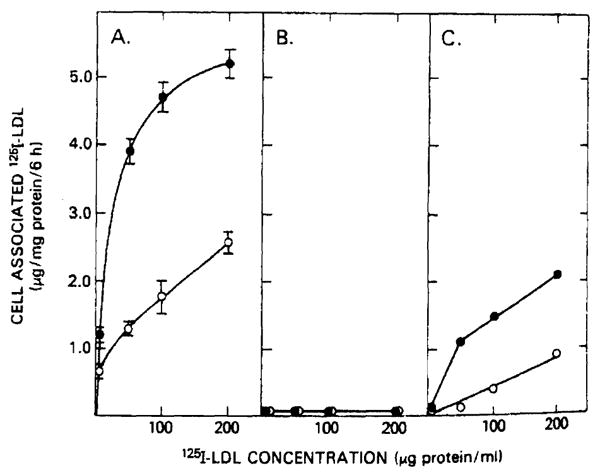
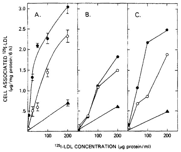
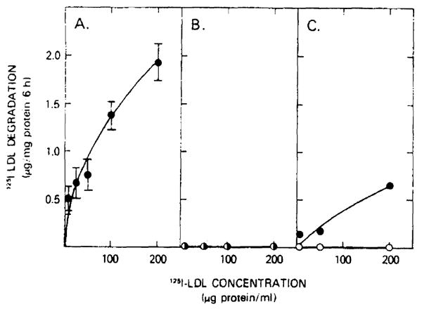
The profoundly elevated concentrations of low-density lipoproteins (LDL) present in homozygous familial hypercholesterolemia lead to symptomatic cardiovascular disease and death by early adulthood. Studies conducted in nonhepatic tissues demonstrated defective cellular recognition and metabolism of LDL in these patients. Since mammalian liver removes at least half of the LDL in the circulation, the metabolism of LDL by cultured hepatocytes isolated from familial hypercholesterolemic homozygotes was compared to hepatocytes from normal individuals. Fibroblast studies demonstrated that the familial hypercholesterolemic subjects studied were LDL receptor-negative (less than 1% normal receptor activity) and LDL receptor-defective (18% normal receptor activity). Cholesterol-depleted hepatocytes from normal subjects bound and internalized 125I-labeled LDL (Bmax = 2.2 micrograms LDL/mg cell protein). Preincubation of normal hepatocytes with 200 micrograms/ml LDL reduced binding and internalization by approx. 40%. In contrast, 125I-labeled LDL binding and internalization by receptor-negative familial hypercholesterolemic hepatocytes was unaffected by cholesterol loading and considerably lower than normal. This residual LDL uptake could not be ascribed to fluid phase endocytosis as determined by [14C]sucrose uptake. The residual LDL binding by familial hypercholesterolemia hepatocytes led to a small increase in hepatocyte cholesterol content which was relatively ineffective in reducing hepatocyte 3-hydroxy-3-methylglutaryl-CoA reductase activity. Receptor-defective familial hypercholesterolemia hepatocytes retained some degree of regulatable 125I-labeled LDL uptake, but LDL uptake did not lead to normal hepatocyte cholesterol content or 3-hydroxy-3-methylglutaryl-CoA reductase activity. These combined results indicate that the LDL receptor abnormality present in familial hypercholesterolemia fibroblasts reflects deranged hepatocyte LDL recognition and metabolism. In addition, a low-affinity, nonsaturable uptake process for LDL is present in human liver which does not efficiently modulate hepatocyte cholesterol content or synthesis.


Figures

Fig. 1
Fig. 1
125I-labeled LDL binding and internalization by fibroblasts from normal and homozygous familial hypercholesterolemia patients. 5–7·105 fibroblasts per 2 cm2 well were incubated for 48 h in serum-free medium (●) or for 24 h in serum-free medium with 200 μg LDL protein/ml (○). After a 6-h, 37°C incubation with the indicated concentrations of 125I-labeled LDL, cells were washed six times with ice-cold phosphate-buffered saline (pH 7.0). Protein and radioactivity determinations were performed on cells solubilized with 1 N NaOH. Values represent specifically bound and internalized 125I-labeled LDL defined as the difference in binding observed in the absence and presence of 10-fold excess of unlabeled LDL. Normal fibroblasts (panel A) and fibroblasts from patient 1 (panel B) and patient 2 (panel C) were evaluated.
Fig. 2
Fig. 2
Typical cells initially isolated from patient 1 (left panel) and patient 2 (right panel) demonstrate the electron microscopic characteristics of hepatocytes.
Fig. 3
Fig. 3
After 7 days in tissue culture, hepatocytes adhered to the tissue culture plates, flattened out and increased in size as shown by phase-contrast microscopy (left panel). The elaboration of cellular lipid, rough endoplasmic reticulum and mitochondria were observed by electron microscopy (right panel).
Fig. 4
Fig. 4
125I-labeled LDL binding and internalization by hepatocytes from normal and homozygous familial hypercholesterolemia patients. Hepatocytes (2–3·105/well) were incubated for 48 h in serum-free medium. After a further 24 h in either serum-free medium (●) of serum-free medium containing 200 μg LDL protein/ml (○), hepatocytes from normal (panel A), patient 1 (panel B) and patient 2 (panel 2) were incubated at 37°C with the indicated concentration of 125I-labeled LDL. After 6 h, the cells were washed six times with ice-cold phosphate-buffered saline (pH 7.0) and solubilized with 1 N NaOH. Total cell-associated binding and binding in the presence of 10-fold excess of unlabeled LDL (nonspecific binding, ▲) is shown. Normal values represent the mean ± S.E. for five different. hepatocyte preparations.
Fig. 5
Fig. 5
125I-labeled LDL degradation by hepatocytes from normal and homozygous familial hypercholesterolemia patients. The media from the normal hepatocytes (panel A), patient 1 hepatocytes (panel B), and from patient 2 (panel C) were harvested after a 6-h incubation with the indicated concentrations of 125I-labeled LDL. The non-chloroform-extractable 125I-label counts in the media (trichloroacetic acid supernatant) defined the degradation as outlined in Methods. Hepatocytes were incubated for 48 h prior to the exposure to 125I-labeled LDL in either the absence (○) or presence (●) of 2 mg unlabeled LDL protein/ml. Values for normal hepatocytes represent the mean ± S.E. for five different hepatocyte preparations.

Similar articles

Cited by

References

    1. Goldstein JL, Brown MS. J Biol Chem. 1974;210:5153–5162. - PubMed
    1. Khachadurian AK. Am J Med. 1964;37:402–407. - PubMed
    1. Brown MS, Goldstein JL. Proc Natl Acad Sci USA. 1974;71:788–792. - PMC - PubMed
    1. Goldstein JL, Dana SE, Brunschede GY, Brown MS. Proc Natl Acad Sci USA. 1975;72:1092–1096. - PMC - PubMed
    1. Toileshaug H, Hobgood KK, Brown MS, Goldstein JL. Cell. 1983;32:941–951. - PubMed

LinkOut - more resources