. 2023 Jul 15:329:121670.
doi: 10.1016/j.envpol.2023.121670. Epub 2023 Apr 18.

Nickel nanoparticles induce autophagy and apoptosis via HIF-1α/mTOR signaling in human bronchial epithelial cells

Affiliations

Nickel nanoparticles induce autophagy and apoptosis via HIF-1α/mTOR signaling in human bronchial epithelial cells

Jiali Yuan et al. Environ Pollut. .

Abstract

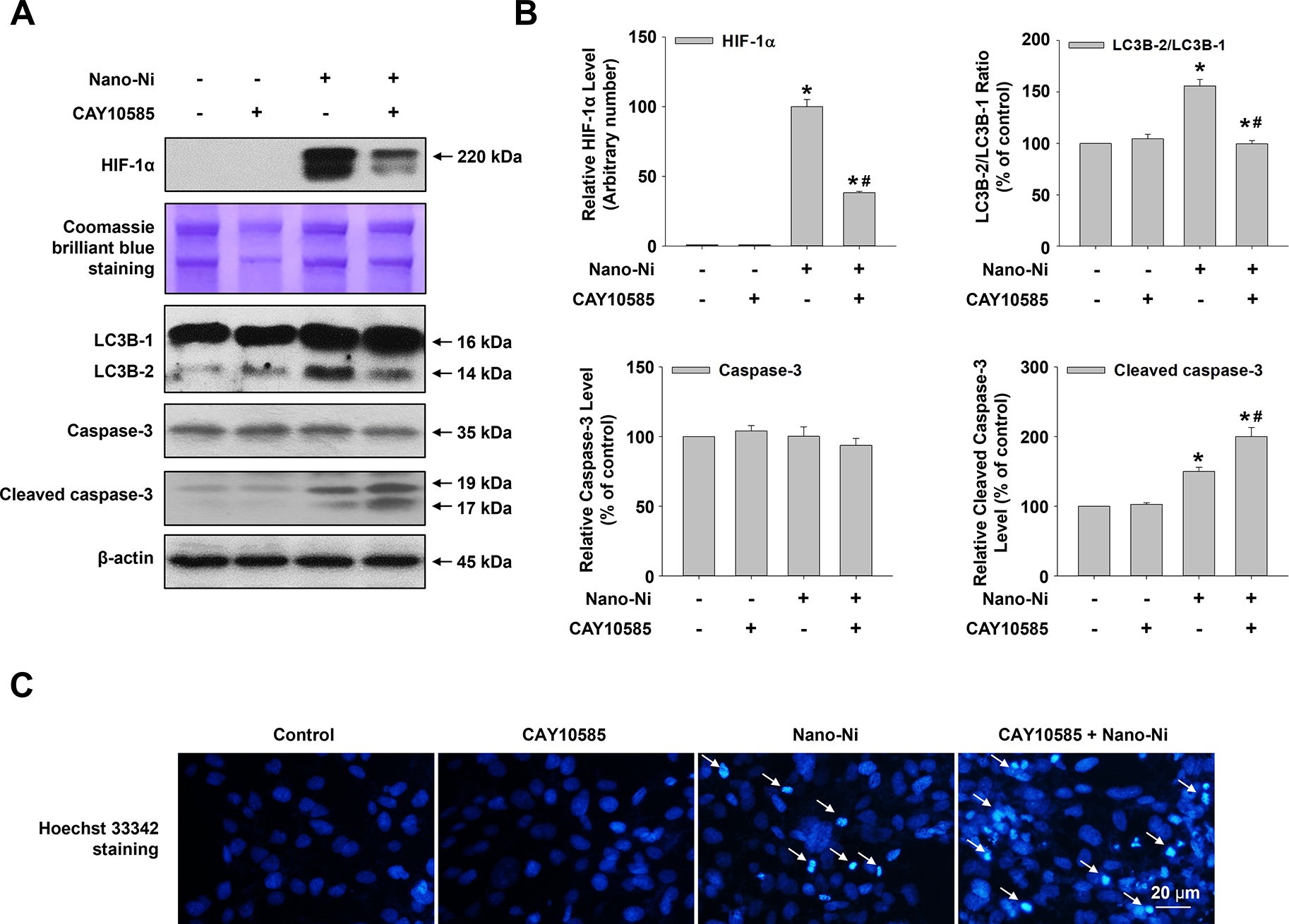
With the rapid development of nanotechnology, the potential adverse health effects of nanoparticles have been caught more attention and become global concerns. However, the underlying mechanisms in metal nanoparticle-induced toxic effects are still largely obscure. In this study, we investigated whether exposure to nickel nanoparticles (Nano-Ni) and titanium dioxide nanoparticles (Nano-TiO2) would alter autophagy and apoptosis levels in normal human bronchial epithelial BEAS-2B cells and the underlying mechanisms involved in this process. Our results showed that the expressions of autophagy- and apoptosis-associated proteins were dysregulated in cells exposed to Nano-Ni. However, exposure to the same doses of Nano-TiO2 had no significant effects on these proteins. In addition, exposure to Nano-Ni, but not Nano-TiO2, led to nuclear accumulation of HIF-1α and decreased phosphorylation of mTOR in BEAS-2B cells. Inhibition of HIF-1α by CAY10585 abolished Nano-Ni-induced decreased phosphorylation of mTOR, while activation of mTOR by MHY1485 did not affect Nano-Ni-induced nuclear accumulation of HIF-1α. Furthermore, both HIF-1α inhibition and mTOR activation abolished Nano-Ni-induced autophagy but enhanced Nano-Ni-induced apoptosis. Blockage of autophagic flux by Bafilomycin A1 exacerbated Nano-Ni-induced apoptosis, while activation of autophagy by Rapamycin effectively rescued Nano-Ni-induced apoptosis. In conclusion, our results demonstrated that Nano-Ni exposure caused increased levels of autophagy and apoptosis via the HIF-1α/mTOR signaling axis. Nano-Ni-induced autophagy has a protective role against Nano-Ni-induced apoptosis. These findings provide us with further insight into Nano-Ni-induced toxicity.

Keywords: Apoptosis; Autophagy; HIF-1α; Nickel nanoparticles; mTOR.


Conflict of interest statement

Declaration of competing interest The authors declare that they have no known competing financial interests or personal relationships that could have appeared to influence the work reported in this paper.

Figures

Fig. 1.
Fig. 1.. Nano-Ni exposure induced apoptosis in BEAS-2B cells.
BEAS-2B cells were exposed to 10 and 20 μg/mL of Nano-Ni or Nano-TiO2 for 24 h (A & C) or exposed to 20 μg/mL of Nano-Ni for 0, 6, 12, 24, and 48 h (B & D). Cells without treatment were used as a control. Western blot (A & B) was performed to determine the expression of apoptosis-associated proteins (Bax, Bcl-2, caspase-3, and cleaved caspase-3) and the normalized grayscale values of the immuno-bands were from at least three independent experiments (C & D). (E) Hoechst 33342 staining was performed to identify the apoptotic cells induced by 20 μg/mL of Nano-Ni or Nano-TiO2 exposure for 24 h. All quantitative results were presented as mean ± SE. * Significant difference as compared with the control group, P < 0.05; # Significant difference as compared with the same dose of Nano-TiO2-treated group, P < 0.05.
Fig. 2.
Fig. 2.. Nano-Ni exposure induced autophagy in BEAS-2B cells.
BEAS-2B cells were exposed to 10 and 20 μg/mL of Nano-Ni or Nano-TiO2 for 24 h (A & C) or exposed to 20 μg/mL of Nano-Ni for 0, 6, 12, 24, and 48 h (B & D). Cells without treatment were used as a control. Western blot (A & B) was performed to measure the expression of autophagy-associated proteins (LC3B-I, LC3B-II, Beclin 1, and p62), and normalized grayscale values of the immuno-bands were from at least three independent experiments (C & D). All quantitative results were presented as mean ± SE. * Significant difference as compared with the control group, P < 0.05; # Significant difference as compared with the same dose of Nano-TiO2-treated group, P < 0.05.
Fig. 3.
Fig. 3.. Nano-Ni exposure caused decreased phosphorylation of mTOR in BEAS-2B cells.
BEAS-2B cells were exposed to 10 and 20 μg/mL of Nano-Ni or Nano-TiO2 for 24 h (A & B) or exposed to 20 μg/mL of Nano-Ni for 0, 6, 12, 24, and 48 h (C & D). Cells without treatment were used as a control. Western blot (A & C) was performed to measure the levels of mTOR and phosphorated mTOR (p-mTOR) and normalized grayscale values of the immuno-bands were from at least three independent experiments (B & D). All quantitative results were presented as mean ± SE. * Significant difference as compared with the control group, P < 0.05; # Significant difference as compared with the same dose of Nano-TiO2-treated group, P < 0.05.
Fig. 4.
Fig. 4.. Activation of mTOR attenuated Nano-Ni-induced autophagy but enhanced Nano-Ni-induced apoptosis in BEAS-2B cells.
BEAS-2B cells were pretreated with 10 μM of mTOR agonist, MHY1485, for 4 h prior to exposure to 20 μg/mL of Nano-Ni for another 24 h. Cells without any treatments were used as a control. Western blot (A) was performed to measure the protein expression levels of mTOR, phosphorated mTOR (p-mTOR), LC3B-I, LC3B-II, caspase-3, and cleaved caspase-3, and normalized grayscale values of the immuno-bands were from at least three independent experiments (B). (C) Hoechst 33342 staining was performed to identify the apoptotic cells. All quantitative results were presented as mean ± SE. * Significant difference as compared with the control group, P < 0.05; # Significant difference as compared with the Nano-Ni only group, P < 0.05.
Fig. 5.
Fig. 5.. Nano-Ni exposure induced nuclear accumulation of HIF-1α in BEAS-2B cells.
BEAS-2B cells were exposed to 10 and 20 μg/mL of Nano-Ni or Nano-TiO2 for 24 h (A & B) or exposed to 20 μg/mL of Nano-Ni for 0, 6, 12, 24, and 48 h (C & D). Cells without any treatment were used as a control. Western blot (A & C) was performed to measure the level of HIF-1α, and normalized grayscale values of the immuno-bands were from at least three independent experiments (B & D). Equal nuclear protein loading was verified by Coomassie Brilliant Blue staining. All quantitative results were presented as mean ± SE. * Significant difference as compared with the control group, P < 0.05; # Significant difference as compared with the same dose of Nano-TiO2-treated group, P < 0.05.
Fig. 6.
Fig. 6.. HIF-1α inhibition suppressed Nano-Ni-induced autophagy but enhanced Nano-Ni-induced apoptosis in BEAS-2B cells.
BEAS-2B cells were pretreated with 30 μM of CAY10585, a HIF-1α inhibitor, for 4 h prior to exposure to 20 μg/mL of Nano-Ni for another 24 h. Cells without treatment were used as a control. Western blot (A) was performed to measure the protein levels of HIF-1α, LC3B-I, LC3B-II, caspase-3, and cleaved caspase-3, and normalized grayscale values of the immuno-bands were from at least three independent experiments (B). Equal protein loading was verified by Coomassie Brilliant Blue staining. (C) Hoechst 33342 staining was performed to identify the apoptotic cells. All quantitative results were presented as mean ± SE. * Significant difference as compared with the control group, P < 0.05; # Significant difference as compared with the Nano-Ni only group, P < 0.05.
Fig. 7.
Fig. 7.. HIF-1α was at the upstream of mTOR.
BEAS-2B cells were pretreated with 30 μM of CAY10585 (a HIF-1α inhibitor) (A & B) or 10 μM of MHY1485 (an mTOR agonist) (C & D) for 4 h prior to exposure to 20 μg/mL of Nano-Ni for another 24 h. Cells without any treatments were used as a control. Western blot (A & C) was performed to measure the levels of mTOR, phosphorated mTOR (p-mTOR), and HIF-1α, and normalized grayscale values of the immuno-bands were from at least three independent experiments (B & D). Equal cytosolic protein loading was verified by β-actin, while equal nuclear protein loading was verified by Coomassie Brilliant Blue staining. All quantitative results were presented as mean ± SE. * Significant difference as compared with the control group, P < 0.05; # Significant difference as compared with the Nano-Ni only group, P < 0.05.
Fig. 8.
Fig. 8.. The efficacy of Bafilomycin A1 and Rapamycin.
BEAS-2B cells were pre-treated with 5 nM of Bafilomycin A1 (Baf A1) (an autophagic flux blocker) or 100 nM of Rapamycin (Rapa) (an autophagy activator) for 4 h prior to exposure to 20 μg/mL of Nano-Ni for another 24 h. Cells without treatment were used as a control. LC3B-I and LC3B-II protein expressions were measured by Western blot (A & C), and normalized grayscale values of the immuno-bands were from at least three independent experiments (B & D). All quantitative results were presented as mean ± SE. * Significant difference as compared with the control group, P < 0.05; # Significant difference as compared with the Nano-Ni only group, P < 0.05.
Fig. 9.
Fig. 9.. Nano-Ni-induced autophagy has a protective role against Nano-Ni-induced apoptosis in BEAS-2B cells.
(A-D) BEAS-2B cells were pre-treated with 5 nM of Bafilomycin A1(Baf A1) (an autophagic flux blocker) or 100 nM of Rapamycin (Rapa) (an autophagy activator) for 4 h prior to exposure to 20 μg/mL of Nano-Ni for another 24 h, and apoptosis-associated proteins (Bax, Bcl-2, caspase-3, and cleaved caspase-3) were measured by Western blot (A & B), and normalized grayscale values of the immuno-bands were from at least three independent experiments (C & D). (E) Hoechst 33342 staining was performed to identify the apoptotic cells in BEAS-2B cells with Baf A1 or Rapa pretreatment for 4 h prior to 20 μg/mL of Nano-Ni exposure for another 24 h. Cells without any treatments were used as a control. All quantitative results were presented as mean ± SE. * Significant difference as compared with the control group, P < 0.05; # Significant difference as compared with the Nano-Ni only group, P < 0.05.
Fig. 10.
Fig. 10.. Schematic graph and potential mechanisms of Nano-Ni-induced autophagy and apoptosis in BEAS-2B cells.
Exposure of BEAS-2B cells to Nano-Ni induced HIF-1α nuclear accumulation, and inhibition of HIF-1α by CAY10585 ameliorated Nano-Ni-induced decreased phosphorylation of mTOR. Inhibition of HIF-1α by CAY10585 or activation of mTOR by MHY1485 abolished Nano-Ni-induced increased autophagy and enhanced Nano-Ni-induced increased apoptosis. Blockade of autophagic flux by Bafilomycin A1 (Baf A1) enhanced Nano-Ni-induced apoptosis, while activation of autophagy by Rapamycin (Rapa) abolished Nano-Ni-induced apoptosis. All these results suggest that Nano-Ni-induced autophagy has a protective role against Nano-Ni-induced apoptosis through the HIF-1α/mTOR pathway.

Similar articles

Cited by

References

    1. Abdrakhmanov A, Yapryntseva MA, Kaminskyy VO, Zhivotovsky B, Gogvadze V, 2021. Receptor-Mediated Mitophagy Rescues Cancer Cells under Hypoxic Conditions. Cancers (Basel) 13. doi:10.3390/cancers13164027. - DOI - PMC - PubMed
    1. Aghapour M, Raee P, Moghaddam SJ, Hiemstra PS, Heijink IH, 2018. Airway Epithelial Barrier Dysfunction in Chronic Obstructive Pulmonary Disease: Role of Cigarette Smoke Exposure. Am J Respir Cell Mol Biol 58, 157–169. doi:10.1165/rcmb.2017-0200TR. - DOI - PubMed
    1. Ashkenazi A, Bento CF, Ricketts T, Vicinanza M, Siddiqi F, Pavel M, Squitieri F, Hardenberg MC, Imarisio S, Menzies FM, Rubinsztein DC, 2017. Polyglutamine tracts regulate beclin 1-dependent autophagy. Nature 545, 108–111. doi:10.1038/nature22078. - DOI - PMC - PubMed
    1. Bajpai R, Roy S, kulshrestha N, Rafiee J, Koratkar N, Misra DS, 2012. Graphene supported nickel nanoparticle as a viable replacement for platinum in dye sensitized solar cells. Nanoscale 4, 926–930. doi:10.1039/c2nr11127f. - DOI - PubMed
    1. Bhattacharjee D, Sheet SK, Khatua S, Biswas K, Joshi S, Myrboh B, 2018. A reusable magnetic nickel nanoparticle based catalyst for the aqueous synthesis of diverse heterocycles and their evaluation as potential anti-bacterial agent. Bioorg Med Chem 26, 5018–5028. doi:10.1016/j.bmc.2018.08.033. - DOI - PubMed

LinkOut - more resources