. 2023 Apr 20;10(4):e200117.
doi: 10.1212/NXI.0000000000200117. Print 2023 Jul.

Effects of a Small-Molecule Perforin Inhibitor in a Mouse Model of CD8 T Cell-Mediated Neuroinflammation

Affiliations

Effects of a Small-Molecule Perforin Inhibitor in a Mouse Model of CD8 T Cell-Mediated Neuroinflammation

Carmen Gonzalez-Fierro et al. Neurol Neuroimmunol Neuroinflamm. .

Abstract

Background and objectives: Alteration of the blood-brain barrier (BBB) at the interface between blood and CNS parenchyma is prominent in most neuroinflammatory diseases. In several neurologic diseases, including cerebral malaria and Susac syndrome, a CD8 T cell-mediated targeting of endothelial cells of the BBB (BBB-ECs) has been implicated in pathogenesis.

Methods: In this study, we used an experimental mouse model to evaluate the ability of a small-molecule perforin inhibitor to prevent neuroinflammation resulting from cytotoxic CD8 T cell-mediated damage of BBB-ECs.

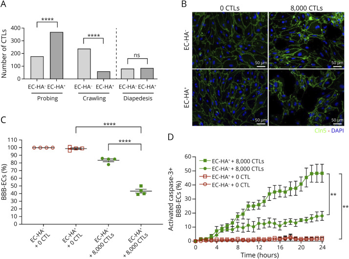
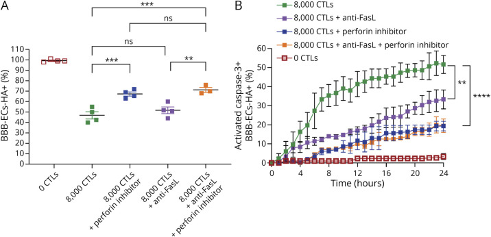
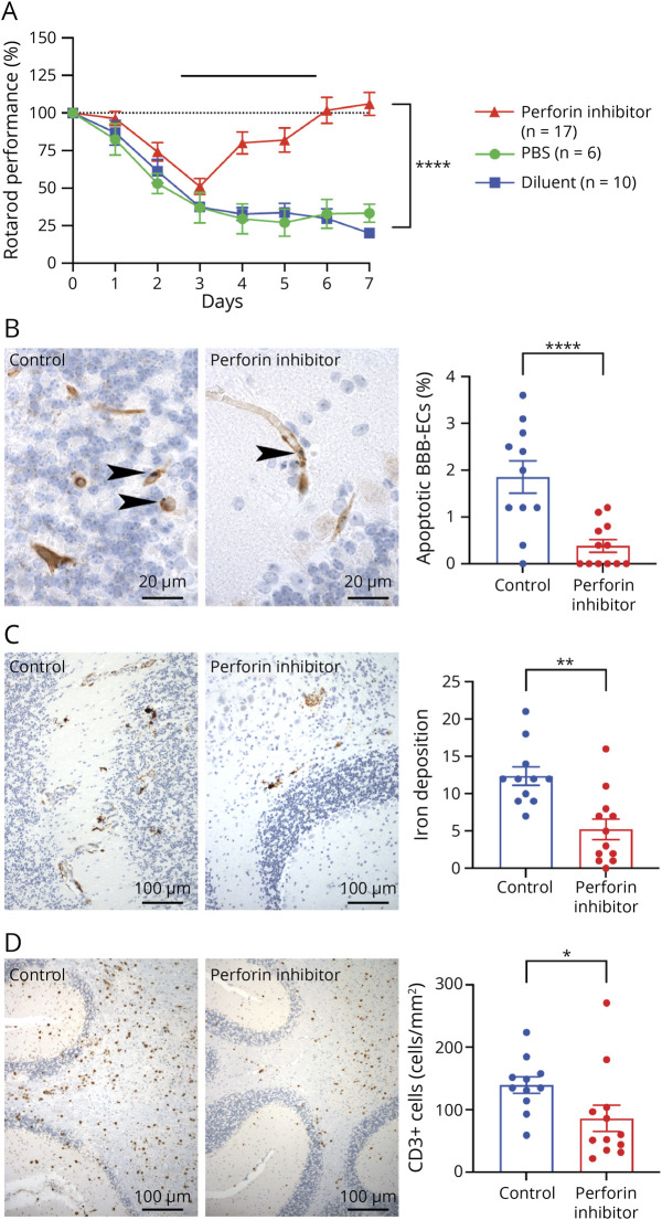
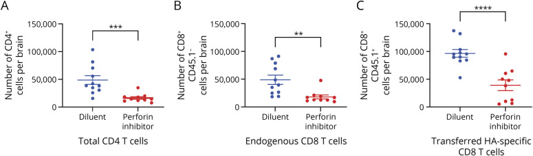
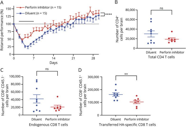
Results: Using an in vitro coculture system, we first identified perforin as an essential molecule for killing of BBB-ECs by CD8 T cells. We then found that short-term pharmacologic inhibition of perforin commencing after disease onset restored motor function and inhibited the neuropathology. Perforin inhibition resulted in preserved BBB-EC viability, maintenance of the BBB, and reduced CD8 T-cell accumulation in the brain and retina.

Discussion: Therefore, perforin-dependent cytotoxicity plays a key role in the death of BBB-ECs inflicted by autoreactive CD8 T cells in a preclinical model and potentially represents a therapeutic target for CD8 T cell-mediated neuroinflammatory diseases, such as cerebral malaria and Susac syndrome.


Conflict of interest statement

C. Fonte, E. Dufourd, V. Cazaentre, S. Aydin, B. Engelhardt, R. R. Caspi, B. Xu, G. Martin-Blondel, J.A. Spicer, and C. Bost report no disclosures relevant to the manuscript. C. Gonzalez-Fierro reports financial support by Fondation pour la Recherche Médicale (FRM). R.S. Liblau reports financial support by Fondation pour la Recherche Médicale (FRM), the French MS society (ARSEP foundation), Agence Nationale pour la Recherche CE17-0014, and BETPSY RHU 18- RHUS-0012. J. Bauer reports financial support by Austrian Science Fund FWF: P34864-B. J.A. Trapani reports financial support by the Strategic Drug Discovery Initiative (SDDI) Program of the Wellcome Trust UK. Go to Neurology.org/NN for full disclosure.

Figures

Figure 1
Figure 1. Coculture of HA-Expressing BBB-ECs With HA-Specific CTLs Leads to an Antigen-Dependent Apoptosis of Endothelial Cells
(A) BBB-ECs from EC-HA+ or EC-HA- mice were superfused for 20 minutes with HA-specific CTLs in a flow chamber. A number of 451 and 444 CTLs were arrested on BBB-ECs from EC-HA+ and EC-HA- mice, respectively, and then 22 and 28 cells, respectively, detached after their arrest. The fractions of durably arrested CTLs probing and crawling were determined as categorical variables (left) and the fractions undergoing diapedesis (right). Results are from 2 independent experiments. The distribution between groups was compared using the χ2 test. (B) BBB-ECs from EC-HA+ or EC-HA- mice were cultured alone or in the presence of 8000 HA-specific CTLs. After 24 hours, BBB-ECs were fixed and stained with DAPI and anticlaudin-5 antibody. (C) DAPI+ nuclei surrounded by claudin-5 staining were counted and, in each experiment, the number of claudin-5–positive ECs in the wells from EC-HA- mice in the absence of CTLs was used as the 100% reference value. Data from 4 independent experiments are shown as mean and SEM, and the p values were determined using one-way ANOVA with post hoc Tukey HSD for multiple comparisons. (D) Kinetic analysis of activated caspase-3 expression in BBB-ECs in culture with or without HA-specific CTLs. Activated caspase-3 activity was determined using a NucView 488 assay and fluorescence microscopy. Data from 3 independent experiments are shown as mean and SEM, and the p values were determined using a two-way ANOVA with post hoc Tukey HSD for multiple comparisons. BBB-EC = endothelial cells of the blood-brain barrier; CTLs = cytotoxic CD8 T cells; EC = endothelial cells; HA = hemagglutinin.
Figure 2
Figure 2. Antigen-Dependent Apoptosis of BBB-ECs Is Inhibited by a Perforin Inhibitor
BBB-ECs from EC-HA+ mice were cultured alone or in the presence of 8000 HA-specific CTLs, in the presence or absence of a perforin inhibitor and/or a neutralizing anti-FasL mAb. (A) After 24 hours, BBB-ECs were fixed and stained with DAPI and claudin-5. Numbers of BBB-ECs nuclei after the 24 hours coculture are shown. The 100% value was set from the number of nuclei in the culture of BBB-ECs from EC-HA- mice without CTLs (see eFigure 2, links.lww.com/NXI/A838). Data represent the mean and SEM of 3–4 independent experiments, and the p values were determined using one-way ANOVA, followed by Sidak correction for multiple comparisons. (B) Kinetics of caspase-3 activation in BBB-ECs from EC-HA+ mice during the coculture with or without perforin inhibitor and/or anti-FasL mAb. Activated caspase-3 activity was determined using a NucView 488 assay and fluorescence microscopy. Data represent the mean and SEM of 3–4 independent experiments, and the p values were determined using two-way ANOVA with post hoc Tukey HSD for multiple comparisons. BBB-EC = endothelial cells of the blood-brain barrier; CTLs = cytotoxic CD8 T cells; EC = endothelial cells; HA = hemagglutinin.
Figure 3
Figure 3. Treatment With the Perforin Inhibitor Alleviates the Clinical Signs and Decreases Damage of the BBB in a Preclinical Model of Neuroinflammatory Disease
(A) Motor performance assessed by Rotarod in EC-HA+ mice after adoptive transfer of HA-specific CTLs. Mice were treated with PBS, diluent or perforin inhibitor every 12 hours, from day 3 until sacrifice at day 7 post-CTL transfer. The 100% was set from the motor performance at day 0, before CTL transfer. Bar shows the treatment period. Data are pooled from 3 independent experiments. Two-way ANOVA was used to determine the p values. (B–D) Frequency of apoptotic BBB-ECs (B), iron deposition (C), and infiltration of CD3+ T cells (D) in the brain of CTL-injected EC-HA+ mice, treated or not with the perforin inhibitor. The control EC-HA+ mice received either diluent or PBS. Data represent the mean and SEM of 11–12 mice per group from 3 independent experiments, and the p values were determined using the Student t test. BBB = blood-brain barrier; BBB-EC = endothelial cells of the blood-brain barrier; EC = endothelial cells; HA = hemagglutinin.
Figure 4
Figure 4. Treatment With the Perforin Inhibitor Reduces Infiltration of the CNS by Immune Cells in a Preclinical Model of Neuroinflammatory Disease
(A) Number of CD4, (B) endogenous, and (C) transferred CD8 T cells infiltrating the CNS of EC-HA+ mice, 7 days after adoptive transfer of CTLs and 4 days after initiation of treatment. Data represent the mean and SEM of 3 independent experiments. For panels A and B, the p values were determined using the Mann-Whitney test, and for panel C, the p values were determined using the Student t test.
Figure 5
Figure 5. Perforin Inhibitor Allows Quicker and Stable Recovery Despite Treatment Discontinuation
(A) Motor performance assessed by Rotarod in EC-HA+ mice after adoptive transfer of HA-specific CTLs. Mice were treated with diluent or perforin inhibitor from day 3 to day 7 after CTL transfer and sacrificed at day 30. The 100% was set from the motor performance at day 0, before CTL transfer. Bar shows the treatment period. Data are pooled from 4 independent experiments. Two-way ANOVA was used to determine the p values. (B) Number of CD4, (C) endogenous, and (D) transferred CD8 T cells infiltrating the CNS of EC-HA+ mice, 30 days after adoptive transfer of CTLs. Data represent the mean and SEM of 2 independent experiments. The p values were determined using the Mann-Whitney test for panels B and C and the Student t test for panel D.

Similar articles

Cited by

References

    1. Waisman A, Liblau RS, Becher B. Innate and adaptive immune responses in the CNS. Lancet Neurol. 2015;14(9):945-955. doi:10.1016/s1474-4422(15)00141-6 - DOI - PubMed
    1. McCandless EE, Zhang B, Diamond MS, Klein RS. CXCR4 antagonism increases T cell trafficking in the central nervous system and improves survival from West Nile virus encephalitis. Proc Natl Acad Sci U S A. 2008;105(32):11270-11275. doi:10.1073/pnas.0800898105 - DOI - PMC - PubMed
    1. Frieser D, Pignata A, Khajavi L, et al. . Tissue-resident CD8+ T cells drive compartmentalized and chronic autoimmune damage against CNS neurons. Sci Transl Med. 2022;14(640):eabl6157. doi:10.1126/scitranslmed.abl6157 - DOI - PubMed
    1. Vincenti I, Page N, Steinbach K, et al. . Tissue-resident memory CD8+ T cells cooperate with CD4+ T cells to drive compartmentalized immunopathology in the CNS. Sci Transl Med. 2022;14(640):eabl6058. doi:10.1126/scitranslmed.abl6058 - DOI - PubMed
    1. Zhang D, Ren J, Luo Y, et al. . T cell response in ischemic stroke: from mechanisms to translational insights. Front Immunol. 2021;12:707972. doi:10.3389/fimmu.2021.707972 - DOI - PMC - PubMed

Publication types