

Impact of topographic localization of corneal ectasia on the outcomes of deep anterior lamellar keratoplasty employing large (9 mm) versus conventional diameter (8 mm) grafts
- PMID: 37081075
- PMCID: PMC10630389
- DOI: 10.1038/s41433-023-02536-6
Impact of topographic localization of corneal ectasia on the outcomes of deep anterior lamellar keratoplasty employing large (9 mm) versus conventional diameter (8 mm) grafts
Abstract
Objectives: Visual and topographic outcomes of large (9.0 mm) versus conventional (8.0 mm) deep anterior lamellar keratoplasty (DALK) for the treatment of keratoconus (KC) were compared in relation to the different localization of the corneal ectasia (within or beyond the central 8.0 mm).
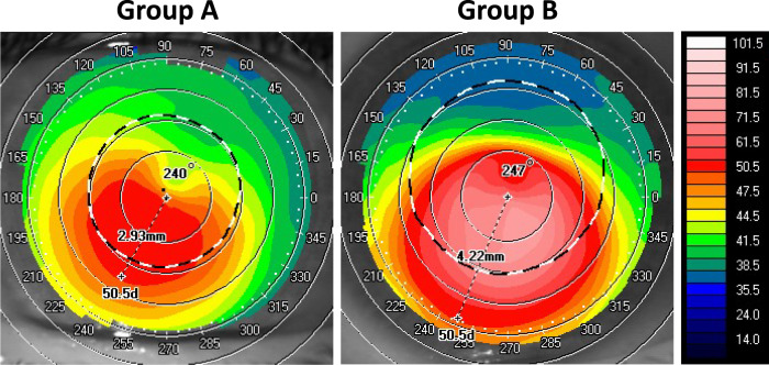
Methods: This is a retrospective, comparative case series. Preoperatively, the topographic extension of the conus was calculated by measuring the distance from the geometric center of the cornea and the outermost point of the corneal ectasia (ectasia <8.0 mm, group A; ectasia ≥8.0 mm, group B). DALK was performed using both small grafts (8.0 mm, group 1) and large grafts (9.0 mm, group 2). Best-corrected visual acuity and topographic astigmatism were evaluated preoperatively (T0) and postoperatively after complete suture removal (1 year, T1).
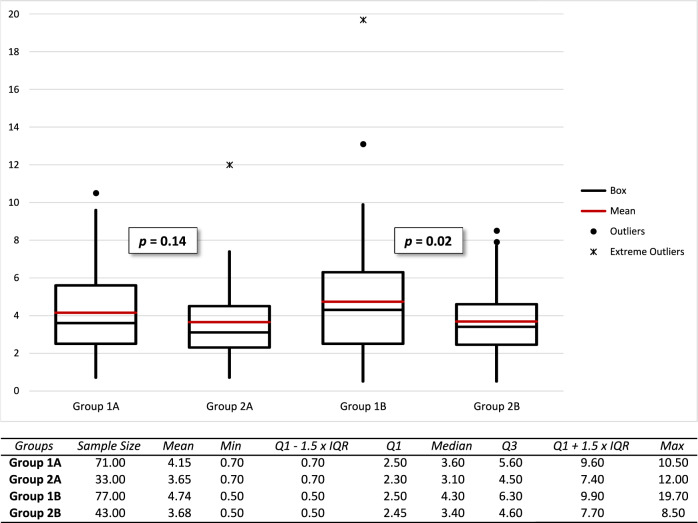
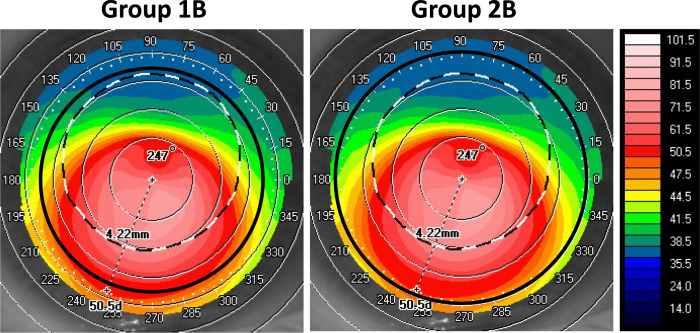
Results: Data from 224 eyes of 196 patients (mean age 37.6 ± 15.1 years) were evaluated. Topographic astigmatism improved from T0 to T1 (4.94 ± 2.92 diopters (D) [95% CI, 4.56-5.33] vs 4.19 ± 2.45 D [95% CI, 3.87-4.51], p = 0.001). There was no significant difference in postoperative topographic cylinder between group 1 and group 2 when considering eyes with corneal ectasia <8.0 mm (group 1 A, 4.15 ± 2.19 D [95% CI, 3.64-4.66] vs group 2 A, 3.65 ± 2.13 D [95% CI, 2.92-4.38], p = 0.14); conversely, the difference was significant considering eyes with corneal ectasia ≥8.0 mm (group 1B, 4.74 ± 2.90 D [95% CI, 4.09-5.38] vs group 2B, 3.68 ± 1.94 D [95% CI, 3.10-4.26], p = 0.02).
Conclusions: Large 9.0-mm DALK provided better anatomical outcomes compared to conventional 8.0-mm DALK, particularly in eyes with corneal ectasia extending beyond the central 8.0 mm.
© 2023. The Author(s), under exclusive licence to The Royal College of Ophthalmologists.
Conflict of interest statement
The authors declare no competing interests.
Figures

Similar articles
-
High Astigmatism After Conventional Diameter Deep Anterior Lamellar Keratoplasty in Keratoconus Can Be Successfully Managed With Repeat Wide Diameter Deep Anterior Lamellar Keratoplasty.Cornea. 2023 Aug 1;42(8):1057-1061. doi: 10.1097/ICO.0000000000003298. Epub 2023 May 1. Cornea. 2023. PMID: 37126842
-
Comparison of outcomes and complications of deep anterior lamellar keratoplasty and penetrating keratoplasty performed in a large group of patients with keratoconus.Int Ophthalmol. 2018 Jun;38(3):985-992. doi: 10.1007/s10792-017-0548-9. Epub 2017 May 22. Int Ophthalmol. 2018. PMID: 28534231
-
Large (9 mm) Deep Anterior Lamellar Keratoplasty with Clearance of a 6-mm Optical Zone Optimizes Outcomes of Keratoconus Surgery.Ophthalmology. 2017 Jul;124(7):1072-1080. doi: 10.1016/j.ophtha.2017.02.011. Epub 2017 Mar 20. Ophthalmology. 2017. PMID: 28336058
-
Excimer Laser-Assisted Deep Anterior Lamellar Keratoplasty Versus Penetrating Keratoplasty for Patients With Keratoconus: A Retrospective Analysis From the Homburg Keratoconus Center.Cornea. 2025 Mar 1;44(3):291-299. doi: 10.1097/ICO.0000000000003703. Cornea. 2025. PMID: 39413248
-
Comparison of refractive outcomes in three corneal transplantation techniques for keratoconus.Graefes Arch Clin Exp Ophthalmol. 2015 Nov;253(11):1947-53. doi: 10.1007/s00417-015-3091-2. Epub 2015 Aug 14. Graefes Arch Clin Exp Ophthalmol. 2015. PMID: 26271303
Cited by
-
Exploring Detection Methods for Synthetic Medical Datasets Created With a Large Language Model.JAMA Ophthalmol. 2025 Jun 1;143(6):517-522. doi: 10.1001/jamaophthalmol.2025.0834. JAMA Ophthalmol. 2025. PMID: 40272814
References
-
- Eye Bank Association of America. 2019 Eye Banking Statistical Report; 2020. Accessed 8 Dec 2022.
MeSH terms
LinkOut - more resources
Full Text Sources
