

Intracranial epidermoid cysts: benign entities with malignant behavior: experience with 36 cases
- PMID: 37081102
- PMCID: PMC10119307
- DOI: 10.1038/s41598-023-33617-x
Intracranial epidermoid cysts: benign entities with malignant behavior: experience with 36 cases
Abstract
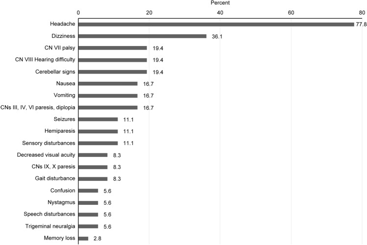
Intracranial epidermoid cysts are benign slow-growing ectodermal inclusions that account for less than 1% of all intracranial tumors. We retrospectively reviewed 36 such cases to evaluate the demographic characteristics, clinical manifestations, anatomical distribution, surgical management, and treatment outcome of these tumors. Additionally, we sought to identify the relationship between median or paramedian cistern tumor localization and clinical parameters, such as recurrence risk, hospitalization duration, and postoperative complication rates. The most frequently observed neurological symptoms were transient headaches (77.8%), dizziness (36.1%), CN VII palsy (19.4%), CN VIII hearing difficulty (19.4%) and cerebellar signs (19.4%). The most common surgical approaches included retrosigmoid (36.1%), subfrontal (19.4%) and telovelar (19.4%) approaches; gross total resection was feasible in 83.3% of cases. The postoperative complication rate was 38.9%. Tumors were more frequently found in the paramedian cisterns (47.2%), followed by the median line cisterns (41.6%). Multivariate analysis revealed that postoperative hydrocephalus and age < 40 years were prognostic factors for tumor recurrence. Median-like tumor location was a risk factor for the presence of symptomatic hydrocephalus both preoperatively and postoperatively, increasing the likelihood of protracted hospitalization (> 10 days). Despite their benign histopathological nature, these tumors have an important clinical resonance, with a high rate of postoperative complications and a degree of recurrence amplified by younger age and hydrocephalus.
© 2023. The Author(s).
Conflict of interest statement
The authors declare no competing interests.
Figures
Similar articles
-
Delayed postoperative hemorrhage in 21 patients with intracranial epidermoid cysts.J Neurosurg. 2011 Jun;114(6):1592-602. doi: 10.3171/2010.12.JNS10325. Epub 2011 Jan 28. J Neurosurg. 2011. PMID: 21275558
-
Whole Course Neuroendoscopic Resection of Cerebellopontine Angle Epidermoid Cysts.J Neurol Surg A Cent Eur Neurosurg. 2016 Sep;77(5):381-8. doi: 10.1055/s-0035-1558818. Epub 2015 Aug 24. J Neurol Surg A Cent Eur Neurosurg. 2016. PMID: 26302403
-
Intraspinal epidermoid and dermoid cysts-tumor resection with multimodal intraoperative neurophysiological monitoring and long-term outcome.Acta Neurochir (Wien). 2020 Nov;162(11):2895-2903. doi: 10.1007/s00701-020-04446-y. Epub 2020 Jun 10. Acta Neurochir (Wien). 2020. PMID: 32524245
-
[Surgery of intracranial epidermoid cysts. Report of 44 patients and review of the literature].Neurochirurgie. 2002 Feb;48(1):5-13. Neurochirurgie. 2002. PMID: 11972145 Review. French.
-
[Epidermoid cyst of the cisterna magna and the fourth ventricle: Report of four cases].Neurochirurgie. 2012 Dec;58(6):358-63. doi: 10.1016/j.neuchi.2011.10.001. Epub 2012 Jun 14. Neurochirurgie. 2012. PMID: 22704404 Review. French.
Cited by
-
A Complicated Case of An Intracranial Epidermoid Cyst.Eur J Case Rep Intern Med. 2025 Jan 27;12(2):005121. doi: 10.12890/2025_005121. eCollection 2025. Eur J Case Rep Intern Med. 2025. PMID: 39926568 Free PMC article.
-
Ossified spinal epidermoid cyst: A systematic review and case report.Heliyon. 2024 Aug 28;10(18):e37093. doi: 10.1016/j.heliyon.2024.e37093. eCollection 2024 Sep 30. Heliyon. 2024. PMID: 39315203 Free PMC article.
-
Cerebellopontine angle epidermoid cyst masquerading as trigeminal neuralgia: A rare presentation and surgical outcome.Radiol Case Rep. 2025 Mar 8;20(5):2294-2299. doi: 10.1016/j.radcr.2025.01.076. eCollection 2025 May. Radiol Case Rep. 2025. PMID: 40129820 Free PMC article.
-
An Anterior Midline Skull Base Epidermoid Cyst Presenting With Spontaneous Intraparenchymal Rupture: A Case Report.Cureus. 2024 Sep 24;16(9):e70076. doi: 10.7759/cureus.70076. eCollection 2024 Sep. Cureus. 2024. PMID: 39449920 Free PMC article.
-
Trigeminal neuralgia secondary to epidermoid cyst and neurovascular conflict: An illustrative case with literature review.Surg Neurol Int. 2024 Feb 9;15:36. doi: 10.25259/SNI_925_2023. eCollection 2024. Surg Neurol Int. 2024. PMID: 38468668 Free PMC article.
References
MeSH terms
LinkOut - more resources
Full Text Sources
Medical
