

Evaluation of retinal microcirculation alterations using OCTA in hyperopic ametropic amblyopia patients before and after treatment
- PMID: 37081133
- PMCID: PMC10400672
- DOI: 10.1007/s10792-023-02707-0
Evaluation of retinal microcirculation alterations using OCTA in hyperopic ametropic amblyopia patients before and after treatment
Abstract
Purpose: We aimed to compare retinal microcirculation in hyperopic ametropic amblyopia patients before and after treatment and in healthy children using optical coherence tomography angiography (OCTA), and to explore the pathogenesis of hyperopic ametropic amblyopia.
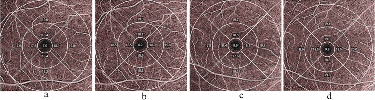
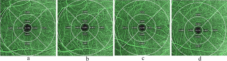
Methods: Eighteen patients with hyperopic ametropic amblyopia aged 4-8 years were selected as the patient group, and 18 age-matched healthy children were randomly selected as controls. The foveal avascular zone (FAZ) area, perimeter and circularity, vessel density (VD) and perfusion density (PD) of macular superficial retinal capillary plexus, macular thickness, peripapillary retinal nerve fiber layer thickness, and ganglion cell-inner plexiform layer thickness were compared between both groups. After 6 months of amblyopia treatment, the same parameters were measured again.
Results: The VD and PD in the central, inner, inner nasal, and inner inferior regions in hyperopic ametropic amblyopia were lower than in the control group after adjustment for axial length. After 6 months of treatment, the VD increased significantly, except in the outer nasal and outer inferior regions. The PD in the central (p < 0.001), inner superior (p = 0.001), inner inferior (p = 0.011) and inner temporal (p = 0.026) regions increased. The FAZ perimeter and circularity significantly differed between the groups. After 6 months of treatment, the FAZ area and perimeter decreased, but circularity increased.
Conclusion: Hyperopic ametropic amblyopia eyes showed a significant decrease in vessel and perfusion densities. After amblyopia treatment, the vessel and perfusion densities of patients with hyperopic ametropic amblyopia increased, suggesting that abnormalities in the microvascular system are a pathogenic factor of amblyopia.
Keywords: Amblyopia treatment; Hyperopic ametropic amblyopia; Optical coherence tomography angiography; Perfusion density; Vessel density.
© 2023. The Author(s).
Conflict of interest statement
The authors have no relevant financial or non-financial interests to disclose.
Figures

Similar articles
-
Evaluation of retinal microcirculation alterations using optical coherence tomography angiography in patients with hyperopia ametropic amblyopia: A case-control study.Medicine (Baltimore). 2023 Mar 10;102(10):e33196. doi: 10.1097/MD.0000000000033196. Medicine (Baltimore). 2023. PMID: 36897692 Free PMC article.
-
Macular superficial vascular density on optical coherence tomography angiography in children with unilateral anisometropic and bilateral hyperopic amblyopia.Sci Rep. 2023 Aug 8;13(1):12879. doi: 10.1038/s41598-023-40025-8. Sci Rep. 2023. PMID: 37553433 Free PMC article.
-
Analysis of Macular Retinal Thickness and Microvascular System Changes in Children with Monocular Hyperopic Anisometropia and Severe Amblyopia.Dis Markers. 2022 Jan 17;2022:9431044. doi: 10.1155/2022/9431044. eCollection 2022. Dis Markers. 2022. PMID: 35082933 Free PMC article.
-
Optical coherence tomography angiography analysis of the fellow eye in unilateral pseudoexfoliation syndrome.BMC Ophthalmol. 2024 Sep 27;24(1):421. doi: 10.1186/s12886-024-03686-1. BMC Ophthalmol. 2024. PMID: 39333925 Free PMC article.
-
Retina and microvascular alterations in migraine: a systemic review and meta-analysis.Front Neurol. 2023 Sep 28;14:1241778. doi: 10.3389/fneur.2023.1241778. eCollection 2023. Front Neurol. 2023. PMID: 37840933 Free PMC article.
References
-
- Chinese Association for Pediatric Ophthalmology and Strabismus, Pediatric Ophthalmology and Strabismus Group of Chinese Ophthalmologist Association Expert consensus on prevention and treatment of amblyopia in children. Chin J Ophthalmol. 2021;57(5):336–540. doi: 10.3760/cma.j.cn112142-20210109-00014. - DOI - PubMed
-
- Rocholz R, Corvi F, Weichsel J, Schmidt S, Staurenghi G. OCT Angiography (OCTA) in retinal diagnostics. In: Bille JF, editor. High resolution imaging in microscopy and ophthalmology: new frontiers in biomedical optics. Cham: Springer; 2019. pp. 135–160. - PubMed
-
- Wong Emily S, Xiu-Juan Z, Nan Y, et al. Association of optical coherence tomography angiography metrics with detection of impaired macular microvasculature and decreased vision in amblyopic eyes: the hong kong children eye study. JAMA Ophthalmol. 2020;138:858–865. doi: 10.1001/jamaophthalmol.2020.2220. - DOI - PMC - PubMed
Publication types
MeSH terms
Grants and funding
LinkOut - more resources
Full Text Sources
Medical
