Review
. 2023 Apr;36(2):159-173.
doi: 10.1007/s10334-023-01080-4. Epub 2023 Apr 20.

Magnetic resonance imaging at 9.4 T: the Maastricht journey

Affiliations
Review

Magnetic resonance imaging at 9.4 T: the Maastricht journey

Dimo Ivanov et al. MAGMA. 2023 Apr.

Abstract

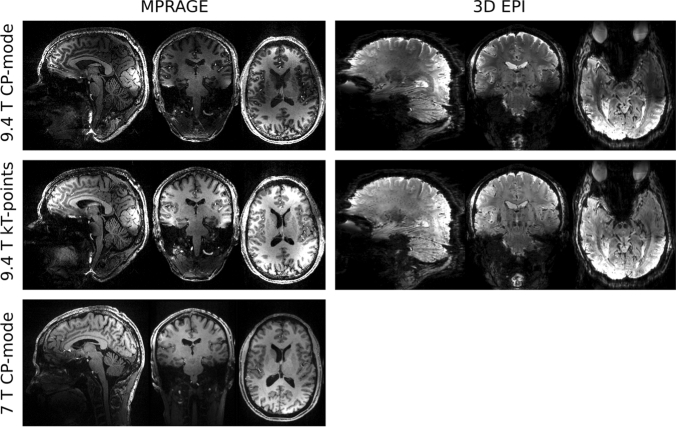
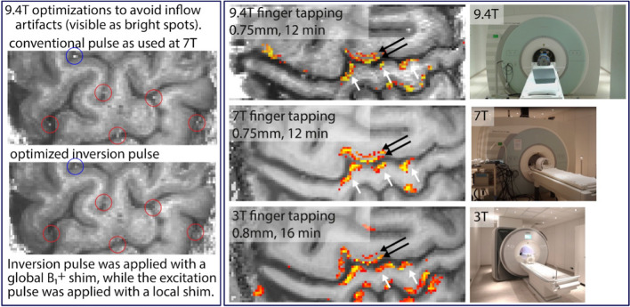
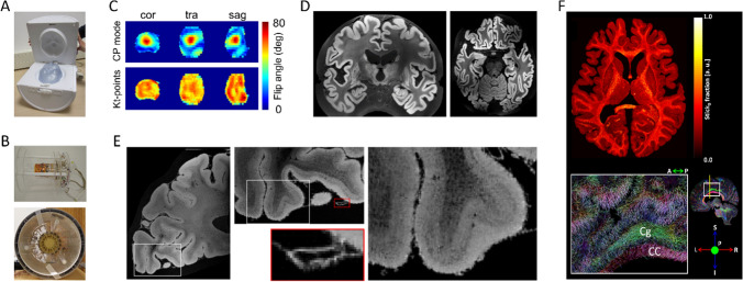
The 9.4 T scanner in Maastricht is a whole-body magnet with head gradients and parallel RF transmit capability. At the time of the design, it was conceptualized to be one of the best fMRI scanners in the world, but it has also been used for anatomical and diffusion imaging. 9.4 T offers increases in sensitivity and contrast, but the technical ultra-high field (UHF) challenges, such as field inhomogeneities and constraints set by RF power deposition, are exacerbated compared to 7 T. This article reviews some of the 9.4 T work done in Maastricht. Functional imaging experiments included blood oxygenation level-dependent (BOLD) and blood-volume weighted (VASO) fMRI using different readouts. BOLD benefits from shorter T2* at 9.4 T while VASO from longer T1. We show examples of both ex vivo and in vivo anatomical imaging. For many applications, pTx and optimized coils are essential to harness the full potential of 9.4 T. Our experience shows that, while considerable effort was required compared to our 7 T scanner, we could obtain high-quality anatomical and functional data, which illustrates the potential of MR acquisitions at even higher field strengths. The practical challenges of working with a relatively unique system are also discussed.

Keywords: 9.4 T; Ultra-high field; fMRI; pTx.


Conflict of interest statement

The authors declare that they have no conflict of interest in relation to the work described in this paper.

Figures

Fig. 1
Fig. 1
MPRAGE (0.6 mm isotropic resolution) and 3D EPI (0.75 mm isotropic resolution) images, obtained using CP-mode (top row) and kT-points (middle row) excitations. MPRAGE (0.6 mm isotropic resolution) from the same participant acquired using the 1Tx/32Rx Nova Medical head coil at 7 T (CP-mode)
Fig. 2
Fig. 2
In vivo high-resolution SMS-GE images from [31] acquired with 3-spoke a single-band, b, c SMS-2 and d, e SMS-3 excitations. Rows b and d show the SMS images before slice-GRAPPA reconstructionṣ
Fig. 3
Fig. 3
A Brain activation of an individual participant evoked by performing three different mental tasks. For every mental task, activations are shown for the left (LH) and the right hemisphere (RH). Every mental task evoked a unique brain-activation pattern that can be differentiated by fMRI. B The visual letter encoding scheme. Combining three mental tasks and nine different time intervals, the letter-encoding technique allows for encoding 3 × 9 = 27 different characters (26 letters and a blank space). Each column is highlighted for 10 s, during which the participant performs the corresponding mental task for the desired letter to be encoded
Fig. 4
Fig. 4
Tonotopic maps in temporal cortex obtained with GE EPI. A The right hemisphere surface of the temporal lobe (top) and tonotopy (bottom) of one of the volunteers. B Tonotopic maps (and cortical surfaces) of eight volunteers
Fig. 5
Fig. 5
The left panel highlights one of the most important challenges of VASO at 9.4 T., namely obtaining a VASO T1 contrast without inflow of fresh (un-inverted blood). We achieve this with an alternating B1+ shim (for inversion and excitation pulses, respectively), and by means of pulse optimizations including phase skips and SAR-optimized lower bandwidth. The right panel depicts representative high-resolution maps of CBV changes. 9.4 T can reveal the same laminar activation features as lower field strengths
Fig. 6
Fig. 6
A Schematic representation of the conventional isotropic and the anisotropic sampling schemes of cortex illustrating the differences for laminar and columnar acquisitions, B example Z-score maps and zoomed panels for the EPI-BOLD and anisotropic multi-echo BOLD data for the finger-tapping experiment overlaid on a registered T1-weighted image at each TE. Bar plot of % signal change with voxels binned according to different T2* for the four TEs of the AVF-BOLD data, (C, left) and comparable TE = 19.7 ms, EPI-BOLD (C, right). D Columnar profiles of the % BOLD signal change at different TEs from the AVF data and the EPI-BOLD data in the GM ROI (zoomed cyan panel of B) overlaid on colourmap corresponding to T2* of the voxel.
Fig. 7
Fig. 7
A The custom-built 8Tx/24Rx 9.4 T whole post-mortem human brain RF-coil. B The custom-built 16Rx 9.4 T medium-sized post-mortem sample RF-coil outside of (top) and inside (bottom) the 16Tx ring C) B1+ maps showing transmit efficiency and homogeneity over the whole post-mortem brain with substantial inhomogeneity in standard circularly polarized (CP) mode, which is improved greatly with kT-points transmit phases optimized for homogeneity. D A coronal (left) and transverse slice (right) through early whole post-mortem brain 3D GE acquisitions at 200 µm isotropic. E Transverse slices through a GE acquisition at 60 μm isotropic of an occipital lobe sample in the medium-sample coil at different zoom levels. F kT-dSTEAM 400 μm diffusion results: a transverse slice through the Stick0 fraction map resulting from a Ball&Stick model fit (top) and direction color coded DTI primary eigenvector in a sagittal slice with zoom-in (bottom) CC corpus callosum, Cg cingulum

References

    1. Pohmann R, Speck O, Scheffler K. Signal-to-noise ratio and MR tissue parameters in human brain imaging at 3, 7, and 9.4 tesla using current receive coil arrays. Magn Reson Med. 2016;75(2):801–809. doi: 10.1002/mrm.25677. - DOI - PubMed
    1. Le Ster C, Grant A, Van de Moortele PF, Monreal-Madrigal A, Adriany G, Vignaud A, Mauconduit F, Rabrait-Lerman C, Poser BA, Ugurbil K, Boulant N. Magnetic field strength dependent SNR gain at the center of a spherical phantom and up to 11.7T. Magn Reson Med. 2022;88(5):2131–2138. doi: 10.1002/mrm.29391. - DOI - PMC - PubMed
    1. Ugurbil K, Adriany G, Andersen P, Chen W, Garwood M, Gruetter R, Henry PG, Kim SG, Lieu H, Tkac I, Vaughan T, Van De Moortele PF, Yacoub E, Zhu XH. Ultrahigh field magnetic resonance imaging and spectroscopy. Magn Reson Imaging. 2003;21(10):1263–1281. doi: 10.1016/j.mri.2003.08.027. - DOI - PubMed
    1. Uludag K, Muller-Bierl B, Ugurbil K. An integrative model for neuronal activity-induced signal changes for gradient and spin echo functional imaging. Neuroimage. 2009;48(1):150–165. doi: 10.1016/j.neuroimage.2009.05.051. - DOI - PubMed
    1. Yacoub E, Duong TQ, Van De Moortele PF, Lindquist M, Adriany G, Kim SG, Ugurbil K, Hu X. Spin-echo fMRI in humans using high spatial resolutions and high magnetic fields. Magn Reson Med. 2003;49(4):655–664. doi: 10.1002/mrm.10433. - DOI - PubMed

MeSH terms