

Ultra-short echo time MR imaging in assessing cartilage endplate damage and relationship between its lesion and disc degeneration for chronic low back pain patients
- PMID: 37081427
- PMCID: PMC10120173
- DOI: 10.1186/s12880-023-01014-5
Ultra-short echo time MR imaging in assessing cartilage endplate damage and relationship between its lesion and disc degeneration for chronic low back pain patients
Abstract
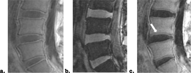
Objective: To investigate the feasibility of ultra-short echo time (UTE) magnetic resonance imaging (MRI) in the assessment of cartilage endplate (CEP) damage and further evaluate the relationship between total endplate score (TEPS) and lumbar intervertebral disc (IVD) degeneration for chronic low back pain patients.
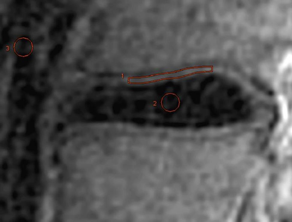
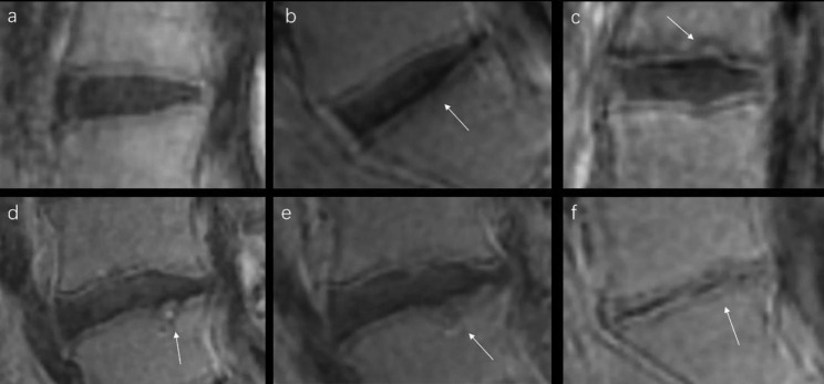
Materials and methods: IVD were measured in 35 patients using UTE imaging at 3T MR. Subtracted UTE images between short and long TEs were obtained to depict anatomy of CEP. The signal-to-noise ratio (SNR) and contrast-to-noise ratio (CNR) were calculated to assess the image quality quantitatively. A new grading criterion for endplate damage evaluation was developed based on Rajasekaran.S grading system in this study. Two radiologists were employed to evaluate CEP and bony vertebral endplates (VEP) using this new grading criterion and assess TEPS, independently. Cohen's kappa analysis was applied to evaluate the inter-observer agreement of endplate damage assessment between two radiologists, and the Kendall's TAU-B analysis was employed to determine the relationship between TEPS and IVD degeneration evaluated with Pfirrmann grading.
Results: Well structural CEP was depicted on subtracted UTE images and confirmed by high SNR (33.06±2.92) and CNR values (9.4±2.08). Qualified subtracted UTE images were used by two radiologists to evaluate the degree of CEP and VEP damage. Excellent inter-observer agreement was confirmed by high value in Cohen's kappa test (0.839, P < 0.001). Ensured by this, 138 endplates from 69 IVDs of 35 patients were classified into six grades based on the new grading criterion and TEPS of each endplate was calculated. In addition, the degeneration degree of IVDs were classified into five grades. Finally, using Kendall's TAU-B analysis, significant relationship was obtained between endplate damage related TEPS and IVD degeneration (r = 0.864, P < 0.001).
Conclusion: Ensured by high image quality, UTE imaging might be considered an effective tool to assess CEP damage. Additionally, further calculated TEPS has shown strong positive association with IVD degeneration, suggesting that the severity of endplate damage is highly linked with the degree of IVD degeneration.
Keywords: Bony vertebral endplate; Cartilage endplate; Chronic low back pain; Disc degeneration; Magnetic resonance imaging; Pfirrmann grade; Total endplate score; Ultra-short echo time.
© 2023. The Author(s).
Conflict of interest statement
The authors declare that they have no competing interests.
Figures
Similar articles
-
Evaluation of the disco-vertebral junction using ultrashort time-to-echo magnetic resonance imaging: inter-reader agreement and association with vertebral endplate lesions.Skeletal Radiol. 2016 Sep;45(9):1249-56. doi: 10.1007/s00256-016-2413-8. Epub 2016 May 30. Skeletal Radiol. 2016. PMID: 27241121 Free PMC article.
-
Cartilage Endplate Thickness Variation Measured by Ultrashort Echo-Time MRI Is Associated With Adjacent Disc Degeneration.Spine (Phila Pa 1976). 2018 May 15;43(10):E592-E600. doi: 10.1097/BRS.0000000000002432. Spine (Phila Pa 1976). 2018. PMID: 28984733 Free PMC article.
-
Ultrashort time-to-echo MRI of the cartilaginous endplate: technique and association with intervertebral disc degeneration.J Med Imaging Radiat Oncol. 2013 Aug;57(4):427-34. doi: 10.1111/1754-9485.12041. Epub 2013 Mar 26. J Med Imaging Radiat Oncol. 2013. PMID: 23870338
-
Diagnostic Role of Magnetic Resonance Imaging in Low Back Pain Caused by Vertebral Endplate Degeneration.J Magn Reson Imaging. 2022 Mar;55(3):755-771. doi: 10.1002/jmri.27858. Epub 2021 Jul 26. J Magn Reson Imaging. 2022. PMID: 34309129 Review.
-
Cartilaginous endplates: A comprehensive review on a neglected structure in intervertebral disc research.JOR Spine. 2023 Oct 21;6(4):e1294. doi: 10.1002/jsp2.1294. eCollection 2023 Dec. JOR Spine. 2023. PMID: 38156054 Free PMC article. Review.
Cited by
-
Ultrashort-Echo-Time MRI of the Disco-Vertebral Junction: Modulation of Image Contrast via Echo Subtraction and Echo Times.Sensors (Basel). 2024 Sep 9;24(17):5842. doi: 10.3390/s24175842. Sensors (Basel). 2024. PMID: 39275753 Free PMC article.
-
Cartilaginous endplate coverage of developmental Schmorl's node and the relevance of this in Schmorl's node etiology-based classification.Quant Imaging Med Surg. 2024 Jun 1;14(6):4288-4303. doi: 10.21037/qims-24-335. Epub 2024 May 9. Quant Imaging Med Surg. 2024. PMID: 38846309 Free PMC article. No abstract available.
-
Intervertebral Disc Degeneration and Regeneration: New Molecular Mechanisms and Therapeutics: Obstacles and Potential Breakthrough Technologies.Cells. 2024 Dec 19;13(24):2103. doi: 10.3390/cells13242103. Cells. 2024. PMID: 39768194 Free PMC article. Review.
-
Qualitative and Quantitative MR Imaging of the Cartilaginous Endplate: A Review.J Magn Reson Imaging. 2025 Apr;61(4):1552-1571. doi: 10.1002/jmri.29562. Epub 2024 Aug 20. J Magn Reson Imaging. 2025. PMID: 39165086 Review.
-
Ultrashort Echo Time Magnetic Resonance Morphology of Discovertebral Junction in Chronic Low Back Pain Subjects.Tomography. 2025 Jan 23;11(2):12. doi: 10.3390/tomography11020012. Tomography. 2025. PMID: 39997995 Free PMC article.
References
MeSH terms
LinkOut - more resources
Full Text Sources
