Review
. 2023 Apr 20;21(1):45.
doi: 10.1186/s12959-023-00484-5.

Venous anomalies and thromboembolism

Affiliations
Review

Venous anomalies and thromboembolism

Caroline Dix et al. Thromb J. .

Abstract

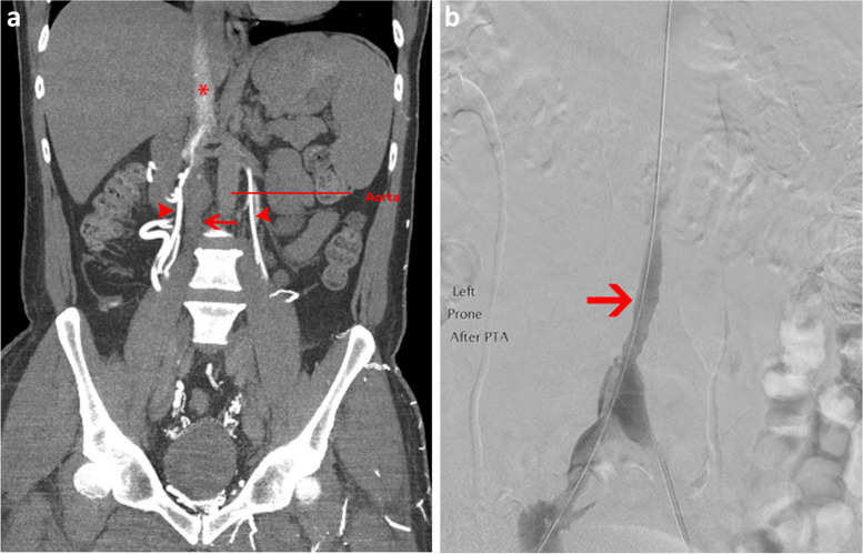
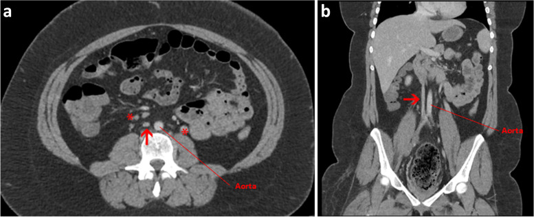
Patients with venous anomalies are at increased risk of developing venous thromboembolism (VTE) and subsequent complications, but they are often under-recognised. While unprovoked VTE may trigger testing for inherited thrombophilias and malignancy screening, anatomic variants are considered less often. Venous anomalies increase the risk due to venous flow disturbance, resulting in hypertension, reduced flow velocity and turbulence. Recognition is important as endovascular or surgical intervention may be appropriate, these patients have a high rate of VTE recurrence if anticoagulation is ceased, and the anomalies can predispose to extensive VTE and severe post-thrombotic syndrome (PTS). In this case series, we present representative cases and radiological images of May-Thurner syndrome (MTS), inferior vena cava (IVC) variants and venous aneurysms, and review the available literature regarding optimal diagnosis and management in each condition.

Keywords: Anatomic variant; Aneurysm; Anticoagulation; Endovascular; IVC agenesis; May-Thurner syndrome; Venous thromboembolism.


Conflict of interest statement

JM is supported by a National Health and Medical Research Council (NHMRC) Early Career Fellowship (APP1161499).

The authors declare no competing interests.

Figures

Fig. 1
Fig. 1
a Anatomy seen in May-Thurner Syndrome: Axial CT venogram of the pelvis shows compression of the left common iliac vein (arrow) between the right common iliac artery (asterisk) and the intervertebral disc of L5-S1. b Anatomy seen in May Thurner Syndrome: Sagittal CT venogram of the pelvis shows compression of the left common iliac vein (arrow) between the right common iliac artery and the intervertebral disc of L5-S1, with acute thrombus in the lower IVC (asterisk). c Frontal iliocaval angiography shows extrinsic compression of the left common iliac vein (arrow). This anatomy is seen in patients with May-Thurner Syndrome. d After placement of self-expanding 14 mm venous stent, improvement in flow is seen as well as resolution of extrinsic compression on the left common iliac vein
Fig. 2
Fig. 2
a Coronal CT venogram shows hypoplastic and acutely thrombosed infrarenal IVC (arrow) with contrast taking path of mature iliolumbar and lumbar collateral veins (arrow head). The suprarenal IVC is patent (asterisk). b Venogram after thrombolysis shows restoration of flow to the infrarenal IVC which remains hypoplastic (arrow)
Fig. 3
Fig. 3
a Axial CT venogram shows small calibre IVC over a long distance (arrow) and established collateral veins (asterisks) compatible with hypoplastic IVC. b Coronal CT venogram shows small calibre IVC over a long distance (arrow) compatible with hypoplastic IVC
Fig. 4
Fig. 4
a axial CT angiogram showing left popliteal vein aneurysm measuring 46.92 mm. b coronal image showing left popliteal vein aneurysm (arrow), with mural thrombus within aneurysmal segment. c Sagittal CT image showing left popliteal vein aneurysm (arrow)

References

    1. Wolberg AS, Rosendaal FR, Weitz JI, Jaffer IH, Agnelli G, Baglin T, et al. Venous thrombosis. Nat Rev Dis Primers. 2015;1:15006. doi: 10.1038/nrdp.2015.6. - DOI - PubMed
    1. Bagot CN, Arya R. Virchow and his triad: a question of attribution. Br J Haematol. 2008;143(2):180–190. doi: 10.1111/j.1365-2141.2008.07323.x. - DOI - PubMed
    1. Anderson FA, Jr, Spencer FA. Risk factors for venous thromboembolism. Circulation. 2003;107(23 Suppl 1):I9–16. - PubMed
    1. Harbin MM, Lutsey PL. May-Thurner syndrome: history of understanding and need for defining population prevalence. J Thromb Haemost. 2020;18(3):534–542. doi: 10.1111/jth.14707. - DOI - PubMed
    1. Ruggeri M, Tosetto A, Castaman G, Rodeghiero F. Congenital absence of the inferior vena cava: a rare risk factor for idiopathic deep-vein thrombosis. Lancet. 2001;357(9254):441. doi: 10.1016/S0140-6736(00)04010-1. - DOI - PubMed

LinkOut - more resources