Case Reports
. 2023 Dec 1;62(23):3495-3500.
doi: 10.2169/internalmedicine.1144-22. Epub 2023 Apr 21.

Cholangiocarcinoma Resembling IgG4-related Sclerosing Cholangitis

Affiliations
Case Reports

Cholangiocarcinoma Resembling IgG4-related Sclerosing Cholangitis

Kazuaki Akahoshi et al. Intern Med. .

Abstract

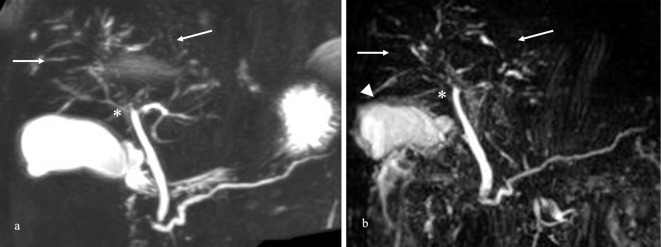
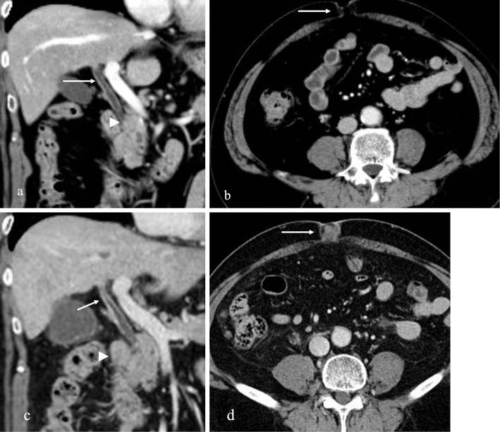
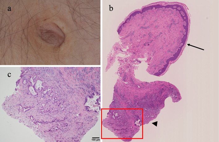
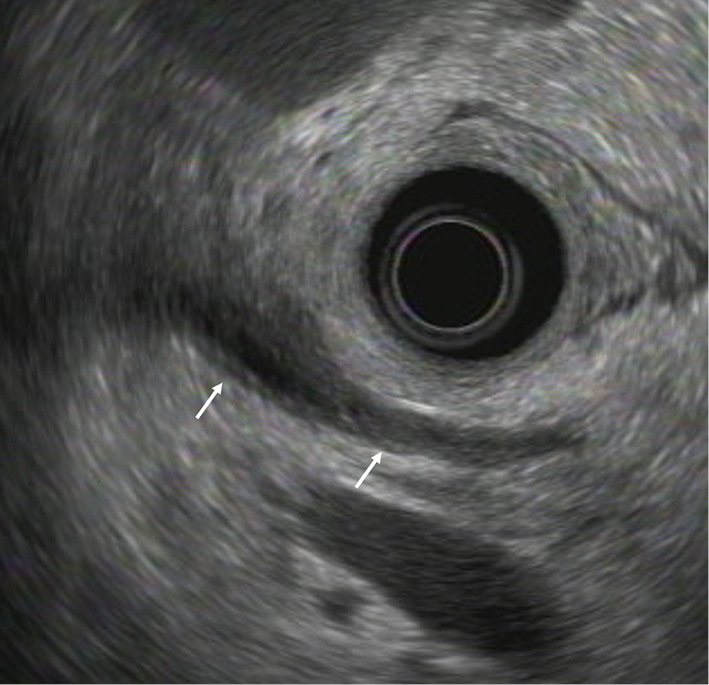
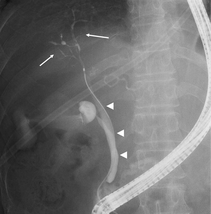
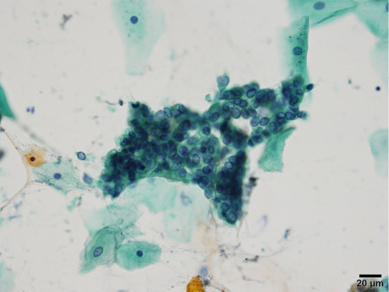
A 66-year-old man diagnosed with immunoglobulin G4-related sclerosing cholangitis (IgG4-SC) with diffuse intrahepatic bile duct stenosis and elevated serum IgG4 levels was referred for a further examination because of elevated serum carbohydrate antigen 19-9 levels despite treatment with corticosteroids. An umbilical nodule was found on a physical examination and a biopsy showed adenocarcinoma. Although several imaging studies revealed no changes from prior studies, bile cytology collected by endoscopic retrograde cholangiopancreatography showed adenocarcinoma. Consequently, the patient was diagnosed with cholangiocarcinoma resembling IgG4-SC after detecting an umbilical metastasis, also known as Sister Mary Joseph's nodule.

Keywords: Sister Mary Joseph's nodule; cholangiocarcinoma; immunoglobulin G4-related disease; immunoglobulin G4-related sclerosing cholangitis.


Conflict of interest statement

The authors state that they have no Conflict of Interest (COI).

Figures

Figure 1.
Figure 1.
Magnetic resonance cholangiopancreatography (MRCP) findings. (a) MRCP images from a year ago showing diffuse stenosis from the hilar to the intrahepatic bile duct (arrows) without diffuse narrowing of the main pancreatic duct. These findings revealed the stenosis of the bile duct in the perihilar portion (asterisk) without a dilated intrahepatic bile duct or cancerous nodules. (b) MRCP on admission revealed images similar to those of the intrahepatic bile duct a year ago and wall thickening of the gallbladder (arrowhead).
Figure 2.
Figure 2.
Abdominal computed tomography (CT) findings. (a) CT revealed the absence of pancreatic enlargement suggestive of autoimmune pancreatitis or swelling of the lymph nodes (arrowhead). The findings revealed wall thickening without a localized solid mass in the perihilar bile ducts (arrow). (b) No solid mass was observed in the umbilical region (arrow). (c) CT revealed worsening wall thickening of the common bile duct with a contrast effect (arrow) and enlarged lymph nodes (arrowhead). (d) A solid mass appeared in the umbilical region (arrow).
Figure 3.
Figure 3.
Histological findings at the umbilicus. (a) The skin is normal, but a 1-cm mass is palpable. (b) An umbilical biopsy was performed from the epidermis to the dermis (macroscopic findings). The sites indicated by the arrow and the arrowhead are the skin-surrounded squamous cells and carcinoma in the subcutaneous tissue, respectively. (c) Histological findings show enlarged nuclei and atypical cells with obscured nucleoli proliferating in an irregular glandular duct structure on the deep side, with fibrosis and inflammatory cell infiltration in the surrounding area, consistent with adenocarcinoma (Hematoxylin and Eosin staining, 200×).
Figure 4.
Figure 4.
Endoscopic ultrasound findings show bile duct wall thickening, mainly at the confluence of the three ducts (arrows). There is no mass or localized stricture.
Figure 5.
Figure 5.
Endoscopic retrograde cholangiopancreatography findings show diffuse stenosis of the intrahepatic bile duct from the hilar bile duct (arrows), with no obvious mass noted in the bile duct (arrowheads). These findings indicate immunoglobulin G4-related sclerosing cholangitis, Nakazawa classification type 2b.
Figure 6.
Figure 6.
Cytological findings show adenocarcinoma (Papanicolaou staining, 200×).

References

    1. Masamune A, Kikuta K, Hamada S, et al. . Nationwide epidemiological survey of autoimmune pancreatitis in Japan in 2016. J Gastroenterol 55: 462-470, 2020. - PubMed
    1. Kamisawa T, Nakazawa T, Tazuma S, et al. . Clinical practice guidelines for IgG4-related sclerosing cholangitis. J Hepatobiliary Pancreat Sci 26: 9-42, 2019. - PMC - PubMed
    1. Nakazawa T, Kamisawa T, Okazaki K, et al. . Clinical diagnostic criteria for IgG4-related sclerosing cholangitis 2020: (Revision of the clinical diagnostic criteria for IgG4-related sclerosing cholangitis 2012). J Hepatobiliary Pancreat Sci 28: 235-242, 2021. - PubMed
    1. Nakazawa T, Naitoh I, Hayashi K, et al. . Diagnostic criteria for IgG4-related sclerosing cholangitis based on cholangiographic classification. J Gastroenterol 47: 79-87, 2012. - PubMed
    1. Mayo WJ. Metastasis in cancer. Proc Staff Meet Mayo Clin 3: 327, 1928.

Publication types

Substances