

The degree and position of phosphorylation determine the impact of toxic and trace metals on phosphoinositide containing model membranes
- PMID: 37082006
- PMCID: PMC10074965
- DOI: 10.1016/j.bbadva.2021.100021
The degree and position of phosphorylation determine the impact of toxic and trace metals on phosphoinositide containing model membranes
Abstract
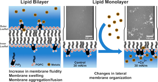
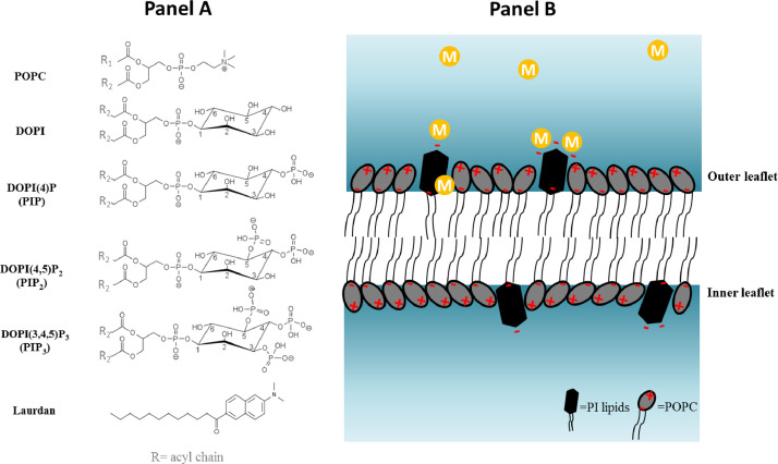
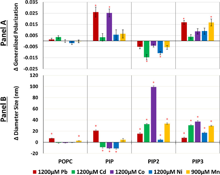
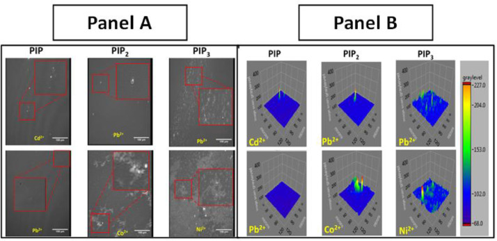
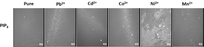
This work assessed effects of metal binding on membrane fluidity, liposome size, and lateral organization in biomimetic membranes composed of 1 mol% of selected phosphorylated phosphoinositides in each system. Representative examples of phosphoinositide phosphate, bisphosphate and triphosphate were investigated. These include phosphatidylinositol-(4,5)-bisphosphate, an important signaling lipid constituting a minor component in plasma membranes whereas phosphatidylinositol-(4,5)-bisphosphate clusters support the propagation of secondary messengers in numerous signaling pathways. The high negative charge of phosphoinositides facilitates electrostatic interactions with metals. Lipids are increasingly identified as toxicological targets for divalent metals, which potentially alter lipid packing and domain formation. Exposure to heavy metals, such as lead and cadmium or elevated levels of essential metals, like cobalt, nickel, and manganese, implicated with various toxic effects were investigated. Phosphatidylinositol-(4)-phosphate and phosphatidylinositol-(3,4,5)-triphosphate containing membranes are rigidified by lead, cobalt, and manganese whilst cadmium and nickel enhanced fluidity of membranes containing phosphatidylinositol-(4,5)-bisphosphate. Only cobalt induced liposome aggregation. All metals enhanced lipid clustering in phosphatidylinositol-(3,4,5)-triphosphate systems, cobalt in phosphatidylinositol-(4,5)-bisphosphate systems, while all metals showed limited changes in lateral film organization in phosphatidylinositol-(4)-phosphate matrices. These observed changes are relevant from the biophysical perspective as interference with the spatiotemporal formation of intricate domains composed of important signaling lipids may contribute to metal toxicity.
Keywords: Lipid domains; Lipid-metal interactions; Liposomes; Membrane fluidity; Model membranes; Phosphatidylinositol.
© 2021 The Authors.
Conflict of interest statement
The authors declare that they have no conflicts of interest with the contents of this article. Supplementary Information accompanies this paper.
Figures
Similar articles
-
Ionization properties of phosphatidylinositol polyphosphates in mixed model membranes.Biochemistry. 2009 Oct 13;48(40):9360-71. doi: 10.1021/bi9008616. Biochemistry. 2009. PMID: 19725516
-
Cobalt and nickel affect the fluidity of negatively-charged biomimetic membranes.Chem Phys Lipids. 2018 Jan;210:28-37. doi: 10.1016/j.chemphyslip.2017.11.016. Epub 2017 Dec 13. Chem Phys Lipids. 2018. PMID: 29247611
-
Inorganic cadmium affects the fluidity and size of phospholipid based liposomes.Biochim Biophys Acta. 2016 Dec;1858(12):3169-3181. doi: 10.1016/j.bbamem.2016.10.005. Epub 2016 Oct 11. Biochim Biophys Acta. 2016. PMID: 27736635
-
Mechanisms of Co, Ni, and Mn toxicity: From exposure and homeostasis to their interactions with and impact on lipids and biomembranes.Biochim Biophys Acta Biomembr. 2020 Aug 1;1862(8):183250. doi: 10.1016/j.bbamem.2020.183250. Epub 2020 Feb 29. Biochim Biophys Acta Biomembr. 2020. PMID: 32126229 Review.
-
Structure and Lateral Organization of Phosphatidylinositol 4,5-bisphosphate.Molecules. 2020 Aug 26;25(17):3885. doi: 10.3390/molecules25173885. Molecules. 2020. PMID: 32858905 Free PMC article. Review.
Cited by
-
Cadmium-cardiolipin disruption of respirasome assembly and redox balance through mitochondrial membrane rigidification.J Lipid Res. 2025 Mar;66(3):100750. doi: 10.1016/j.jlr.2025.100750. Epub 2025 Jan 27. J Lipid Res. 2025. PMID: 39880166 Free PMC article.
-
Impact of Biogenic and Chemogenic Selenium Nanoparticles on Model Eukaryotic Lipid Membranes.Langmuir. 2023 Aug 1;39(30):10406-10419. doi: 10.1021/acs.langmuir.3c00718. Epub 2023 Jul 18. Langmuir. 2023. PMID: 37462214 Free PMC article.
-
Differential interactions of essential and toxic metal ions with biologically relevant phosphatidic acid and phosphatidylserine membranes.Biometals. 2024 Jun;37(3):631-648. doi: 10.1007/s10534-023-00576-9. Epub 2024 Jan 30. Biometals. 2024. PMID: 38289415
-
Disease-associated metabolic pathways affected by heavy metals and metalloid.Toxicol Rep. 2023 Apr 24;10:554-570. doi: 10.1016/j.toxrep.2023.04.010. eCollection 2023. Toxicol Rep. 2023. PMID: 37396849 Free PMC article. Review.
-
Gastrointestinal-Ocular Toxicity Following Systematic Ingestion of Nickel-Cadmium Contaminated Water in Wistar Rats Demonstrates Possible Health Complications Characterized by Oxidative Stress, Inflammatory and Histological Changes.Biol Trace Elem Res. 2025 Apr 23. doi: 10.1007/s12011-025-04622-0. Online ahead of print. Biol Trace Elem Res. 2025. PMID: 40268769
References
-
- Berridge M.J., Irvine R.F. Inositol phosphates and cell signalling. Nature. 1989;341(6239):197–205. - PubMed
-
- Michell R.H., Kirk C., Maccallum S., Hunt P. Inositol lipids: receptor-stimulated hydrolysis and cellular lipid pools. Philos. Trans. R. Soc. Lond. B Biol. Sci. 1988;320(1199):239–246. - PubMed
-
- Bushby R.J., Byard S.J., Hansbro P.M., Reid D.G. The conformational behaviour of phosphatidylinositol. Biochimica et Biophysica Acta (BBA)-Lipids and Lipid Metabolism. 1990;1044(2):231–236. - PubMed
-
- D'Souza K., Epand R.M. Enrichment of phosphatidylinositols with specific acyl chains. Biochimica et Biophysica Acta (BBA)-Biomembranes. 2014;1838(6):1501–1508. - PubMed
-
- Capelluto D.G. Springer; 2013. Lipid-mediated protein signaling.
LinkOut - more resources
Full Text Sources
