Expression sites of immunohistochemistry markers in oral diseases - A scoping review
- PMID: 37082070
- PMCID: PMC10112098
- DOI: 10.4103/jomfp.jomfp_364_22
Expression sites of immunohistochemistry markers in oral diseases - A scoping review
Abstract
Introduction: Immunohistochemistry (IHC) has not always been an easy field for the research beginners like postgraduates, research fellows and scientists. Meaningful interpretation of IHC positivity needs expertise. This could be made easier for beginners by developing a conceptual framework of markers. The literature review revealed a lack of qualitative evidence on the hitherto IHC studies on oral diseases about the overall expression of IHC markers and its comparison with pathology and normal tissues.
Aim: This scoping review aimed to examine the literature and classify the various immunohistochemistry markers of oral diseases based on the tissue, cell and site of expression.
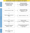
Materials and methods: The review was in accordance with Preferred Reporting Items for scoping reviews (PRISMA -ScR). Electronic databases such as PubMed and Cochrane were searched for relevant articles till 2021.
Results: We included 43 articles. We found five different possibilities of the site of expression of a marker in a cell. They are the nucleus, cytoplasm, cell membrane, extracellular matrix or any of the above combinations. Based on the tissue of expression, we also mapped the markers expressed in oral diseases to their tissue of origin as ectoderm, endoderm, mesoderm and markers with multiple tissues of expression. Based on our results, we derived two classifications that give an overview of the expression of IHC markers in oral diseases.
Conclusion: This scoping review derived new insight into the classification of IHC markers based on cell lineage, tissue and site of expression. This would enable a beginner to better understand a marker with its application and the interpretation of the staining in research. This could also serve as a beginner's guide for any researcher to thrive and explore the IHC world.
Keywords: Biomarkers; classification; immunohistochemistry; markers; oral disease.
Copyright: © 2022 Journal of Oral and Maxillofacial Pathology.
Conflict of interest statement
There are no conflicts of interest.
Figures
Similar articles
-
Beyond the black stump: rapid reviews of health research issues affecting regional, rural and remote Australia.Med J Aust. 2020 Dec;213 Suppl 11:S3-S32.e1. doi: 10.5694/mja2.50881. Med J Aust. 2020. PMID: 33314144
-
The Use of Diagnostic Tumor Markers in Detecting Tobacco- and Betel Quid-Induced Oral Squamous Cell Carcinoma: A Scoping Review of Empirical Evidence.Health Sci Rep. 2025 Apr 18;8(4):e70650. doi: 10.1002/hsr2.70650. eCollection 2025 Apr. Health Sci Rep. 2025. PMID: 40256126 Free PMC article. Review.
-
Immunohistochemical biomarkers in oral submucous fibrosis: A scoping review.J Oral Pathol Med. 2022 Aug;51(7):594-602. doi: 10.1111/jop.13280. Epub 2022 Feb 22. J Oral Pathol Med. 2022. PMID: 35102645
-
The distribution of work-related musculoskeletal disorders among nurses in sub-Saharan Africa: a scoping review protocol.Syst Rev. 2021 Aug 13;10(1):229. doi: 10.1186/s13643-021-01774-7. Syst Rev. 2021. PMID: 34389051 Free PMC article.
-
Migration of oral health professionals: a scoping review protocol.BMJ Open. 2023 Apr 12;13(4):e069954. doi: 10.1136/bmjopen-2022-069954. BMJ Open. 2023. PMID: 37045578 Free PMC article.
References
-
- Destro Rodrigues MF, Sedassari BT, Esteves CM, de Andrade NP, Altemani A, de Sousa SC, et al. Embryonic stem cells markers Oct4 and Nanog correlate with perineural invasion in human salivary gland mucoepidermoid carcinoma. J Oral Pathol Med. 2017;46:112–20. - PubMed
Publication types
LinkOut - more resources
Full Text Sources


