

Modulation of the IKS channel by PIP2 requires two binding sites per monomer
- PMID: 37082259
- PMCID: PMC10074941
- DOI: 10.1016/j.bbadva.2023.100073
Modulation of the IKS channel by PIP2 requires two binding sites per monomer
Abstract
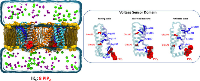
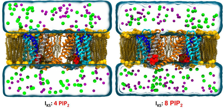
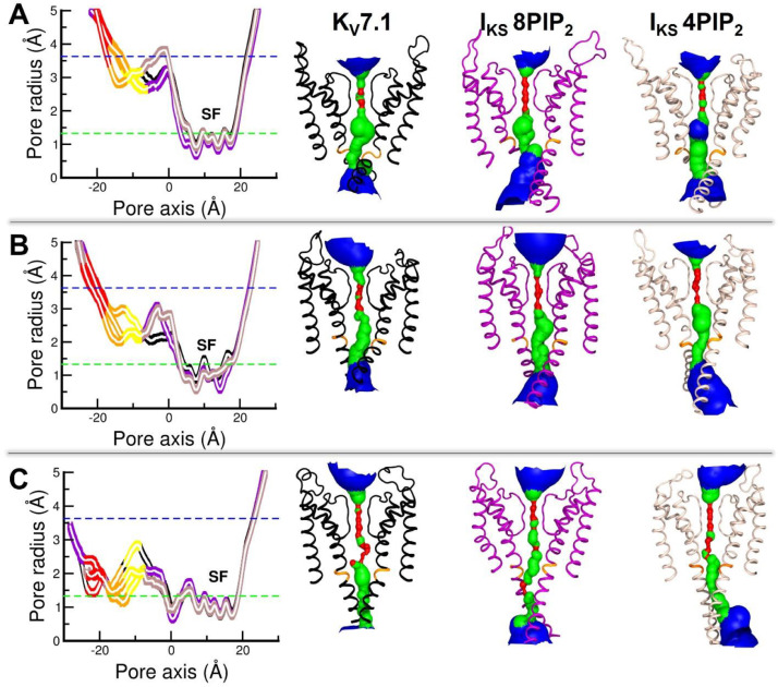
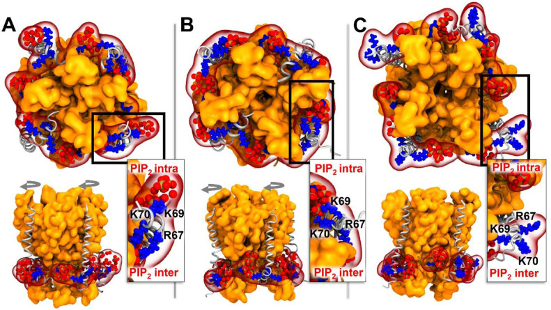
The phosphatidyl-inositol-4,5-bisphosphate (PIP2) lipid has been shown to be crucial for the coupling between the voltage sensor and the pore of the potassium voltage-gated KV7 channel family, especially the KV7.1 channel. Expressed in the myocardium membrane, KV7.1 forms a complex with KCNE1 auxiliary subunits to generate the IKS current. Here we present molecular models of the transmembrane region of this complex in its three known states, namely the Resting/Closed (RC), the Intermediate/Closed (IC), and the Activated/Open (AO), robustness of which is assessed by agreement with a range of biophysical data. Molecular Dynamics (MD) simulations of these models embedded in a lipid bilayer including phosphatidyl-inositol-4,5-bisphosphate (PIP2) lipids show that in presence of KCNE1, two PIP2 lipids are necessary to stabilize each state. The simulations also show that KCNE1 interacts with both PIP2 binding sites, forming a tourniquet around the pore and preventing its opening. The present investigation provides therefore key molecular elements that govern the role of PIP2 in KCNE1 modulation of IKS channels, possibly a common mechanism by which auxiliary KCNE subunits might modulate a variety of other ion channels.
© 2023 The Author(s).
Conflict of interest statement
The authors declare that they have no known competing financial interests or personal relationships that could have appeared to influence the work reported in this paper.
Figures
Similar articles
-
Competition of calcified calmodulin N lobe and PIP2 to an LQT mutation site in Kv7.1 channel.Proc Natl Acad Sci U S A. 2017 Jan 31;114(5):E869-E878. doi: 10.1073/pnas.1612622114. Epub 2017 Jan 17. Proc Natl Acad Sci U S A. 2017. PMID: 28096388 Free PMC article.
-
Novel Kv7.1-phosphatidylinositol 4,5-bisphosphate interaction sites uncovered by charge neutralization scanning.J Biol Chem. 2014 Aug 15;289(33):22749-22758. doi: 10.1074/jbc.M114.589796. Epub 2014 Jun 19. J Biol Chem. 2014. PMID: 24947509 Free PMC article.
-
Gating and Regulation of KCNQ1 and KCNQ1 + KCNE1 Channel Complexes.Front Physiol. 2020 Jun 4;11:504. doi: 10.3389/fphys.2020.00504. eCollection 2020. Front Physiol. 2020. PMID: 32581825 Free PMC article. Review.
-
PIP2-dependent coupling of voltage sensor and pore domains in Kv7.2 channel.Commun Biol. 2021 Oct 14;4(1):1189. doi: 10.1038/s42003-021-02729-3. Commun Biol. 2021. PMID: 34650221 Free PMC article.
-
PIP2 regulation of KCNQ channels: biophysical and molecular mechanisms for lipid modulation of voltage-dependent gating.Front Physiol. 2014 May 27;5:195. doi: 10.3389/fphys.2014.00195. eCollection 2014. Front Physiol. 2014. PMID: 24904429 Free PMC article. Review.
References
-
- Barhanin J., Lesage F., Guillemare E., Fink M., Lazdunski M., Romey G. KvLQT1 and IsK (minK) proteins associate to form the IKS cardiac potassium current. Nature. 1996;384:78–80. - PubMed
-
- Beckstein O., Tai K., Sansom M.S.P. Not ions alone: barriers to ion permeation in nanopores and channels. J. Am. Chem. Soc. 2004;126:14694–14695. - PubMed
-
- Catterall W.A. Molecular properties of voltage-sensitive sodium channels. New Insights Cell Membr. Transp. Process. 1986:3–20. - PubMed
LinkOut - more resources
Full Text Sources
