

Tru-cut needle biopsy: A novel approach in the diagnosis of solid oral pathologies
- PMID: 37082287
- PMCID: PMC10112698
- DOI: 10.4103/jomfp.jomfp_212_22
Tru-cut needle biopsy: A novel approach in the diagnosis of solid oral pathologies
Abstract
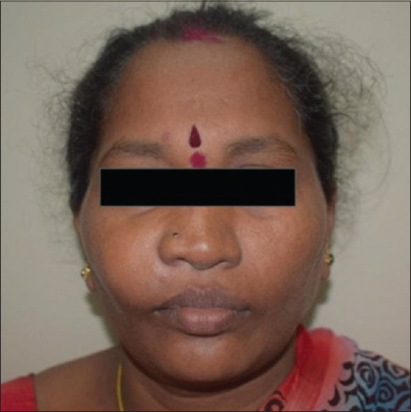
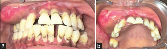
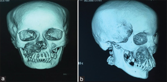
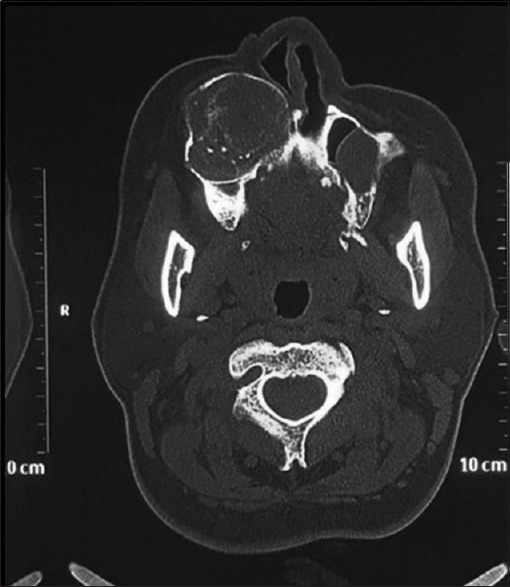
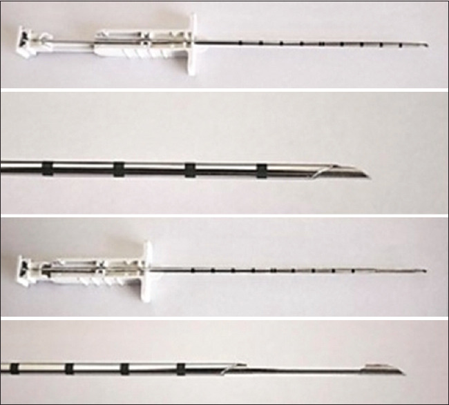
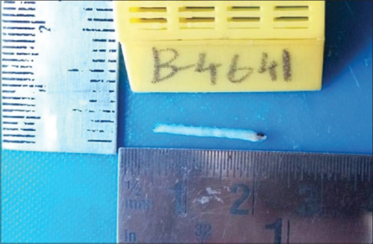
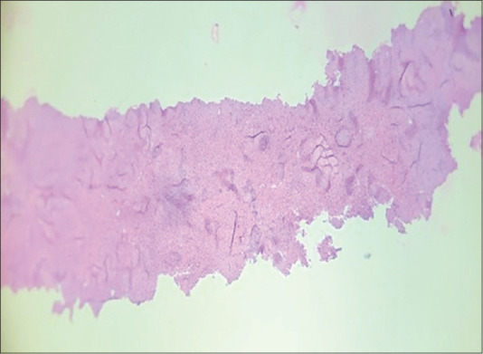
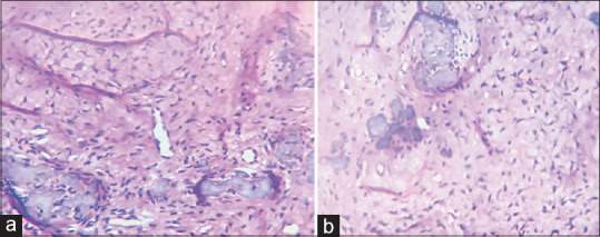
Nowadays, new biopsy techniques such as fine and wide needles are now employed instead of invasive biopsy techniques. Compared to open biopsy, true-cut needle biopsy has a number of advantages. It's quick and simple to do, can be done in an outpatient department, avoids incisions into previously irradiated skin, and has few risks. In order to examine malignant and benign tumours, there had been a debate in the past century on the utilisation and efficiency of tru-cut biopsy over Fine needle aspiration cytology (FNAC) and surgical biopsy in solid tumours. A non-odontogenic tumour that occurs in jaws, also classified as a fibro-osseous lesion of the jaw, is Cemento-Ossifying Fibroma. Clinically, these lesions occur as gradually growing, reaching an enormous size if not treated. In this article, a case of cemento-ossifying fibroma noticed in the maxilla with facial swelling is discussed and the diagnosis was done using a tru-cut needle biopsy.
Keywords: Newer biopsy techniques; solid tumour biopsy; tru-cut biopsy.
Copyright: © 2023 Journal of Oral and Maxillofacial Pathology.
Conflict of interest statement
There are no conflicts of interest.
Figures
Similar articles
-
Comparison of Tru-Cut Biopsy and Incisional Biopsy in Achieving Prompt Diagnosis of Maxillofacial Pathology.J Maxillofac Oral Surg. 2021 Sep;20(3):479-485. doi: 10.1007/s12663-021-01557-6. Epub 2021 Apr 10. J Maxillofac Oral Surg. 2021. PMID: 34408377 Free PMC article.
-
Comparison of tru-cut biopsy and fine-needle aspiration cytology in an experimental alcoholic liver disease model.Rev Assoc Med Bras (1992). 2020 Aug;66(8):1030-1035. doi: 10.1590/1806-9282.66.8.1030. Rev Assoc Med Bras (1992). 2020. PMID: 32935794
-
A comparison of the use of the "Tru-Cut" needle and fine needle aspiration cytology in the pre-operative diagnosis of carcinoma of the breast.Histopathology. 1978 Jul;2(4):239-54. doi: 10.1111/j.1365-2559.1978.tb01717.x. Histopathology. 1978. PMID: 700601
-
[Is the Tru-Cut needle more efficient than the fine needle in the diagnosis of hepatic lesions? Comparative study of 45 echography-guided punctures].Gastroenterol Clin Biol. 1990;14(1):62-6. Gastroenterol Clin Biol. 1990. PMID: 2179009 Review. French.
-
Cemento ossifying fibroma.Indian J Dent Res. 2004 Jan-Mar;15(1):35-9. Indian J Dent Res. 2004. PMID: 15682795 Review.
Cited by
-
Bone Biopsies: Practical Considerations and Technical Tips.Semin Intervent Radiol. 2024 Dec 10;41(5):444-454. doi: 10.1055/s-0044-1791720. eCollection 2024 Oct. Semin Intervent Radiol. 2024. PMID: 39664228 Review.
References
-
- Yildirim E, Kirbas I, Harman A, Ozyer U, Tore HG, Aytekin C, et al. CT-guided cutting needle lung biopsy using modified coaxial technique: Factors effecting risk of complications. Eur J Radiol. 2009;70:57–60. - PubMed
-
- Cevik FC, Aykin N, Naz H. Complications and efficiency of liver biopsies using the Tru-Cut biopsy Gun. J Infect Dev Ctries. 2010;4:91–5. - PubMed
-
- Yuan J, Li X-H. Evaluation of pathological diagnosis using ultrasonography-guided lymph node core-needle biopsy. Chin Med J. 2010;123:690–4. - PubMed
Publication types
LinkOut - more resources
Full Text Sources
Miscellaneous