

Gradual and Remarkable Tumor Shrinkage Following Seven-Fraction Stereotactic Radiosurgery Alone With a Marginal Dose of 48.3 Gy for Large Lobar Possibly Intra-sulcal Brain Metastasis From Renal Cell Carcinoma
- PMID: 37082500
- PMCID: PMC10111507
- DOI: 10.7759/cureus.36346
Gradual and Remarkable Tumor Shrinkage Following Seven-Fraction Stereotactic Radiosurgery Alone With a Marginal Dose of 48.3 Gy for Large Lobar Possibly Intra-sulcal Brain Metastasis From Renal Cell Carcinoma
Abstract
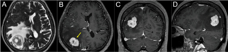
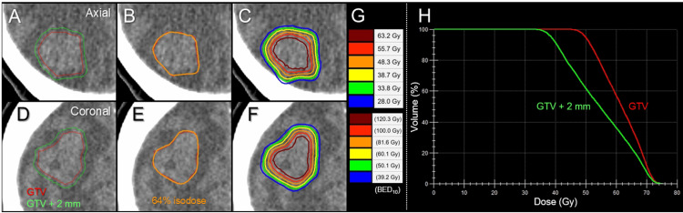
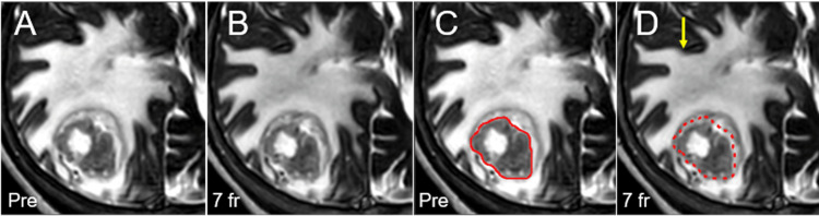
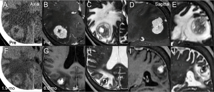
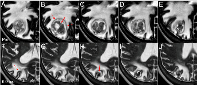
Brain metastases (BMs) from renal cell carcinoma (RCC) have the tendency of slow and insufficient tumor shrinkage along with prolongation of massive peritumoral edema following stereotactic radiosurgery (SRS). Herein, we describe a case of large lobar RCC-BM, with possible intra-sulcal location, treated with 7-fraction (fr) SRS without subsequent anti-cancer medication, which resulted in gradual and remarkable tumor shrinkage with extrication from the mass effect. A 59-year-old woman was incidentally diagnosed with bilateral RCC associated with multiple lung metastases and subsequently presented with symptomatic single BM of 32 mm in the maximum diameter (9.54 cm3) two months later while vacillating. A biopsy of the kidney showed clear cell carcinoma. The patient was deemed medically inoperable for BM due to unfit conditions, including severe deep venous thromboses and thrombocytopenia. Considering the tumor volume, irregular tumor configuration, non-superficial location, and mass effect, 98% of the gross tumor volume (GTV D98%) was covered by 48.3 Gy in 7 fr with 64% isodose. Dose distribution was optimized with volumetric modulated arcs with the affirmative allowance of very inhomogeneous GTV dose. Anti-cancer medication was limited to nivolumab plus ipilimumab followed by everolismus 12 days before and during SRS, respectively. Subsequently, the patient transitioned to palliative care due to a declining general condition. Although long-term administration of steroids was required, gradual and marked tumor shrinkage (1.25 cm3, 13.1% of the initial volume) and mitigation of the peritumoral edema was observed during six months after SRS. The main location of the initial BM was deemed as intra-sulcal in the intraparietal sulcus and originated in the cerebral cortex. The patient died nine months after SRS. The gradual but remarkable tumor response obtained with 7-fr SRS alone, in this case, provides a basis to further optimize fractionated SRS dosage to enhance efficacy and safety for large and/or symptomatic RCC-BMs not amenable to immediate surgical removal, in combination with anti-cancer pharmacotherapy, if feasible, including tyrosine kinase inhibitors, which may enhance efficacy against BM and mitigate adverse effects relevant to high dose SRS.
Keywords: brain metastasis; coagulopathy; fractionation; immune ckeckpoint inhibitor; large tumor; renal cell carcinoma; stereotactic radiosurgery; tyrosine kinase inhibitor; volumetric modulated arc-based radiosurgery.
Copyright © 2023, Ohtakara et al.
Conflict of interest statement
The authors have declared that no competing interests exist.
Figures
Similar articles
-
Local Control Failure After Five-Fraction Stereotactic Radiosurgery Alone for Symptomatic Brain Metastasis From Squamous Cell Lung Carcinoma Despite 43 Gy to Gross Tumor Margin With Internal Steep Dose Increase and Tumor Shrinkage During Irradiation.Cureus. 2023 May 6;15(5):e38645. doi: 10.7759/cureus.38645. eCollection 2023 May. Cureus. 2023. PMID: 37284398 Free PMC article.
-
Ten-Fraction Stereotactic Radiosurgery With Different Gross Tumor Doses and Inhomogeneities for Brain Metastasis of >10 cc: Treatment Responses Suggesting Suitable Biological Effective Dose Formula for Single and 10 Fractions.Cureus. 2023 Feb 4;15(2):e34636. doi: 10.7759/cureus.34636. eCollection 2023 Feb. Cureus. 2023. PMID: 36895545 Free PMC article.
-
Five-Year Sustained Complete Remission With Minimal Adverse Effects Following Radiosurgery for 2-cm Brain Metastasis With Deep Eloquent Location From Lung Adenocarcinoma Despite Low Marginal Dose and High 12 Gy Volume.Cureus. 2023 Mar 25;15(3):e36680. doi: 10.7759/cureus.36680. eCollection 2023 Mar. Cureus. 2023. PMID: 37113354 Free PMC article.
-
Stereotactic radiosurgery for treatment of brain metastases. A report of the DEGRO Working Group on Stereotactic Radiotherapy.Strahlenther Onkol. 2014 Jun;190(6):521-32. doi: 10.1007/s00066-014-0648-7. Epub 2014 Apr 9. Strahlenther Onkol. 2014. PMID: 24715242 Review.
-
Single- and Multifraction Stereotactic Radiosurgery Dose/Volume Tolerances of the Brain.Int J Radiat Oncol Biol Phys. 2021 May 1;110(1):68-86. doi: 10.1016/j.ijrobp.2020.08.013. Epub 2020 Sep 11. Int J Radiat Oncol Biol Phys. 2021. PMID: 32921513 Free PMC article. Review.
Cited by
-
Proposal of an Alternative Near-Minimum Isodose Surface DV-0.01 cc Equally Minimizing Gross Tumor Volume Below the Relevant Dose as the Basis for Dose Prescription and Evaluation of Stereotactic Radiosurgery for Brain Metastases.Cureus. 2024 Apr 4;16(4):e57580. doi: 10.7759/cureus.57580. eCollection 2024 Apr. Cureus. 2024. PMID: 38707120 Free PMC article.
-
Determining Simple and Effective Cost Functions for an Efficient Volumetric-Modulated Arcs-Based Stereotactic Radiosurgery for Single Brain Metastases Using Monaco® Planning System.Cureus. 2024 Sep 30;16(9):e70560. doi: 10.7759/cureus.70560. eCollection 2024 Sep. Cureus. 2024. PMID: 39479057 Free PMC article.
-
Consideration of Optimal Evaluation Metrics for Internal Gross Tumor Dose Relevant to Tumor Response in Multi-fraction Stereotactic Radiosurgery of Brain Metastasis.Cureus. 2024 Jul 25;16(7):e65338. doi: 10.7759/cureus.65338. eCollection 2024 Jul. Cureus. 2024. PMID: 39184769 Free PMC article.
-
Non-coplanar Arc-Involved Beam Arrangement With Sufficient Arc Rotations Is Suitable for Volumetric-Modulated Arc-Based Radiosurgery for Single Brain Metastasis.Cureus. 2024 Aug 20;16(8):e67265. doi: 10.7759/cureus.67265. eCollection 2024 Aug. Cureus. 2024. PMID: 39301366 Free PMC article.
-
Appropriateness of Dose Attenuation Margin Outside the Gross Tumor Volume (GTV) in Volumetric-Modulated Arc-Based Radiosurgery for Brain Metastasis With the Steepest Dose Gradient Outside the GTV and Biologically Effective Dose 80 Gy to GTV Boundary.Cureus. 2024 Jun 20;16(6):e62784. doi: 10.7759/cureus.62784. eCollection 2024 Jun. Cureus. 2024. PMID: 39036259 Free PMC article.
References
-
- Pathologic complete response in renal cell carcinoma brain metastases treated with stereotactic radiosurgery. Teh BS, Bloch C, Paulino AC, et al. Clin Genitourin Cancer. 2007;5:334–337. - PubMed
-
- Gamma knife surgery for metastatic brain tumors from renal cell carcinoma. Shuto T, Inomori S, Fujino H, Nagano H. J Neurosurg. 2006;105:555–560. - PubMed
-
- Treatment strategy for metastatic brain tumors from renal cell carcinoma: selection of gamma knife surgery or craniotomy for control of growth and peritumoral edema. Shuto T, Matsunaga S, Suenaga J, Inomori S, Fujino H. J Neurooncol. 2010;98:169–175. - PubMed
-
- Stereotactic radiation therapy for renal cell carcinoma brain metastases in the tyrosine kinase inhibitors era: Outcomes of 120 patients. Klausner G, Troussier I, Biau J, et al. Clin Genitourin Cancer. 2019;17:191–200. - PubMed
Publication types
LinkOut - more resources
Full Text Sources