Review
. 2022 Mar 20:2:100050.
doi: 10.1016/j.bbadva.2022.100050. eCollection 2022.

Subcellular localization of X-linked inhibitor of apoptosis protein (XIAP) in cancer: Does that matter?

Affiliations
Review

Subcellular localization of X-linked inhibitor of apoptosis protein (XIAP) in cancer: Does that matter?

B S Mendonça et al. BBA Adv. .

Abstract

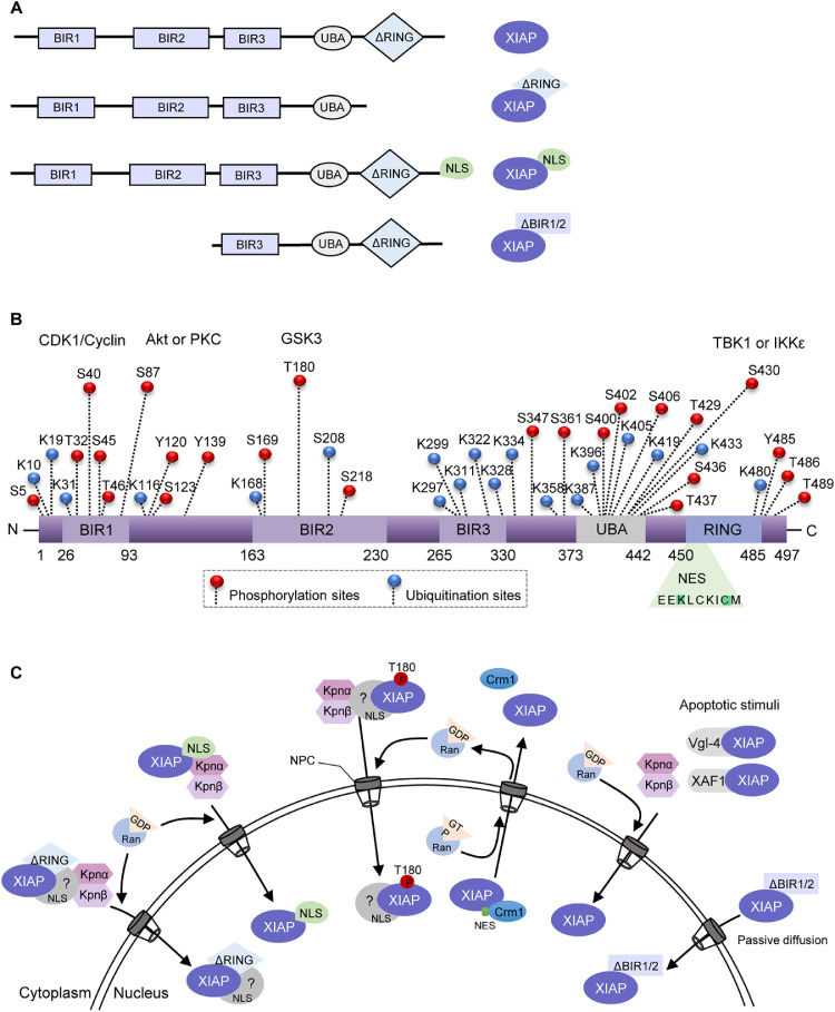
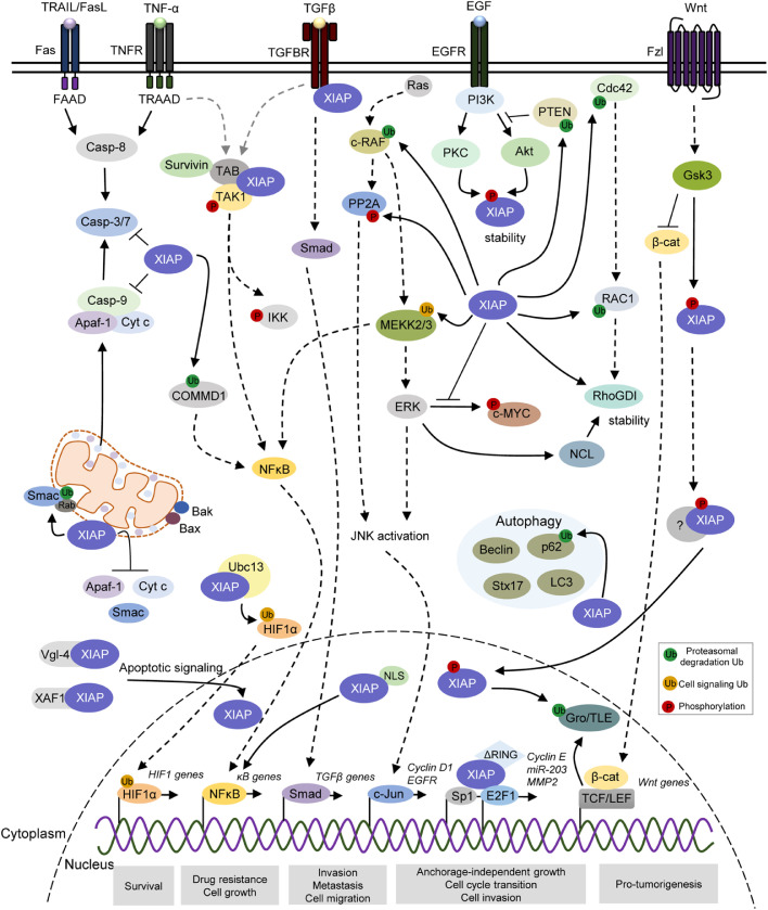
X-linked inhibitor of apoptosis protein (XIAP) finely tunes the balance between survival and death to control homeostasis. XIAP is found aberrantly expressed in cancer, which has been shown to promote resistance to therapy-induced apoptosis and confer poor outcome. Despite its predominant cytoplasmic localization in human tissues, growing evidence implicates the expression of XIAP in other subcellular compartments in sustaining cancer hallmarks. Herein, we review our current knowledge on the prognostic role of XIAP localization and discuss molecular mechanisms underlying differential biological functions played in each compartment. The comprehension of XIAP subcellular shuttling and functional dynamics might provide the rationale for future anticancer therapeutics.

Keywords: Cancer; Molecular functional mechanisms; Prognostic marker; Resistance to apoptosis; XIAP subcellular localization.


Conflict of interest statement

The authors declare no conflict of interest.

Figures

Fig 1:
Fig. 1
Schematic representation of the XIAP structure and dynamic intracellular localization. (A) Structural representation of XIAP variants that have been shown to translocate into the nucleus. XIAP wild type is composed of three baculoviral IAP repeat (BIR) domains, a ubiquitin-binding domain (UBA) and a RING finger domain. XIAPΔRING is a RING domain-deleted form and XIAPNLS is the variant with a nuclear localization signal (NLS) insertion at the carboxi-terminal end. XIAPΔBIR1/2 is a protein fragment shown to be generated following chemotherapeutic treatment. (B) Predicted phosphorylation, ubiquitination, and nuclear export signal (NES) sites on XIAP have been curated by PhosphoSitePlus and NetNES 1.1 online resources tools. XIAP is a substrate for CDK-cyclin B1, Akt, PKC, GSK3, TBK1 and IKKε phosphorylation at different sites and presents an NES located within the RING domain. (C) A model of XIAP nuclear-cytoplasmatic shuttling regulation. XIAPΔRING, XIAPNLS and T108-phosphorylated forms might complex with cargo proteins at the cytoplasm and translocate to the nuclear compartment through the nuclear pore complex (NPC). Under apoptotic stimuli, Vgl-4 and XAF1 can directly bind to and sequester XIAP to the nuclear compartment. Also, the XIAPΔBIR1/2 fragment might transport to the nucleus by passive diffusion. Image abbreviations: P, phosphorylation-modified protein; Akt, Protein kinase B; CDK1, Cyclin-dependent kinase 1; Crm1, Chromosomal maintenance 1; GDP, Guanosine diphosphate, GSK-3, Glycogen synthase kinase 3; GTP, Guanosine triphosphate; IKKε, IκB kinase epsilon; IKKβ: IκB kinase beta; Kpnα, karyopherin-alpha; Kpnβ: karyopherin-alpha; PKC, Protein kinase C; Ran, Ras-related nuclear protein; TBK1, TANK-biding kinase 1; Vgl-4, Vestigial-like family member 4; XAF1, XIAP-associated factor 1.
Fig 2:
Fig. 2
XIAP differentially regulates apoptotic and pro-oncogenic signaling pathways in distinct subcellular compartments. XIAP is a multi-functional protein involved in multiple cellular signaling pathways associated with cellular processes that favor tumor features, including survival, drug resistance, autophagy, anchorage-independent cell growth, cell proliferation, migration, invasion and metastasis. XIAP blocks the intrinsic and extrinsic apoptotic pathways through binding to and inhibiting caspases-3/7/9. XIAP also acts on apoptosis signaling by catalyzing ubiquitination of its mitochondrial regulator Smac, as well as limit the release of Cyt C (Cytochrome c), Smac and Apaf-1 during antiapoptotic response. During hypoxia, the XIAP/Ubc13 complex ubiquitinates HIF1, subsequently upregulating the expression of HIF target genes. XIAP binds to TAB1/TAK1 complex and interact with survivin to stimulate NFκB-dependent signaling. Indirectly, XIAP modulates NFκB activity by ubiquitination of COMMD1 and MEKK2/3. XIAP physically interacts with TGF-β type I receptor through Smad-dependent signaling, promoting TGF-β signaling pathway. XIAP has also a role in cell migration via ubiquitination of c-RAF, CDC42 and RAC1, as well as regulation of ERK activity to modulate c-MYC, nucleolin and RhoGDI stability. XIAP also induces PP2A phosphorylation, further contributing to c-Jun/AP-1 activation signaling. XIAP plays a role in the autophagy, in which it interacts and suppresses SQSTM1 (p62) by ubiquitination-dependent proteasomal degradation. Also, XIAP indirectly modulates the levels of LC3-II, Beclin 1 and Syntax 17 during the autophagic flux. XIAP can ubiquitinate PTEN, promoting Akt activation, which in turn leads to XIAP stability. XIAP is phosphorylated by GSK3 and, thus, recruited to TCF/Lef transcriptional complexes where it promotes monoubiquitination of co-repressor Groucho/TLE, further leading to the transcription of Wnt-regulated genes. Also, intranuclear XIAP binds to transcriptional factor Sp1 and E2F1, increasing Cyclin E, miR-203 and MMP2. The black and dotted arrows represent direct and indirect interactions, respectively. P, phosphorylation-modified protein; Ub, ubiquitin-modified protein. Akt, Protein kinase B; Apaf-1, Apoptotic protease activating factor 1; Cdc42, Cell division control protein 42 homologue; CDK1, Cyclin-dependent kinase 1; c-Jun/AP-1: Jun proto-oncogene/Activator protein 1; c-Myc: Cellular Myc; COMMD1, copper metabolism MURR1 domain-containing; c-Raf: Raf proto-oncogene serine/threonine-protein kinase; Crm1, Chromosomal maintenance 1; Cyt C, Cytochrome c; E2F1, E2F transcription factor 1; EGFR: Epidermal growth factor receptor; ERK, Extracellular signal-regulated kinase; GDP, Guanosine diphosphate, GSK-3, Glycogen synthase kinase 3; GTP, Guanosine triphosphate; HIF1; Hypoxia-inducible factor 1; IKKε, IκB kinase epsilon; IKKβ: IκB kinase beta; Kpnα, karyopherin-alpha; Kpnβ: karyopherin-alpha; LC3-II: Microtubule associated protein 1 light chain 3; p62/SQSTM1: Sequestosome 1; PKC, Protein kinase C; PP2A, Protein phosphatase 2; LC3-II: Microtubule associated protein 1 light chain 3; MEKK, Mitogen-activated protein kinase kinase kinase; MMP2, Matrix metalloproteinase-2; NFκB, Nuclear factor kappa B; PI3K, Phosphoinositide 3-kinase; Rac1, Ras-related C3 botulinum toxin substrate 1; Ran, Ras-related nuclear protein; RhoGDI: Rho GDP-dissociation inhibitor; Smac, Second mitochoncrial-derived activator of caspases; Sp1: Specificity protein 1; TAB1/TAK:TAK1-binding protein 1/Transforming growth factor β-activated protein kinase 1; TBK1, TANK-biding kinase 1; TCF/Lef, T cell factor/lymphoid enhancer factor family; TGF-β: Transforming growth factor beta; Vgl-4, Vestigial-like family member 4; XAF1, XIAP-associated factor 1.

Similar articles

Cited by

References

    1. Kocab A.J., Duckett C.S. Inhibitor of apoptosis proteins as intracellular signaling intermediates. FEBS J. 2016;283:221–231. doi: 10.1111/febs.13554. - DOI - PMC - PubMed
    1. Tamm I., Wang Y., Sausville E., Scudiero D.A., Vigna N., Oltersdorf T., Reed J.C. IAP-family protein survivin inhibits caspase activity and apoptosis induced by Fas (CD95), Bax, caspases, and anticancer drugs. Cancer Res. 1998;58:5315–5320. - PubMed
    1. Takahashi R., Deveraux Q., Tamm I., Welsh K., Assa-Munt N., Salvesen G.S., Reed J.C. A single BIR domain of XIAP sufficient for inhibiting caspases. J. Biol. Chem. 1998;273:7787–7790. doi: 10.1074/jbc.273.14.7787. - DOI - PubMed
    1. Scott F.L., Denault J.-.B., Riedl S.J., Shin H., Renatus M., Salvesen G.S. XIAP inhibits caspase-3 and -7 using two binding sites: evolutionarily conserved mechanism of IAPs. EMBO J. 2005;24:645–655. doi: 10.1038/sj.emboj.7600544. - DOI - PMC - PubMed
    1. Galbán S., Duckett C.S. XIAP as a ubiquitin ligase in cellular signaling. Cell Death Differ. 2010;17:54–60. doi: 10.1038/cdd.2009.81. - DOI - PMC - PubMed

LinkOut - more resources