. 2023 Oct;243(4):630-647.
doi: 10.1111/joa.13877. Epub 2023 Apr 21.

Neurochemical alterations of intrinsic cardiac ganglionated nerve plexus caused by arterial hypertension developed during ageing in spontaneously hypertensive and Wistar Kyoto rats

Affiliations

Neurochemical alterations of intrinsic cardiac ganglionated nerve plexus caused by arterial hypertension developed during ageing in spontaneously hypertensive and Wistar Kyoto rats

Neringa Pauziene et al. J Anat. 2023 Oct.

Abstract

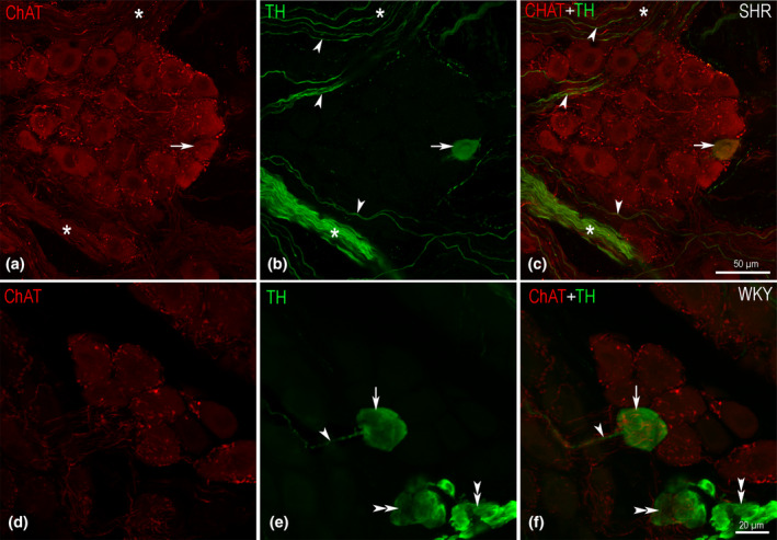
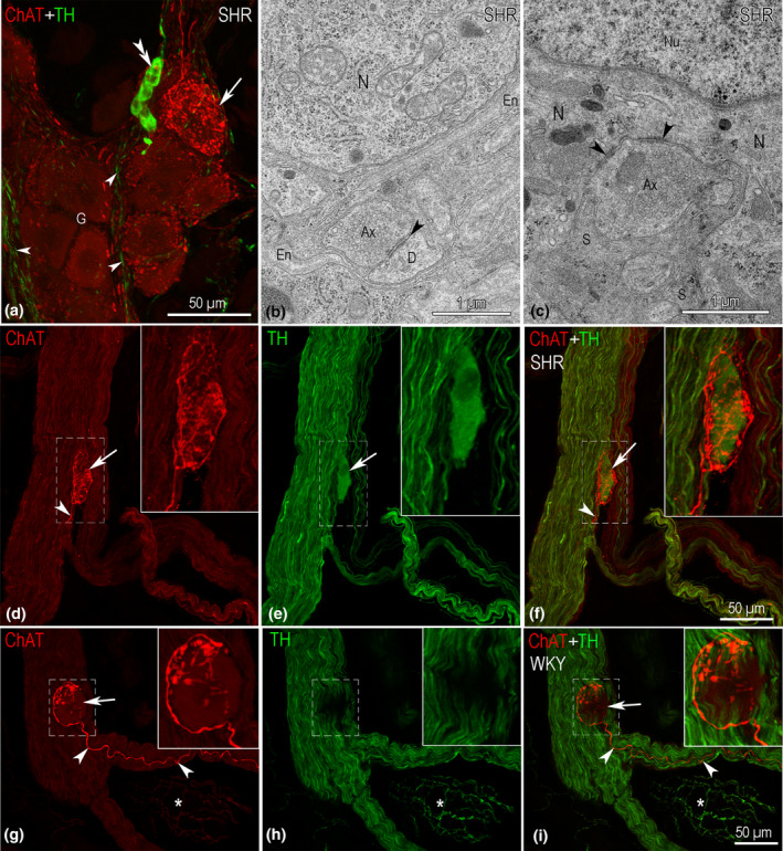
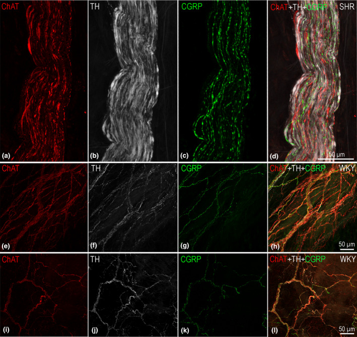
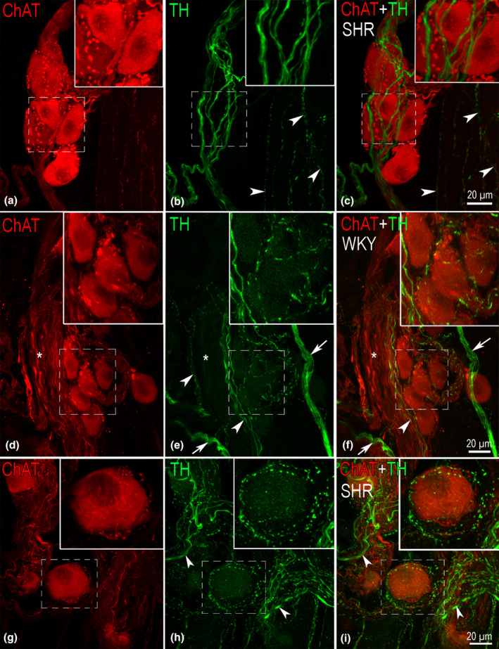
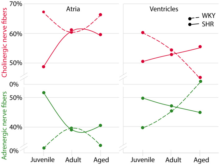
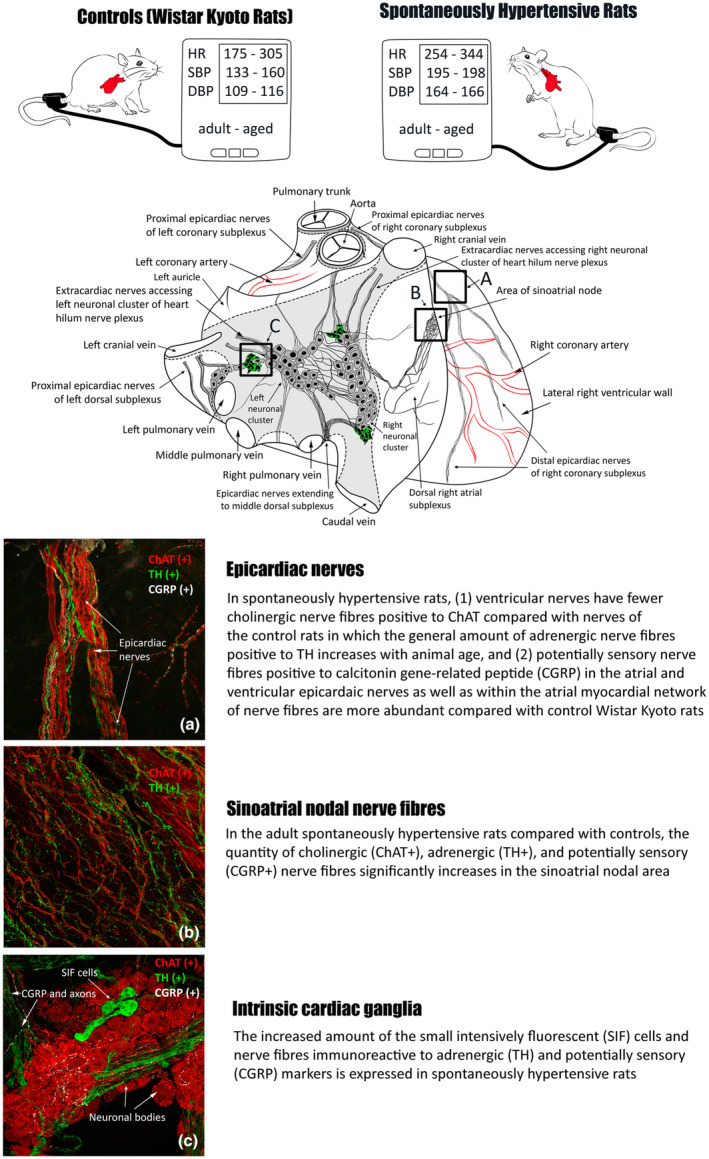
The acknowledged hypothesis of the cause of arterial hypertension is the emerging disbalance in sympathetic and parasympathetic regulations of the cardiovascular system. This disbalance manifests in a disorder of sustainability of endogenous autonomic and sensory neural substances including calcitonin gene-related peptide (CGRP). This study aimed to examine neurochemical alterations of intrinsic cardiac ganglionated nerve plexus (GP) triggered by arterial hypertension during ageing in spontaneously hypertensive rats of juvenile (prehypertensive, 8-9 weeks), adult (early hypertensive, 12-18 weeks) and elderly (persistent hypertensive, 46-60 weeks) age in comparison with the age-matched Wistar-Kyoto rats as controls. Parasympathetic, sympathetic and sensory neural structures of GP were analysed and evaluated morphometrically in tissue sections and whole-mount cardiac preparations. Both the elevated blood pressure and the evident ultrasonic signs of heart failure were identified for spontaneously hypertensive rats and in part for the aged control rats. The amount of adrenergic and immunoreactive to CGRP neural structures was increased in the adult group of spontaneously hypertensive rats along with the significant alterations that occurred during ageing. In conclusion, the revealed chemical alterations of GP support the hypothesis about the possible disbalance of efferent and afferent heart innervation and may be considered as the basis for the emergence and progression of arterial hypertension and perhaps even as a consequence of hypertension in the aged spontaneously hypertensive rats. The determined anatomical changes in the ageing Wistar-Kyoto rats suggest this breed being as inappropriate for its use as control animals for hypertension studies in older animal age.

Keywords: CGRP; ChAT; SIF cells; TH; heart ganglia, nerves and plexus; hypertension; immunohistochemistry.


Figures

FIGURE 1
FIGURE 1
Immunofluorescent images of intracardiac ganglia in spontaneously hypertensive and control rats. (a–c): Single cholinergic biphenotypic adrenergic neuronal body (arrow) located between purely cholinergic neuronal somata. Numerous axonal terminals strongly positive for choline acetyltransferase (ChAT) seen as bright red buttons are interspersed between neuronal bodies in the ganglion. The ganglion is located at the intersection of two nerves (*), the lower of which is composed mainly of adrenergic nerve fibres, while in the upper one, just a few adrenergic nerve fibres proceed. Few adrenergic nerve fibres (arrowheads) extend close to ganglionic cells, yet they have no varicosities and other specific signs of synaptic contacts. (d–f) Lesser ganglion in which a single non‐cholinergic adrenergic neuronal body (arrow) is located between purely cholinergic ganglionic cells. The axon of this adrenergic neuron is indicated by the arrowhead. Double arrows point to two clusters of SIF cells that differ from principal neurons by evidently smaller size and intensive fluorescent labelling.
FIGURE 2
FIGURE 2
Cholinergic terminals in contact with intracardiac neuronal somata. (a) Flattened from a z‐stack of optical sections immunofluorescent image of the intracardiac ganglion (G) composed of cholinergic neurons. Note, the variable densities of cholinergic terminals surrounding neuronal somata. The arrow indicates neuronal soma that is unusually densely bounded by axonal varicosities. Several adrenergic nerve fibres (arrowheads) pass through the ganglion. The cluster of SIF cells is indicated by double arrowheads. (b, c) Electron micrographs of axodendritic (B) and axosomatic (C) synapses within intracardiac ganglia. Ax, terminal of axon; En, endoneurium; N, neuronal cytoplasm; Nu, nucleus of neuron; S, satellite cells; (d–f) Solitary non‐cholinergic adrenergic neuronal body (arrow) inside intracardiac nerve that involves almost entirely adrenergic nerve fibres. Note the cholinergic nerve fibre (arrowhead) trailing to this neuronal body, bounteously ramifying, containing numerous varicosities that wrap the neuronal body from all sides. (g–i) Adrenergic nerve fibres involving intracardiac nerve with the single cholinergic neuronal body (arrow). Arrowheads indicate cholinergic nerve fibre trailing through the nerve branch, surrounding the neuronal body and containing plentiful varicosities that encompass the neuron body. Asterisks in (g–i) indicate a blood vessel with a network of adrenergic nerve fibres.
FIGURE 3
FIGURE 3
(a–d) immunofluorescent images demonstrating the mixed intracardiac nerve in which the majority of nerve fibres are either cholinergic (ChAT), adrenergic (TH) or immunoreactive for CGRP. Note, an almost equal number of ChAT and CGRP‐positive nerve fibres. (e–h) Cholinergic (ChAT), adrenergic (TH) and CGRP immuno‐positive nerve fibres within dense tiny meshwork spread between invisible (non‐labelled) cells of the sinoatrial node (SAN) area. (i–l) a sparse meshwork of adrenergic (TH), cholinergic (ChAT) and peptidergic (CGRP) nerve fibres in contractile myocardium.
FIGURE 4
FIGURE 4
Immunofluorescent images demonstrating adrenergic nerve fibres inside intracardiac ganglia of spontaneously hypertensive and control rats. (a–c) Minor ganglion with cholinergic neuronal somata and no varicosities containing adrenergic nerve fibres passing through the ganglion in transit. Note the numerous cholinergic varicosities that are well visible around and between the neuronal somata in (a) and (c), while adrenergic nerve fibres (immunoreactive to TH) just pass the ganglion and have no varicosities (b and c). Note the varicosities containing adrenergic nerve fibres visible within the myocardial nerve meshwork in the left lower corner of the image (arrowheads). (d–f) Minor ganglion nearby the nerve (*) composed of cholinergic neuronal somata. Note (i) the nerve involving mainly cholinergic nerve fibres and just a few adrenergic ones (arrowheads), (ii) two thin bundles of adrenergic nerve fibres (arrows) pass nearby the ganglion, (iii) the varicosities containing adrenergic nerve fibres within the ganglion intervening between neuronal somata and surrounding them (e and f). (g–i) Fragment of intracardiac ganglion from spontaneously hypertensive rat with abundant adrenergic (immunoreactive to TH) nerve fibres. Note that location of some adrenergic fibres is not linked to the location of the neuronal body, and this indicates that these axons presumably pass in transit through the ganglion avoiding synaptic contacts (arrowheads). However, other adrenergic nerve fibres with varicosities are interspersed between neuronal somata and even encircle those somata. The latter type of adrenergic axons suggests that synaptic contact between adrenergic nerve fibres and cholinergic neurons exists within intracardiac ganglia in the rat heart.
FIGURE 5
FIGURE 5
Histogram to demonstrate the proportion of cholinergic (ChAT) and adrenergic (TH) nerve fibres in the atrial and ventricular nerves of spontaneously hypertensive and control rats during ageing.
FIGURE 6
FIGURE 6
Immunofluorescent and electron microscope images illustrating the morphologic pattern of SIF cells within intracardiac ganglia and nerves. (a) Clusters of SIF cells (arrowheads) scattered between cholinergic neuronal somata within intracardiac ganglion (G). (b) a cluster of SIF cells that occupies a larger area than the ganglion (G) to which it is nestled. Note the intensive immunopositivity to TH of SIF cells in comparison with the TH‐positive nerve fibres in the nerve (N). (c) Electronogram of SIF cell inside the intracardiac nerve. SIF cell is in part ensheathed with the glial cell (arrowheads), yet in sites where the glial sheath is broken its cytoplasm is exposed to endoneurial space (double arrowheads). Like other SIF cells, this SIF cell is located nearby a blood vessel (endothelial cells are indicated by arrows). Ax, axons; En, endoneurium; F, the process of fibroblast.
FIGURE 7
FIGURE 7
Immunofluorescent images demonstrating peptidergic (CGRP‐positive) nerve fibres and buttons within the intracardiac ganglion. The majority of CGRP immunoreactive nerve fibres are particularly thin nerve fibres with numerous small varicosities (arrowheads) and distribute evenly over the entire area of the ganglion. These nerve fibres morphologically look like a meandering network in the ganglion between the cholinergic neuronal somata. Moreover, some peptidergic nerve fibres are significantly thicker and exhibit terminal boutons that are strongly positive to calcitonin gene‐related peptide (CGRP, arrows) and distribute widely within intracardiac ganglia.
FIGURE 8
FIGURE 8
Schematic summing‐up of the main findings of the present study. In the upper part, the significantly elevated heart rate (HR), systolic (SBP) and diastolic (DBP) blood pressure of the adult and aged spontaneously hypertensive rats in comparison with controls Wistar–Kyoto rats was confirmed employing the non‐invasive tail‐cuff blood pressure measurements. The drawing in the middle of the schema depicts the principal morphologic pattern of the rat intrinsic cardiac ganglionated plexus from a right posterolateral view of the rat heart. The boxed areas (a–c) in the drawing point to locations of diverse intrinsic cardiac structures ((a), ventricular epicardiac nerves; (b), dense network of nerve fibres within the sinoatrial nodal area; (c) intrinsic cardiac ganglia) in that the most significant differences between the spontaneously hypertensive and control rats were determined. The boxed areas (a–c) of the schematic drawing are correspondingly illustrated below by three typical immunofluorescent images specifying the principal structural alterations in spontaneously hypertensive rats compared with control Wistar–Kyoto rats. The dashed lines around the aorta‐pulmonary trunk and orifices of pulmonary‐caval veins outline the limits of the arterial and venous parts of heart hilum. The venous part of heart hilum encompasses two largest clusters of intracardiac neuronal bodies between which numerous small intensively fluorescent (SIF) cells are distributed.

Similar articles

References

    1. Adams, M.A. , Bobik, A. & Korner, P.I. (1989) Differential development of vascular and cardiac hypertrophy in genetic hypertension. Relation to sympathetic function. Hypertension (Dallas, Tex: 1979), 14(2), 191–202. - PubMed
    1. Aksu, T. , Gopinathannair, R. , Gupta, D. & Pauza, D.H. (2021) Intrinsic cardiac autonomic nervous system: what do clinical electrophysiologists need to know about the “heart brain”? Journal of Cardiovascular Electrophysiology, 32(6), 1737–1747. - PubMed
    1. Aksu, T. , Gupta, D. & Pauza, D.H. (2021) Anatomy and physiology of intrinsic cardiac autonomic nervous system: da Vinci anatomy card #2. Case Reports, 3(4), 625–629. - PMC - PubMed
    1. Ashton, J.L. , Argent, L. , Smith, J.E.G. , Jin, S. , Sands, G.B. , Smaill, B.H. et al. (2020) Evidence of structural and functional plasticity occurring within the intracardiac nervous system of spontaneously hypertensive rats. American Journal of Physiology. Heart and Circulatory Physiology, 318(6), H1387–H1400. - PubMed
    1. Atanasova, D.Y. , Iliev, M.E. & Lazarov, N.E. (2011) Morphology of the rat carotid body. Biomedical Reviews, 22, 41–55.

Publication types

Substances