. 2023 Jun;49(6):115.
doi: 10.3892/or.2023.8552. Epub 2023 Apr 21.

Paclitaxel induces pyroptosis by inhibiting the volume‑sensitive chloride channel leucine‑rich repeat‑containing 8a in ovarian cancer cells

Affiliations

Paclitaxel induces pyroptosis by inhibiting the volume‑sensitive chloride channel leucine‑rich repeat‑containing 8a in ovarian cancer cells

Xiurou Yang et al. Oncol Rep. 2023 Jun.

Abstract

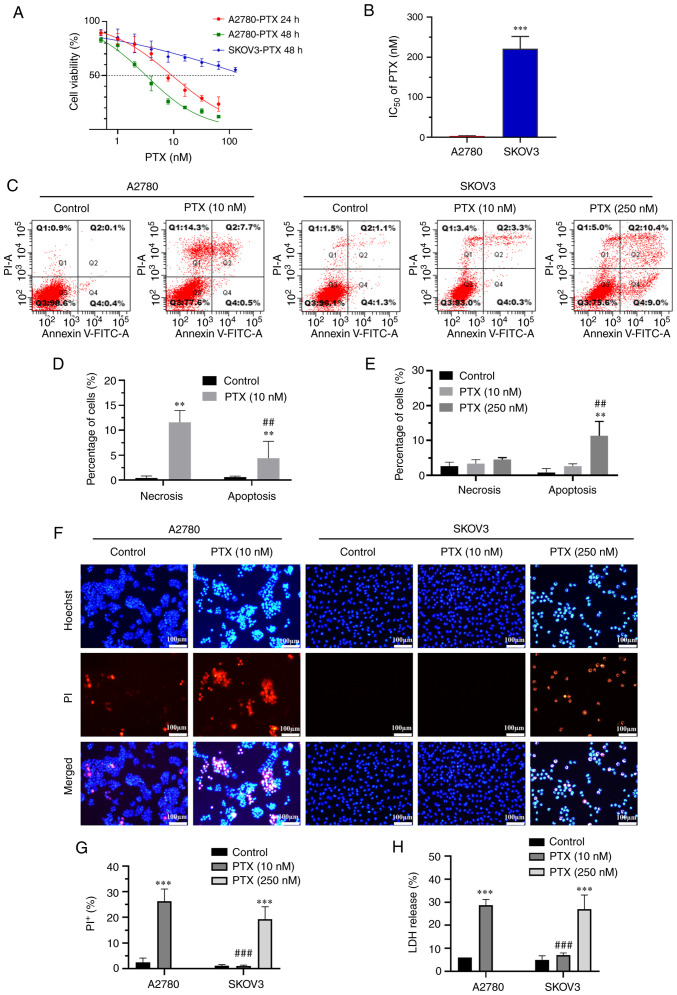
Pyroptosis is a newly identified form of cell death, morphologically characterized by excessive cell swelling. In the present study, paclitaxel (PTX) combined with platinum were used as first‑line chemotherapy, against ovarian cancer cells by inducing multiple types of cell death. However, it remains unclear whether PTX can induce pyroptosis in ovarian cancer cells. It was recently reported that PTX inhibited chloride channels, an inhibition known to cause cell swelling. In the present study, it was first verified that pyroptosis‑like cell death, as well as cleaved‑caspase‑3 and cleaved‑gasdermin E (GSDME) were induced by PTX in A2780 ovarian cancer cells. PTX inhibited the background‑ and hypotonicity‑activated chloride currents, promoted intracellular chloride ion accumulation, those manifestations are similar to those of the classic volume‑regulatory anion channel (VRAC) blocker, 4‑(2‑butyl‑6,7‑dichloro‑2‑cy-clopentyl‑indan‑1‑on5‑yl) oxobutyric acid (DCPIB). Of note, both DCPIB and the downregulation of VRAC constituent protein leucine‑rich repeat‑containing 8a themselves could not induce persisted cell swelling and pyroptosis‑like phenotypes. However, they could enhance the effects of PTX in inducing pyroptosis‑like phenotypes, such as marked cell swelling, cell membrane rupture and excessive activation of caspase‑3 and GSDME N‑terminal fragment, which ultimately caused marked pyroptosis in A2780 cells. These findings revealed a potential mechanism of PTX and offered new insights into the effects of a synergistical combination of PTX and VRACs blockers in ovarian cancer chemotherapy.

Keywords: chloride channel; ovarian cancer; paclitaxel; pyroptosis.


Conflict of interest statement

The authors declare that they have no competing interests.

Figures

Figure 1.
Figure 1.
PTX induces pyroptosis-like cell death in A2780 ovarian cancer cells. (A and B) Different effects of PTX on the viability of A2780 ovarian cancer cells and SKOV3 cells treated with PTX for 24 and 48 h, respectively (n=4 for both; ***P<0.001 vs. A2780 cells). (C-E) Representative analysis of flow cytometry and percentage of apoptosis (stained with Annexin V+/PI) or necrosis (stained with Annexin V+/PI+) induced by 10 nM PTX in A2780 cells at 24 h, and 10 and 250 nM PTX in SKOV3 cells at 48 h (n=4; **P<0.01 vs. Control; and ##P<0.01 vs. necrosis). (F and G) Photomicrographs of double-fluorescent staining with Hoechst 33342 (blue) and PI (red) was acquired with or without PTX for 48 h (n=3; ***P<0.001 vs. Control; and ###P<0.001 vs. A2780 cells treated with 10 nM PTX). (H) Effects of PTX on LDH release in the supernatant was determined by LDH release assay (n=3; ***P<0.001 vs. Control; and ###P<0.001 vs. A2780 cells treated with 10 nM PTX). PTX, paclitaxel; PI, propidium iodide; LDH lactate dehydrogenase.
Figure 2.
Figure 2.
PTX induces pyroptosis in ovarian cancer cells through the caspase-3/GSDME pathway. (A-C) Flow cytometry was used to detect the effect of PTX on the volume of A2780 and SKOV3 ovarian cancer cells. The A2780 ovarian cancer cell line was treated with 10 nM PTX and the SKOV3 ovarian cancer cell line with 10 or 250 nM PTX for different time points (n=4; *P<0.05 and **P<0.01; ns>0.05). (D) Scanning electron micrographs of A2780 cells treated with 10 nM PTX for 12 and 24 h (scale bar, 10 µm). (E and F) Relative fluorescence units of caspases-1/3/4/5, GSDMD, GSDME and GAPDH mRNA in A2780 cells in the control and PTX treatment groups, as detected by reverse transcription-quantitative PCR (n=3; **P<0.01 vs. Control). (G and H) Protein expression levels of full-length GSDME, GSDME-N, caspase-3 and cl-caspase-3 were assessed using western blotting (n=3; **P<0.01 vs. Control). PTX, paclitaxel; GSDMD, gasdermin D; GSDME, gasdermin E; GSDME N-terminal fragment; ns, not significant; cl-caspase-3, cleaved-caspase-3.
Figure 3.
Figure 3.
PTX inhibits volume-sensitive chloride channel current and increases intracellular chloride ion accumulation. (A and B) Whole cell recording was used to investigate the function of the chloride channel. The voltage was held at 0 mV and then stepped repeatedly to 0, ±40 and ±80 mV with an interval of 4 sec between steps. The hollow black dots are the chlorine current at +80 and +40 mV voltages, and the solid black dots are the chlorine current at −80 and −40 mV voltages. (Aa-b) The typical time-course of background chloride currents was recorded and influenced by (Aa) PTX or (Ab) DCPIB in A2780 cells. (Ac) The inhibition of background currents by PTX and DCPIB was calculated in A2780 cells (n=4; *P<0.05 vs. the PTX group at +80 mV). (Ba-b) The typical time-course of 47% hypotonicity-activated chloride currents was recorded in A2780 cells inhibited by (Ba) PTX or (Bb) DCPIB. (Bc) The inhibition of 47% hypotonicity-activated chloride currents by PTX and DCPIB was calculated in A2780 cells (n=4; *P<0.05 vs. the PTX group at +80 mV). (C and D) Micrographs of Hoechst 33342 (blue) and MQAE (green) double fluorescence staining were captured using an inverted fluorescence microscope under control conditions, in A2780 cells treated with PTX or DCPIB for 6 h (n=3; *P<0.05 and **P<0.01 vs. Control). (E and F) The fluorescence intensity of MQAE was assessed using a real-time fluorescence assay in A2780 cells treated with PTX and DCPIB. (G) Statistical results of relative fluorescence intensity (n=3; **P<0.01 vs. Control). PTX, paclitaxel; DCPIB, 4-(2-butyl-6,7-dichloro-2-cyclopentyl-indan-1-on5-yl) oxobutyric acid; MQAE, N-(ethoxycarbonylmethyl)-6-methoxyquinolinium bromide.
Figure 4.
Figure 4.
LRRC8A is involved in PTX-induced pyroptosis in A2780 cells. A2780 ovarian cancer cells were treated with 10 nM PTX for 24 h. There were two treatement groups: A control group without PTX (Control) and a PTX-treated group (PTX). (A and B) The relative fluorescence units of ClC-2, ClC-3, ANO1, LRRC8A and GAPDH mRNA in A2780 cells were detected using reverse transcription-quantitative PCR. (n=3; *P<0.05 vs. Control). (C-G) The protein expression levels of ClC-2, ClC-3, ANO1 and LRRC8A were assessed using western blotting (n=3; *P<0.05 vs. Control). LRRC8A, leucine-rich repeat-containing 8a; PTX, paclitaxel; ClC, chloride channel; ANO1, anoctamin-1.
Figure 5.
Figure 5.
DCPIB and si-LRRC8A enhance PTX-induced pyroptosis. The effect of (A) DCPIB or (B) si-LRRC8A combined with PTX treatment on LDH release was determined using an LDH release assay (n=3; **P<0.01 vs. Control; and #P<0.05 vs. the PTX group). Flow cytometry was used to determine the cell volume of A2780 cells. (C and D) The combination of PTX and DCPIB or (E and F) si-LRRC8A led to significant cell swelling compared with PTX alone in A2780 cells (n=3; **P<0.01 vs. Control; and #P<0.05 vs. the PTX group). (G) Cell morphology was detected using scanning electron microscopy following treatment with DCPIB or si-LRRC8A combined with PTX treatment in A2780 cells (scale bar, 10 µm). The experiments were analyzed using Dunnett's t-test. DCPIB, 4-(2-butyl-6,7-dichloro-2-cyclopentyl-indan-1-on5-yl) oxobutyric acid; si-, small interfering; LRRC8A, leucine-rich repeat-containing 8a; PTX, paclitaxel; LDH, lactate dehydrogenase.
Figure 6.
Figure 6.
Effects of VRAC inhibitors and silencing of LRRC8A combined with PTX on pyroptosis-related proteins. (A) Representative western blotting and (C and D) analyzed data was used to detect the effect of PTX combined with DCPIB on the expression of GSDME-N and cl-caspase-3 proteins (n=3; **P<0.01 and ***P<0.001 vs. Control; #P<0.05 and ###P<0.001 vs. the PTX group). (B) Representative western blotting results and (E-G) analyzed data was used to detect the effects of PTX on the expression of LRRC8A, GSDME-N and cl-caspase-3 proteins by LRRC8A siRNA treatment (n=3; **P<0.01 and ***P<0.001 vs. Control; #P<0.05 and ##P<0.01 vs. the PTX group). The experiments were analyzed using Dunnett's t-test. VRAC, volume-regulatory anion channel; LRRC8A, leucine-rich repeat-containing 8a; PTX, paclitaxel; GSDME-N, cleaved-gasdermin E N-terminal fragment; DCPIB, 4-(2-butyl-6,7-dichloro-2-cyclopentyl-indan-1-on5-yl) oxobutyric acid; siRNA or si-, small interfering RNA.

Similar articles

Cited by

References

    1. Shao F. Gasdermins: Making pores for pyroptosis. Nat Rev Immunol. 2021;21:620–621. doi: 10.1038/s41577-021-00602-2. - DOI - PubMed
    1. Shi J, Zhao Y, Wang K, Shi X, Wang Y, Huang H, Zhuang Y, Cai T, Wang F, Shao F. Cleavage of GSDMD by inflammatory caspases determines pyroptotic cell death. Nature. 2015;526:660–665. doi: 10.1038/nature15514. - DOI - PubMed
    1. Kayagaki N, Stowe IB, Lee BL, O'Rourke K, Anderson K, Warming S, Cuellar T, Haley B, Roose-Girma M, Phung QT, et al. Caspase-11 cleaves gasdermin D for non-canonical inflammasome signalling. Nature. 2015;526:666–671. doi: 10.1038/nature15541. - DOI - PubMed
    1. Rogers C, Fernandes-Alnemri T, Mayes L, Alnemri D, Cingolani G, Alnemri ES. Cleavage of DFNA5 by caspase-3 during apoptosis mediates progression to secondary necrotic/pyroptotic cell death. Nat Commun. 2017;8:14128. doi: 10.1038/ncomms14128. - DOI - PMC - PubMed
    1. Kovacs SB, Miao EA. Gasdermins: Effectors of Pyroptosis. Trends Cell Biol. 2017;27:673–684. doi: 10.1016/j.tcb.2017.05.005. - DOI - PMC - PubMed