. 2023 Jun;49(6):114.
doi: 10.3892/or.2023.8551. Epub 2023 Apr 21.

TFAP2A promotes cervical cancer via a positive feedback pathway with PD‑L1

Affiliations

TFAP2A promotes cervical cancer via a positive feedback pathway with PD‑L1

Junyuan Yang et al. Oncol Rep. 2023 Jun.

Abstract

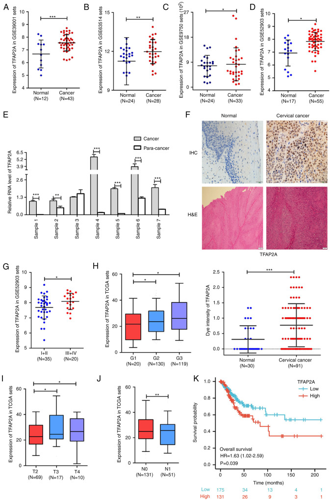
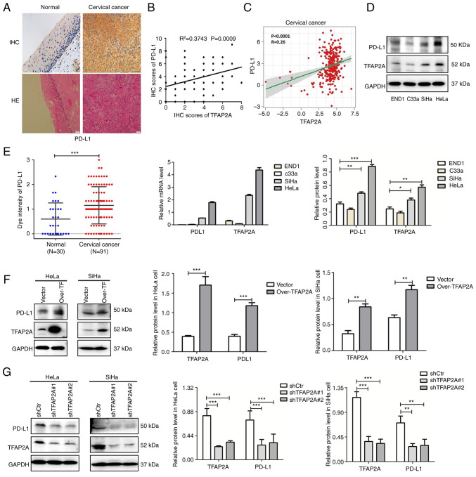
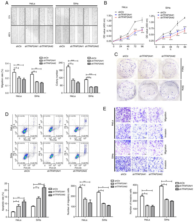
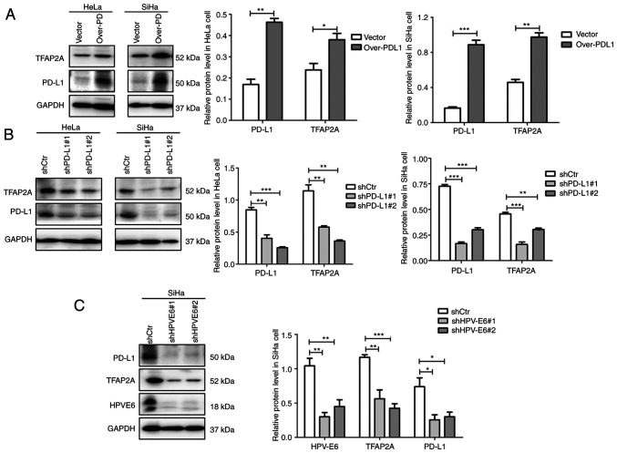
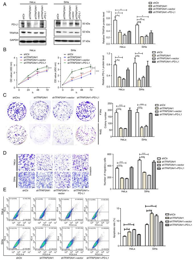
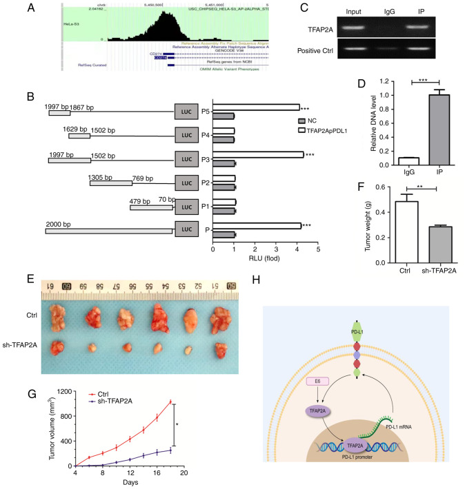
Transcription factor AP‑2 alpha (TFAP2A) is a critical cell growth regulator that is overexpressed in various tumor tissues. However, its role in the development of cervical cancer remains unknown. In the present study, public databases were thus explored and a higher expression of TFAP2A was found in cervical cancer. A total of 131 clinical samples were collected and it was also identified that TFAP2A was highly expressed in cervical tumor tissues. TFAP2A was also found to be associated with a higher tumor stage, lymph node metastasis and a poor patient survival. In vitro experiments revealed that the knockdown of TFAP2A inhibited the proliferation and migration of cervical cancer cells and promoted apoptosis. Furthermore, it was observed that TFAP2A could bind the programmed death‑ligand 1 (PD‑L1) promoter region and PD‑L1 rescued TFAP2A expression. In vivo experiments also revealed that TFAP2A promoted tumor growth. Collectively, in the present study it was demonstrated that TFAP2A is a transcription factor of PD‑L1 and a prognostic factor with clinical value, identifying a positive feedback loop of TFAP2A/PD‑L1.

Keywords: cervical cancer; migration; programmed death‑ligand 1; proliferation; transcription factor AP‑2 alpha.


Conflict of interest statement

The authors declare that they have no competing interests.

Figures

Figure 1.
Figure 1.
TFAP2A expression is increased in cervical cancer and is associated with clinicopathological features. (A-D) Analysis of TFAP2A mRNA expression in different datasets. (E) Expression of TFAP2A mRNA in six sets of paired cancer-paraneoplastic tissue samples. (F) IHC staining of TFAP2A expression and H&E staining in normal and cancer tissues. (G) Analysis of TFAP2A mRNA expression in different FIGO grades. (H) The expression level of the TFAP2A gene in different histopathology grades. (I) The expression level of TFAP2A gene in different FIGO T Stages. (J) The expression level of the TFAP2A gene according to lymph node metastasis. (K) The expression level of TFAP2A gene in association with patient survival. in each group. *P<0.05, **P<0.01 and ***P<0.001. TFAP2A, transcription factor AP-2 alpha; H&E, hematoxylin-eosin; IHC, immunohistochemical.
Figure 2.
Figure 2.
TFAP2A positively regulates PD-L1 expression in cervical cancer. (A) IHC staining of PD-L1 expression and hematoxylin-eosin staining in normal and cancer tissues. (B) The association between PD-L1 and TFAP2A protein (semi-quantitative analysis using IHC staining). (C) The association between PD-L1 and TFAP2A protein in The Cancer Genome Atlas database. (D) Western blot analysis of TFAP2A and PD-L1 expression in normal (END1) and cervical epithelial (HeLa, C33a and SiHa) cell lines. (E) Reverse transcription-quantitative PCR of TFAP2A and PD-L1 expression in normal (END1) and cancer) cervical epithelial (HeLa, C33a and SiHa) cell lines. (F) Western blot analysis revealed that PD-L1 expression was reduced in TFAP2A-overexpressing cervical cancer cell lines. (G) Western blot analysis revealed that PD-L1 expression was reduced in cervical cancer cell lines subjected to TFAP2A knockdown. n=3 for cell culture analysis. *P<0.05, **P<0.01 and ***P<0.001. TFAP2A, transcription factor AP-2 alpha; PD-L1, programmed death-ligand 1; H&E, hematoxylin-eosin; IHC, immunohistochemical.
Figure 3.
Figure 3.
Knockdown of TFAP2A inhibits the proliferation, migration and invasion, and promotes the apoptosis of cervical cancer cells. (A) Wound healing assay revealed that the downregulation of TFAP2A significantly reduced the cell migration rate. Representative images were obtained at 48 h. Scale bars, 100 µm. (B) Cell Counting Kit-8 assays revealed that the downregulation of TFAP2A reduced the growth rate. (C) Colony formation assay revealed that the downregulation of TFAP2A reduced the growth rate. (D) Apoptosis detection using flow cytometric analysis revealed that downregulation of TFAP2A promoted the apoptotic rate. (E) Transwell assay revealed that the downregulation of TFAP2A reduced the migration and invasion rate. n=3 in each group. *P<0.05, **P<0.01 and ***P<0.001. TFAP2A, transcription factor AP-2 alpha; sh-, short hairpin.
Figure 4.
Figure 4.
TFAP2A and PD-L1 form a positive feedback pathway, which may be regulated by HPV E6 protein. (A) Western blot analysis revealed that TFAP2A expression was reduced in PD-L1-overexpressing cervical cancer cell lines. (B) Western blot analysis revealed that TFAP2A expression was reduced in cervical cancer cell lines in which PD-L1 was knocked down. (C) Western blot analysis revealed that TFAP2A and PD-L1 expression was reduced in cervical cancer cells in which HPV E6 was knocked down. n=3 in each group. *P<0.05, **P<0.01 and ***P<0.001. TFAP2A, transcription factor AP-2 alpha; PD-L1, programmed death-ligand 1; sh-, short hairpin.
Figure 5.
Figure 5.
TFAP2A promotes the proliferation and migration, and inhibits the apoptosis of cervical cancer cells via PD-L1. (A) Western blot analysis of TFAP2A, PD-L1 expression in the shctr, shTFAP2A#1, shTFAP2A #1 + Vector and shTFAP2A #1 + PD-L1 groups. (B) Cell Counting Kit-8 assays revealed the growth rate of the cells. (C) Colony formation assay revealed the proliferation of the cells. (D) Transwell assay revealed the migration and invasion of cells. (E) Apoptosis detection using flow cytometric analysis revealed apoptosis rate of the cells. n=3 in each group. *P<0.05, **P<0.01 and ***P<0.001. TFAP2A, transcription factor AP-2 alpha; PD-L1, programmed death-ligand 1; sh-, short hairpin.
Figure 6.
Figure 6.
TFAP2A targets PD-L1 and the knockdown of TFAP2A inhibits the progression of cervical cancer. (A) Enrichment of TFAP2A in the PD-L1 promoter region in RNA-Seq Atlas [RNA-Seq Atlas-Database Commons (cncb.ac.cn)]. (B) PD-L1 promoter transient reporter assay, including deletions of the putative binding sites. Results are represented as normalized RLUs. (C and D) Chromatin Immunoprecipitation assay was used to determine the physical interaction of TFAP2A protein with the -PD-L1 promoter region and reverse transcription-quantitative PCR was used to analyze immune complexes. (E) Gross images of excised tumors in the control, Ctrl, sh-TFAP2A groups. (F) Tumor volume and (G) tumor weight. (H) Experimental hypothesis diagram; n=6 in each group. *P<0.05, **P<0.01 and ***P<0.001. TFAP2A, transcription factor AP-2 alpha; PD-L1, programmed death-ligand 1; RLUs, relative luciferase units; sh-, short hairpin.

Similar articles

Cited by

References

    1. Martínez-Rodríguez F, Limones-González JE, Mendoza-Almanza B, Esparza-Ibarra EL, Gallegos-Flores PI, Ayala-Luján JL, Ayala-Luján JL, Godina-González S, Salinas E, Mendoza-Almanza G. Understanding cervical cancer through proteomics. Cells. 2021;10:1854. doi: 10.3390/cells10081854. - DOI - PMC - PubMed
    1. Revathidevi S, Murugan AK, Nakaoka H, Inoue I, Munirajan AK. APOBEC: A molecular driver in cervical cancer pathogenesis. Cancer Lett. 2021;496:104–116. doi: 10.1016/j.canlet.2020.10.004. - DOI - PMC - PubMed
    1. Sung H, Ferlay J, Siegel RL, Laversanne M, Soerjomataram I, Jemal A, Bray F. Global Cancer Statistics 2020: GLOBOCAN estimates of incidence and mortality worldwide for 36 cancers in 185 countries. CA Cancer J Clin. 2021;71:209–249. doi: 10.3322/caac.21660. - DOI - PubMed
    1. Yamashita H, Kawasawa YI, Shuman L, Zheng Z, Tran T, Walter V, Warrick JI, Chen G, Al-Ahmadie H, Kaag M, et al. Repression of transcription factor AP-2 alpha by PPARγ reveals a novel transcriptional circuit in basal-squamous bladder cancer. Oncogenesis. 2019;8:69. doi: 10.1038/s41389-019-0178-3. - DOI - PMC - PubMed
    1. Wu J. Pancreatic cancer-derived exosomes promote the proliferation, invasion, and metastasis of pancreatic cancer by the miR-3960/TFAP2A Axis. J Oncol. 2022;2022:3590326. doi: 10.1155/2022/3590326. - DOI - PMC - PubMed