. 2023 Apr 21;19(4):e1011346.
doi: 10.1371/journal.ppat.1011346. eCollection 2023 Apr.

A modified Agrobacterium-mediated transformation for two oomycete pathogens

Affiliations

A modified Agrobacterium-mediated transformation for two oomycete pathogens

Luyao Wang et al. PLoS Pathog. .

Abstract

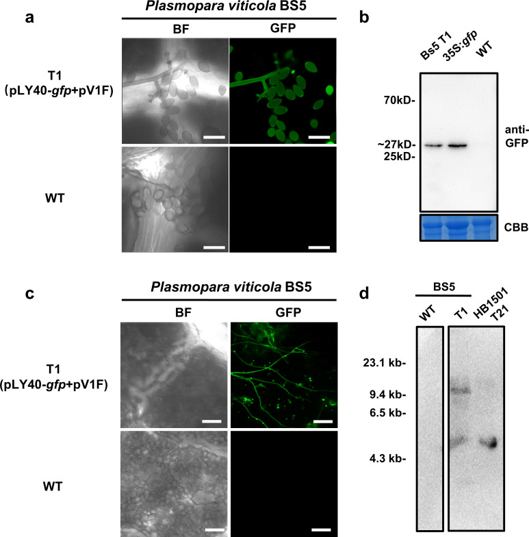
Oomycetes are a group of filamentous microorganisms that include some of the biggest threats to food security and natural ecosystems. However, much of the molecular basis of the pathogenesis and the development in these organisms remains to be learned, largely due to shortage of efficient genetic manipulation methods. In this study, we developed modified transformation methods for two important oomycete species, Phytophthora infestans and Plasmopara viticola, that bring destructive damage in agricultural production. As part of the study, we established an improved Agrobacterium-mediated transformation (AMT) method by prokaryotic expression in Agrobacterium tumefaciens of AtVIP1 (VirE2-interacting protein 1), an Arabidopsis bZIP gene required for AMT but absent in oomycetes genomes. Using the new method, we achieved an increment in transformation efficiency in two P. infestans strains. We further obtained a positive GFP transformant of P. viticola using the modified AMT method. By combining this method with the CRISPR/Cas12a genome editing system, we successfully performed targeted mutagenesis and generated loss-of-function mutations in two P. infestans genes. We edited a MADS-box transcription factor-encoding gene and found that a homozygous mutation in MADS-box results in poor sporulation and significantly reduced virulence. Meanwhile, a single-copy avirulence effector-encoding gene Avr8 in P. infestans was targeted and the edited transformants were virulent on potato carrying the cognate resistance gene R8, suggesting that loss of Avr8 led to successful evasion of the host immune response by the pathogen. In summary, this study reports on a modified genetic transformation and genome editing system, providing a potential tool for accelerating molecular genetic studies not only in oomycetes, but also other microorganisms.


Conflict of interest statement

The authors have declared that no competing interests exist.

Figures

Fig 1
Fig 1
(a) The protein similarity analysis of plant proteins, important for AMT of Arabidopsis, and their homologues in other plant and oomycete species. Representative plants with an established Agrobacterium-mediated transformation protocol and eight oomycete species were selected for phylogenetic analysis. The left phylogenetic tree was generated by the OrthoFinder software based on variations of single-copy orthologous genes in released genomic data of different species using IQ-tree with JTT + G4 model of evolution. The left bar indicates amino acid substitutions per site. The right heatmap indicates sequence similarity of known plant proteins envolved in Arabidopsis AMT process and their homologues in selected plants and oomycete species. The right bar indicates sequence similarity. (b) Schematic representation of the main idea of our improved transformation strategy. The use of the helper plasmid pV1F results in expression of virFΔN42 tagged AtVIP1. The recombinant AtVIP1 is delivered into oomycete zoospores via VirB/VirD4 T4SS, and thereby facilitates the process of T-DNA complex (T-complex) targeting the nucleus of an infected zoospore or an encysted zoospore with a germ tube.
Fig 2
Fig 2. Generating P. infestans transformants expressing gfp using the modified AMT method.
(a) Schematic representation of the constructs used in this experiment. A. tumefaciens strain EHA105 with pV1F and pLY40-gfp was used for P. infestans transformation. (b) Quantification of colonies that acquired G418 resistance as a result of transforming gfp into P. infestans strains JH19 and HB1501 using our modified AMT procedure. EHA105 with the helper plasmid pEV was used as control in this experiment. All data represent average values from three independent experiments with the indicated standard deviations. Statistical differences among the samples were analyzed with Šídák’s multiple comparisons test (P< 0.0021: **, P< 0.0001: ****). (c) Two representative plates from the transformation experiment shown in (b). (d) Immunoblot of P. infestans JH19 transformant T1, expressing free gfp, probed with an anti-GFP antibody. Protein extracted from N. benthamiana leaves transiently expressing gfp driven by the CaMV35s promoter was used as positive control in lane 1. (e) The P. infestans JH19 transformant T1 expressing a detectable GFP signal was obtained by AMT using A. tumefaciens carrying constructs described in (a). Scale bars = 40 μm. T3, a randomly selected empty vector transformant was used as negative control. GFP expression in the transformant was analysed by confocal microscopy. The protein blot was stained with Coomassie Blue to confirm equal loading. (f) Southern blot analysis of representative gfp transformants of P. infestans strain HB1501. Genomic DNA (4 μg) was digested with HindIII and all blots were probed with a fragment containing the nptII gene to detect the presence of T-DNA. Numbers on the left indicate the positions of molecular weight markers (kb).
Fig 3
Fig 3. Obtaining Plasmopara viticola BS5 stable transformant T1 using the modified AMT protocol.
(a) P. viticola BS5 strain was used for transformation assay and was obtained by AMT using A. tumefaciens carrying constructs pLY40-gfp and pV1F. Scale bars = 40 μm. The confocal microscopy images were taken 7 days post inoculation with the zoospores from the third sub-generation of T1; wild type BS5 was used as negative control. (b) Immunoblot of P. viticola BS5 transformant T1 expressing free GFP, probed with an anti-GFP antibody. Protein extracted from N. benthamiana leaves transiently expressing gfp driven by the CaMV35s promoter was used as positive control in lane 2. (c) Transformant T1 of P. viticola BS5 expresses detectable GFP signal when infecting grapevine leaves (Zitian seedless, A17). Scale bars = 100 μm. Images were taken 7 days post inoculation and wild type BS5 was used as negative control. (d) Southern blot analysis of transformant T1 of P. viticola BS5. Genomic DNA (4 μg) was digested with HindIII and all blots were probed with a fragment containing the nptII gene to detect the presence of T-DNA. Numbers on the left indicate the positions of molecular weight markers (kb).
Fig 4
Fig 4. Integration of LbCas12a in P. infestans.
(a) Schematic representation of the constructs used in the experiment. A. tumefaciens EHA105 carrying pLY40-Cas12a-gfp and either pV1F or pEV was used for P. infestans transformation. (b) Confocal micrograph of T5 and T6 transformants expressing PsNLS-lbCas12-GFP. The nuclear localization pattern of the fusion protein, indicated by white arrows, revealed mycelia and sporangia. T3, an empty vector pLY40 transformed line, is used as negative control. Bright field and GFP channels are presented. Scale bars = 40 μm. (c) Immunoblot of two representative P. infestans transformants expressing PsNLS-lbCas12-GFP. The gfp tagged hlbCas12a driven by CaMV35S promoter was expressed in N. benthamiana as positive control. The expected size of the protein is 176.6 kDa. The protein blot was stained with Coomassie Blue to confirm equal loading. (d) Quantification of positive P. infestans transformants expressing GFP tagged Cas12a (JH19 and HB1501 backgrounds) generated using the modified AMT procedure. A. tumefaciens EHA105 carrying the helper plasmid pEV was used as control in this experiment. Statistical differences among the samples were analyzed with Šídák’s multiple comparisons test (P< 0.0021: **, P< 0.0001: ****).
Fig 5
Fig 5. Editing a MADS-box transcription factor coding gene in P. infestans strain JH19.
(a) Target sites of two gRNAs in the MADS-box coding sequence. The MEF2-like domain was predicted by InterproScan (version 5.52–86.0). The MEF2-like domain locates at 2–77 aa sites. (b) Detecting of editing events in MADS-box in JH19 transformants. The PCR assay on T8 and T16 revealed two homozygous editing events. (c) Analysis of PCR amplicon sequencing results from (b). The PAM sequences are marked in red, the target sequences are marked in blue, and the stop codons are indicated by red arrowheads. Each dash line represents a deleted nucleotide. (d) Culture scrapings from 10 days Pea broth cultures of wild type JH19, T5, T8 and T16. Scale bars = 0.4 mm. (e) Infection phenotypes of wild type JH19 (WT), transformants T5, T8 and T16 on leaves of susceptible cultivar potato cv. Désirée. Detached potato leaves were inoculated with mycelia medium discs, and images were recorded 5 days post inoculation. Scale bars = 2 cm. (f) Quantification of sporangia numbers in 10 μL PEA broth culture of wild type JH19, T5, T8 and T16 in (d). (g) Quantification of lesion size in detached leaves of potato cv. Désirée inoculated with wild type JH19 (WT), T5, T8 and T16 in (e). All data represent average values from three independent experiments with the indicated standard deviations. Statistical differences among the samples were analyzed with Šídák’s multiple comparisons test (P< 0.0021: **, P< 0.0002: ***).
Fig 6
Fig 6. Editing an avirulence gene Avr8 in P. infestans strain HB1501.
(a) Target sites of two gRNAs in the Avr8 coding sequence. The LWY motifs were predicted by InterproScan (version 5.52–86.0). The LWY1 domain locates at 63–107 aa sites, the LWY2 domain locates at 105–162 aa sites, and the LWY3 domain locates at 165–218 aa sites. (b) Detecting editing of Avr8 in HB1501 transformants. The PCR assay in T3 and T10 revealed two homozygous editing events. (c) Analysis of PCR amplicon sequencing results from (b). The PAM sequences are marked in red, the target sequences are marked in blue, and stop codons are indicated by red arrowheads. Each dash line represents a deleted nucleotide. (d) Infection phenotypes of wild type HB1501 (WT), T3, T10 and T22 on detached leaves of R8 transgenic potato. Detached potato leaves were inoculated with zoospores of selected strains, and images were taken 5 days post inoculation. Scale bars = 2 cm. (e) Quantification of lesion sizes in (d). All data represent average values from three independent experiments with the indicated standard deviations. Statistical differences among the samples were analyzed with Šídák’s multiple comparisons test (P< 0.0002: ***).

Similar articles

Cited by

References

    1. Dubresson R, Kravchuk Z, Neuhaus J-M, Mauch-Mani B. Optimisation and comparison of transient expression methods to express the green fluorescent protein in the obligate biotrophic oomycete Plasmopara viticola. Vitis. 2008;47(4):235–40.
    1. Fry W. Phytophthora infestans: the plant (and R gene) destroyer. Molecular plant pathology. 2008;9(3):385–402. doi: 10.1111/j.1364-3703.2007.00465.x - DOI - PMC - PubMed
    1. Chen H, Shu H, Wang L, Zhang F, Li X, Ochola SO, et al.. Phytophthora methylomes are modulated by 6mA methyltransferases and associated with adaptive genome regions. Genome biology. 2018;19(1):1–16. - PMC - PubMed
    1. Sabbadin F, Urresti S, Henrissat B, Avrova AO, Welsh LR, Lindley PJ, et al.. Secreted pectin monooxygenases drive plant infection by pathogenic oomycetes. Science. 2021;373(6556):774–9. doi: 10.1126/science.abj1342 - DOI - PubMed
    1. Luo X, Tian T, Feng L, Yang X, Li L, Tan X, et al.. Pathogenesis-related protein 1 suppresses oomycete pathogen by targeting against AMPK kinase complex. Journal of Advanced Research. 2022. doi: 10.1016/j.jare.2022.02.002 - DOI - PMC - PubMed

Publication types