Presumed pituitary apoplexy in 26 dogs: Clinical findings, treatments, and outcomes
- PMID: 37084035
- PMCID: PMC10229324
- DOI: 10.1111/jvim.16703
Presumed pituitary apoplexy in 26 dogs: Clinical findings, treatments, and outcomes
Abstract
Background: Pituitary apoplexy refers to hemorrhage or infarction within the pituitary gland resulting in acute neurological abnormalities. This condition is poorly described in dogs.
Objectives: To document presenting complaints, examination findings, endocrinopathies, magnetic resonance imaging (MRI), treatments, and outcomes of dogs with pituitary apoplexy.
Animals: Twenty-six client-owned dogs with acute onset of neurological dysfunction.
Methods: Retrospective case series. Dogs were diagnosed with pituitary apoplexy if MRI or histopathology documented an intrasellar or suprasellar mass with evidence of hemorrhage or infarction in conjunction with acute neurological dysfunction. Clinical information was obtained from medical records and imaging reports.
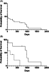
Results: Common presenting complaints included altered mentation (16/26, 62%) and gastrointestinal dysfunction (14/26, 54%). Gait or posture changes (22/26, 85%), mentation changes (18/26, 69%), cranial neuropathies (17/26, 65%), cervical or head hyperpathia (12/26, 46%), and hyperthermia (8/26, 31%) were the most frequent exam findings. Ten dogs (38%) lacked evidence of an endocrinopathy before presentation. Common MRI findings included T1-weighted hypo- to isointensity of the hemorrhagic lesion (21/25, 84%), peripheral enhancement of the pituitary mass lesion (15/25, 60%), brain herniation (14/25, 56%), and obstructive hydrocephalus (13/25, 52%). Fifteen dogs (58%) survived to hospital discharge. Seven of these dogs received medical management alone (median survival 143 days; range, 7-641 days) and 8 received medications and radiation therapy (median survival 973 days; range, 41-1719 days).
Conclusions and clinical importance: Dogs with pituitary apoplexy present with a variety of acute signs of neurological disease and inconsistent endocrine dysfunction. Dogs that survive to discharge can have a favorable outcome.
Keywords: adenoma; carcinoma; endocrionopathy; hemorrhage; magnetic resonance imaging; suprasellar; survival.
© 2023 The Authors. Journal of Veterinary Internal Medicine published by Wiley Periodicals LLC on behalf of American College of Veterinary Internal Medicine.
Conflict of interest statement
Authors declare no conflicts of interest.
Figures


References
-
- Barkhoudarian G, Kelly DF. Pituitary apoplexy. Neurosurg Clin N Am. 2019;30:457‐463. - PubMed
-
- Briet C, Salenave S, Bonneville JF, Laws ER, Chanson P. Pituitary apoplexy. Endocr Rev. 2015;36:622‐645. - PubMed
-
- Briet C, Salenave S, Chanson P. Pituitary apoplexy. Endocrinol Metab Clin North Am. 2015;44:199‐209. - PubMed
-
- Mayol Del Valle M, De Jesus O. Pituitary apoplexy. StatPearls [Internet]. Treasure Island: StatPearls Publishing; 2022. - PubMed
MeSH terms
Grants and funding
LinkOut - more resources
Full Text Sources
Medical

