. 2023 May 3;14(9):1672-1685.
doi: 10.1021/acschemneuro.3c00135. Epub 2023 Apr 21.

Discovery of a Potent and Selective CDKL5/GSK3 Chemical Probe That Is Neuroprotective

Affiliations

Discovery of a Potent and Selective CDKL5/GSK3 Chemical Probe That Is Neuroprotective

Han Wee Ong et al. ACS Chem Neurosci. .

Abstract

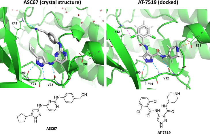
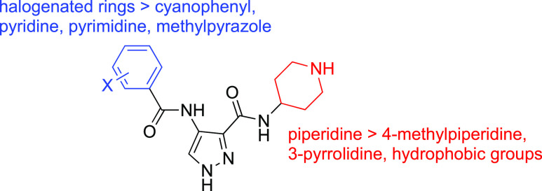
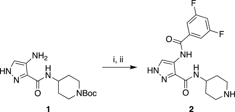
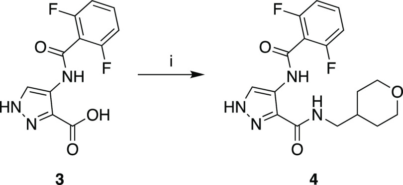
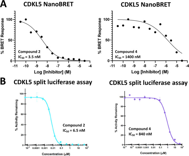
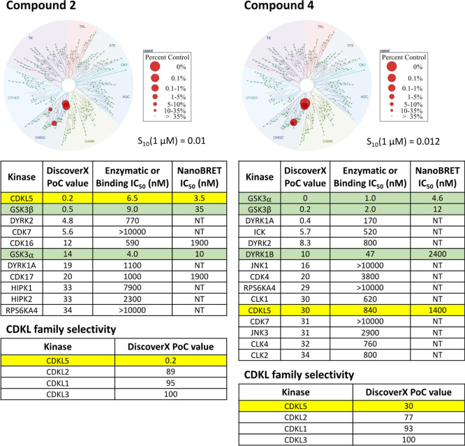
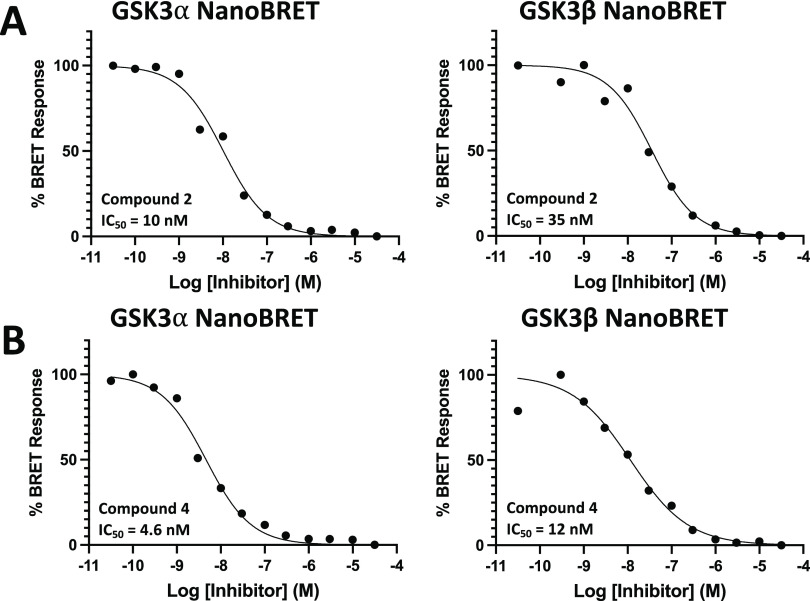
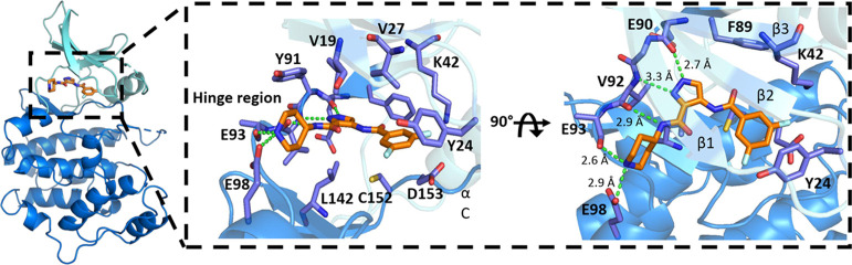
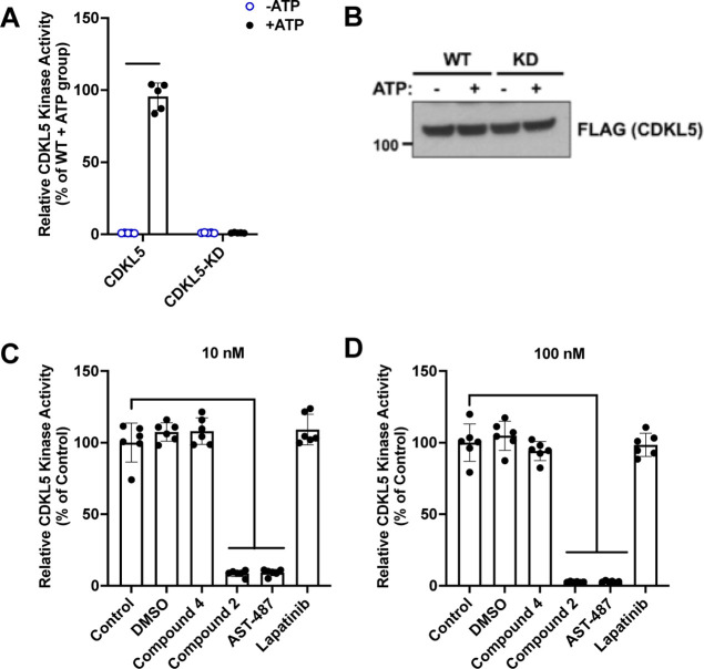
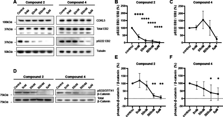
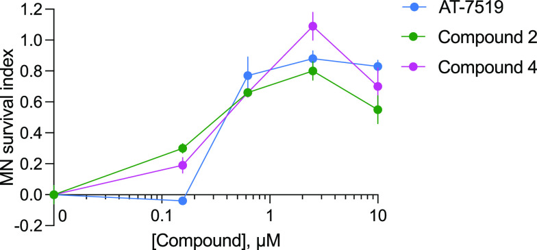
Despite mediating several essential processes in the brain, including during development, cyclin-dependent kinase-like 5 (CDKL5) remains a poorly characterized human protein kinase. Accordingly, its substrates, functions, and regulatory mechanisms have not been fully described. We realized that availability of a potent and selective small molecule probe targeting CDKL5 could enable illumination of its roles in normal development as well as in diseases where it has become aberrant due to mutation. We prepared analogs of AT-7519, a compound that has advanced to phase II clinical trials and is a known inhibitor of several cyclin-dependent kinases (CDKs) and cyclin-dependent kinase-like kinases (CDKLs). We identified analog 2 as a highly potent and cell-active chemical probe for CDKL5/GSK3 (glycogen synthase kinase 3). Evaluation of its kinome-wide selectivity confirmed that analog 2 demonstrates excellent selectivity and only retains GSK3α/β affinity. We next demonstrated the inhibition of downstream CDKL5 and GSK3α/β signaling and solved a co-crystal structure of analog 2 bound to human CDKL5. A structurally similar analog (4) proved to lack CDKL5 affinity and maintain potent and selective inhibition of GSK3α/β, making it a suitable negative control. Finally, we used our chemical probe pair (2 and 4) to demonstrate that inhibition of CDKL5 and/or GSK3α/β promotes the survival of human motor neurons exposed to endoplasmic reticulum stress. We have demonstrated a neuroprotective phenotype elicited by our chemical probe pair and exemplified the utility of our compounds to characterize the role of CDKL5/GSK3 in neurons and beyond.

Keywords: CDKL5; GSK3α; GSK3β; chemical probe; crystal structure; kinase; neuroprotective.


Conflict of interest statement

The authors declare no competing financial interest.

Figures

Figure 1
Figure 1
Comparison of CDKL5 co-crystal structure with ASC67 (left, PDB code: 4BGQ) and docking of AT-7519 (right). Key interactions, including the critical hinge binding interactions, are highlighted. Hydrogen bonds are indicated as light blue lines. An NH–Cl interaction, which is shown as a pink line, was observed in the docked structure of AT-7519 with Lys42 of CDKL5. An ionic interaction, illustrated with an orange line, was observed in the docked structure of AT-7519 with Glu98 of CDKL5. For clarity of presentation, only key amino acids are labeled.
Figure 2
Figure 2
Structural modifications of AT-7519. A summary of different groups that were appended at the termini of the AT-7519 scaffold and how they augmented CDKL5 affinity. The greater than symbol (>) indicates that some groups are preferred over others and result in greater affinity for CDKL5.
Scheme 1
Scheme 1. Synthesis of Chemical Probe Compound (2)
(i) 3,5-Difluorobenzoic acid, n-propanephosphonic acid anhydride, DIPEA, THF, 0–25 °C, 1 h and (ii) HCl, dioxane, 0–25 °C, 1 h.
Scheme 2
Scheme 2. Synthesis of Negative Control Compound (4)
(i) 4-Aminomethyltetrahydropyran, n-propanephosphonic acid anhydride, DIPEA, THF, 0–25 °C, 1 h.
Figure 3
Figure 3
(A) CDKL5 cellular and (B) CDKL5 biochemical potency for compounds 2 and 4. Normalized dose–response curves generated using the CDKL5 NanoBRET assay are shown in panel (A). Dose–response curves produced using the CDKL5 split luciferase assay are included in panel (B). Data generated using these orthogonal assay formats and included in Figure 4 support that compound 2 binds with high affinity to CDKL5, while compound 4 shows limited affinity for CDKL5.
Figure 4
Figure 4
Selectivity data for compounds 2 and 4. The circular kinome tree diagrams illustrate the selectivity of these compounds when profiled against 403 WT human kinases at 1 μM in the Eurofins DiscoverX scanMAX panel. The percent control legend classifies that red circles of different sizes on the trees correspond with the PoC value with which each kinase binds compound 2 or 4. Selectivity scores (S10, 1 μM) were calculated using the PoC values for only WT human kinases in the scanMAX panel. An S10 score also conveys selectivity and corresponds with the percent of the kinases screened that bind with a PoC value < 10. Kinases in the embedded tables are listed by their gene names and ranked by their PoC value generated in the scanMAX panel. While rows containing CDKL5 data are colored yellow, rows in the top tables colored green are kinases that demonstrate enzymatic IC50 values within a 30-fold window of the CDKL5 binding IC50 value for compound 2 and within a 30-fold window of the GSK3β enzymatic IC50 value for compound 4. All IC50 values in the enzymatic or binding IC50 column were generated using enzymatic assays, except for CDKL5, which was produced using the CDKL5 split luciferase assay. NT = not tested.
Figure 5
Figure 5
Cellular target engagement of GSK3α/β by compounds 2 and 4. Normalized dose–response curves generated using the GSK3α and GSK3β NanoBRET assays are shown for compound 2 in panel (A) and for compound 4 in panel (B). Data produced using these assays are included in Figure 4 and support that both compounds 2 and 4 bind with high affinity to GSK3α and GSK3β.
Figure 6
Figure 6
Overview of the CDKL5 co-crystal structure with compound 2 (PDB code: 8CIE). N- and C-terminal lobes colored in aquamarine and blue, respectively. Compound 2 binds to the ATP pocket between the two lobes and is shown as orange sticks. Hydrogen bonds are indicated as green dashed lines, including interactions between the hinge region (Glu90 and Val92, colored purple) and the pyrazole shown in the cutout. The hinge region is labeled for clarity.
Figure 7
Figure 7
Compound 2 potently inhibits CDKL5 kinase activity. (A) Representative graph from a kinase assay showing that purified WT human CDKL5 retains kinase activity, while the KD (CDKL5 K42R) human protein is functionally inactive. n = 3 replicates. (B) Representative western blot showing equal levels of WT and KD proteins in the kinase assay experiments. (C,D) Purified WT human CDKL5 was used for kinase assays as described above in the presence of indicated compounds at 10 or 100 nM concentrations. n = 3 replicates. One-way ANOVA with Dunnett’s multiple comparison test was done. *** = p < 0.0001. Non-significant comparisons are not illustrated.
Figure 8
Figure 8
Analysis of phospho-EB2 and phospho-β-catenin expression following treatment with compound 2 or 4 confirms inhibition of downstream signaling mediated by CDKL5 and GSK3α/β. (A) Western blots showing the expression of CDKL5, EB2, and tubulin and the level of S222 EB2 phosphorylation following 1 h treatment of DIV11–14 rat primary cortical neurons with 5 nM, 50 nM, 500 nM, or 5 μM of compound 2 or 4. (B) Quantification of S222 EB2 phosphorylation following treatment of DIV11–14 rat primary cortical neurons with compound 2. Two-way ANOVA with pairwise comparisons of each condition against the control condition. n = 3 replicates. (C) Quantification of S222 EB2 phosphorylation following treatment of DIV11–14 rat primary cortical neurons with compound 4. Two-way ANOVA with pairwise comparisons of each condition against the control condition. n = 3 replicates. (D) Western blots showing the expression of β-catenin and the level of S33/37/T41 β-catenin phosphorylation following 1 h treatment of DIV11–14 rat primary cortical neurons with 5 nM, 50 nM, 500 nM or 5 μM of compound 2 or 4. (E) Quantification of S33/37/T41 β-catenin phosphorylation following treatment of DIV11–14 rat primary cortical neurons with compound 2. Two-way ANOVA with pairwise comparisons of each condition against the control condition. n = 3 replicates. (F) Quantification of S33/37/T41 β-catenin phosphorylation following treatment of DIV11–14 rat primary cortical neurons with compound 4. Two-way ANOVA with pairwise comparisons of each condition against the control condition. n = 3 replicates. * = p ≤ 0.05, ** = p ≤ 0.01, *** = p ≤ 0.001, and **** = p ≤ 0.0001. Non-significant (p > 0.05) comparisons are not illustrated.
Figure 9
Figure 9
Compounds 2 and 4 promote survival of motor neurons in response to ER stress. Differentiated human motor neurons derived from human stem cells were pharmacologically stressed using CPA. At low concentrations (<200 nM), compounds 2 and 4 preserved the viability of these vulnerable cells. Error bars represent standard error of the mean (SEM) calculated using two-way ANOVA. MN: motor neuron.

Update of

Similar articles

Cited by

References

    1. Ricciardi S.; Ungaro F.; Hambrock M.; Rademacher N.; Stefanelli G.; Brambilla D.; Sessa A.; Magagnotti C.; Bachi A.; Giarda E.; et al. CDKL5 ensures excitatory synapse stability by reinforcing NGL-1-PSD95 interaction in the postsynaptic compartment and is impaired in patient iPSC-derived neurons. Nat. Cell Biol. 2012, 14, 911–923. 10.1038/ncb2566. - DOI - PMC - PubMed
    1. Berginski M. E.; Moret N.; Liu C.; Goldfarb D.; Sorger P. K.; Gomez S. M. The Dark Kinase Knowledgebase: an online compendium of knowledge and experimental results of understudied kinases. Nucleic Acids Res. 2021, 49, D529–D535. 10.1093/nar/gkaa853. - DOI - PMC - PubMed
    1. Chen Q.; Zhu Y.-C.; Yu J.; Miao S.; Zheng J.; Xu L.; Zhou Y.; Li D.; Zhang C.; Tao J.; Xiong Z.-Q. CDKL5, a protein associated with Rett Syndrome, regulates neuronal morphogenesis via Rac1 signaling. J. Neurosci. 2010, 30, 12777–12786. 10.1523/jneurosci.1102-10.2010. - DOI - PMC - PubMed
    1. Fuchs C.; Trazzi S.; Torricella R.; Viggiano R.; De Franceschi M.; Amendola E.; Gross C.; Calzà L.; Bartesaghi R.; Ciani E. Loss of CDKL5 impairs survival and dendritic growth of newborn neurons by altering AKT/GSK-3β signaling. Neurobiol. Dis. 2014, 70, 53–68. 10.1016/j.nbd.2014.06.006. - DOI - PMC - PubMed
    1. Barbiero I.; Valente D.; Chandola C.; Magi F.; Bergo A.; Monteonofrio L.; Tramarin M.; Fazzari M.; Soddu S.; Landsberger N.; Rinaldo C.; Kilstrup-Nielsen C. CDKL5 localizes at the centrosome and midbody and is required for faithful cell division. Sci. Rep. 2017, 7, 6228.10.1038/s41598-017-05875-z. - DOI - PMC - PubMed

Publication types

Substances