. 2023 May 11;41(20):3233-3246.
doi: 10.1016/j.vaccine.2023.04.020. Epub 2023 Apr 14.

Intranasal administration of adenoviral vaccines expressing SARS-CoV-2 spike protein improves vaccine immunity in mouse models

Affiliations

Intranasal administration of adenoviral vaccines expressing SARS-CoV-2 spike protein improves vaccine immunity in mouse models

Tobias L Freitag et al. Vaccine. .

Abstract

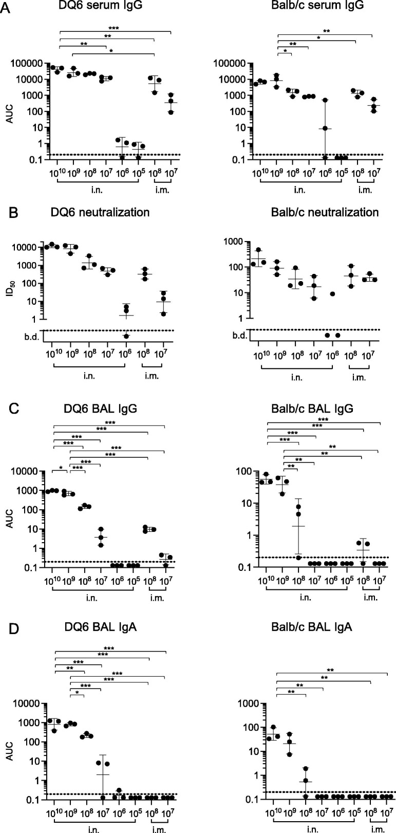
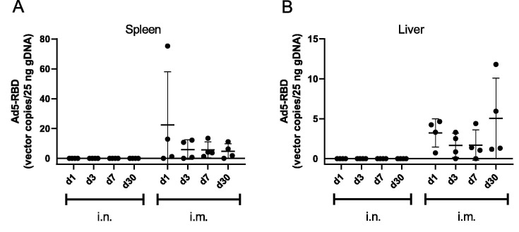
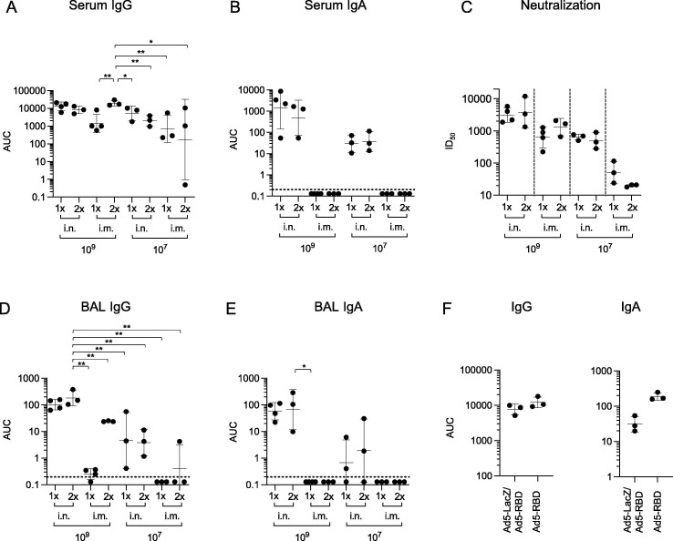
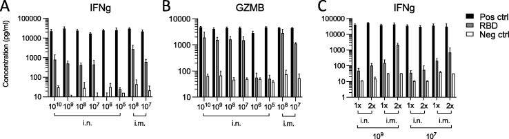
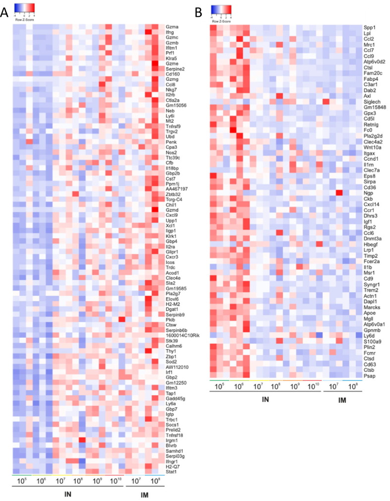
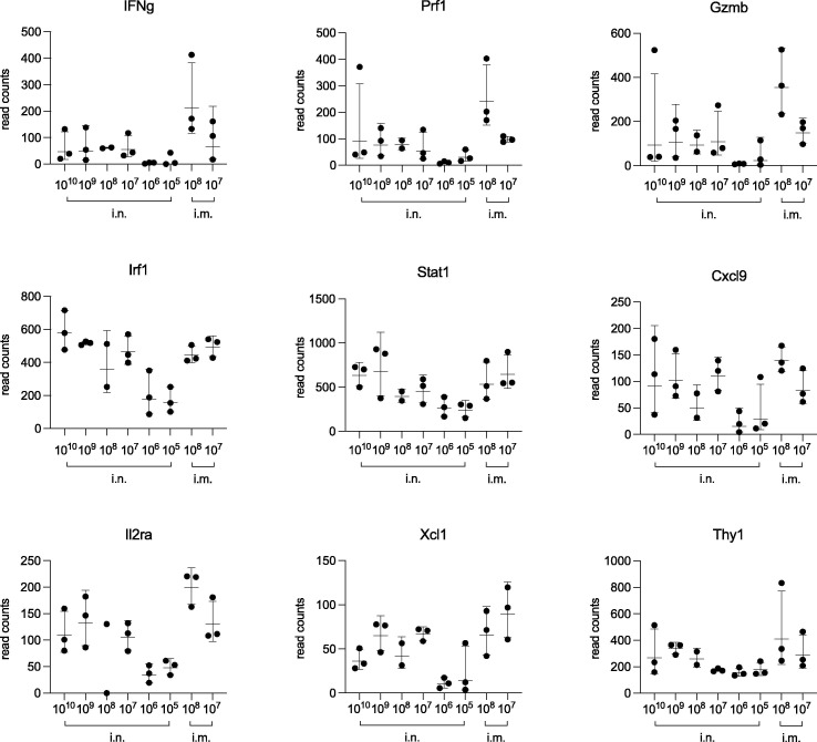
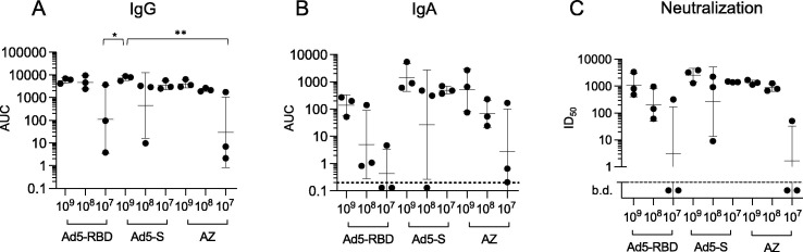
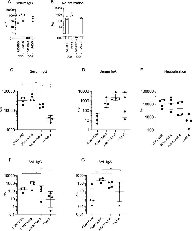
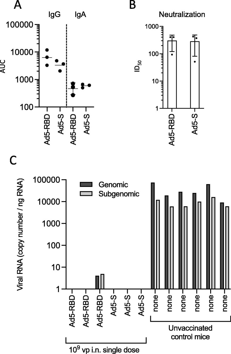
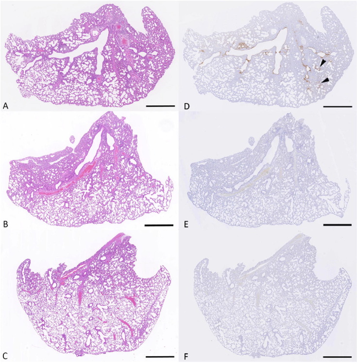
The ongoing SARS-CoV-2 pandemic is controlled but not halted by public health measures and mass vaccination strategies which have exclusively relied on intramuscular vaccines. Intranasal vaccines can prime or recruit to the respiratory epithelium mucosal immune cells capable of preventing infection. Here we report a comprehensive series of studies on this concept using various mouse models, including HLA class II-humanized transgenic strains. We found that a single intranasal (i.n.) dose of serotype-5 adenoviral vectors expressing either the receptor binding domain (Ad5-RBD) or the complete ectodomain (Ad5-S) of the SARS-CoV-2 spike protein was effective in inducing i) serum and bronchoalveolar lavage (BAL) anti-spike IgA and IgG, ii) robust SARS-CoV-2-neutralizing activity in the serum and BAL, iii) rigorous spike-directed T helper 1 cell/cytotoxic T cell immunity, and iv) protection of mice from a challenge with the SARS-CoV-2 beta variant. Intramuscular (i.m.) Ad5-RBD or Ad5-S administration did not induce serum or BAL IgA, and resulted in lower neutralizing titers in the serum. Moreover, prior immunity induced by an intramuscular mRNA vaccine could be potently enhanced and modulated towards a mucosal IgA response by an i.n. Ad5-S booster. Notably, Ad5 DNA was found in the liver or spleen after i.m. but not i.n. administration, indicating a lack of systemic spread of the vaccine vector, which has been associated with a risk of thrombotic thrombocytopenia. Unlike in otherwise genetically identical HLA-DQ6 mice, in HLA-DQ8 mice Ad5-RBD vaccine was inferior to Ad5-S, suggesting that the RBD fragment does not contain a sufficient collection of helper-T cell epitopes to constitute an optimal vaccine antigen. Our data add to previous promising preclinical results on intranasal SARS-CoV-2 vaccination and support the potential of this approach to elicit mucosal immunity for preventing transmission of SARS-CoV-2.

Keywords: Adenoviral vector; COVID-19; Intranasal vaccination; Mucosal immunity; SARS-CoV-2.


Conflict of interest statement

Declaration of Competing Interest The authors declare the following financial interests/personal relationships which may be considered as potential competing interests: Tobias Freitag reports a relationship with Rokote Laboratories Finland Ltd that includes: employment. Kalle Saksela reports a relationship with Rokote Laboratories Finland Ltd that includes: board membership and equity or stocks. Seppo Yla-Herttuala reports a relationship with Rokote Laboratories Finland Ltd that includes: board membership and equity or stocks. Kari Alitalo reports a relationship with Rokote Laboratories Finland Ltd that includes: equity or stocks.

Figures

Fig. 1
Fig. 1
Humoral responses to single treatment with Ad5-RBD administered intranasally (i.n.) or intramuscularly (I.m.) in HLA-DQ6 vs. Balb/c mice. Groups of adult female mice (n = 3) were inoculated with Ad5-RBD at doses ranging from 105-1010 viral particles/mouse. Anti-spike IgG serum antibodies (a; area under the curve (AUC)), SARS-CoV-2 pseudovirus neutralization (b; calculated serum dilution producing 50% inhibition (ID50)), and anti-spike IgG and IgA bronchoalveolar lavage fluid antibodies (c, d; AUC) were measured after 25 days. For statistical comparisons, one-way ANOVA and Tukey’s test for multiple comparisons (a, c, d) or Kruskal-Wallis test and Dunn’s test for multiple comparisons (b) were used. Adjusted p-values are displayed as * <0.05, ** <0.01 and *** <0.001. To improve readability, adjusted p-values calculated in comparisons involving groups treated with doses 105 or 106 are not shown. Lines and bars represent geometric means and standard deviations. Dotted line represents the limit of detection of the assay.
Fig. 2
Fig. 2
Distribution of Ad5 vector DNA to the spleen and liver. Spleen (A) and liver (B) samples were collected from C57BL/6 mice (n = 4) at 1, 3, 7 or 30 days following either intranasal (i.n.) or intramuscular (i.m.) administration of Ad-RBD (109 virus particles). Ad5-RBD vector was quantified from genomic DNA samples (25 ng) by real-time PCR using a specific probe-based assay to detect the RBD. Vector copy numbers were determined by comparison to plasmid standard (pAdapt-RBD). Individual values and means ± SEM are plotted.
Fig. 3
Fig. 3
Humoral responses to one vs. two treatments with Ad5-RBD administered intranasally (i.n.) or intramuscularly (i.m.) in HLA-DQ6 mice, and lack of effect of pre-treatment with Ad5 vector on priming with Ad5-RBD. Groups of adult male and female mice (n = 3) were inoculated either once or twice (14 days apart) with Ad5-RBD at doses 107 or 109 viral particles/mouse (a-e). Anti-spike IgG or IgA serum antibodies (a, b; area under the curve (AUC)), SARS-CoV-2 pseudovirus neutralization (c; calculated serum dilution producing 50% inhibition (ID50)), and anti-spike IgG and IgA bronchoalveolar lavage fluid antibodies (d, e; AUC) were measured after 28 days. In a different experiment, one group of HLA-DQ6 mice was pre-treated i.n. with Ad5-vector encoding β-galactosidase (Ad5-LacZ; 109 vp/mouse), while a second group remained untreated (n = 3; 3f). After 21 days, a priming dose of Ad5-RBD i.n. was administered to both groups (109 vp/mouse). Serum was collected after 42 days, and anti-spike IgG or IgA serum antibodies were measured (f; AUC). For statistical comparisons, one-way ANOVA and Tukey’s test for multiple comparisons (a, b, d, e, f) or Kruskal-Wallis test and Dunn’s test for multiple comparisons (c) were used. Adjusted p-values are displayed as * <0.05, ** <0.01 and *** <0.001. Lines and bars represent geometric means and standard deviations. Dotted line represents the limit of detection of the assay.
Fig. 4
Fig. 4
Cellular responses to treatment with Ad5-RBD administered intranasally (i.n.) or intramuscularly (i.m.) in HLA-DQ6 mice. Groups of female HLA-DQ6 mice (n = 3) were inoculated once with Ad5-RBD at doses ranging from 105-1010 viral particles/mouse (a, b). Groups of male or female HLA-DQ6 mice were inoculated once or twice (14 days apart) with Ad5-RBD at doses 107 or 109 viral particles/mouse (c). After 25–28 days, spleen cells were restimulated in vitro with recombinant SARS-CoV-2 spike protein receptor binding domain (RBD). Supernatants were harvested after 3 days, and the effector proteins IFNg and granzyme B were measured by ELISA. Results expressed as concentrations (pg/ml). Statistical comparisons were performed using Kruskal-Wallis test and Dunn’s test for multiple comparisons. Group differences were non-significant (adjusted p-values > 0.05).
Fig. 5
Fig. 5
Transcriptomic profiles of RBD-stimulated spleen cells from mice treated with Ad5-RBD administered intranasally (i.n.) or intramuscularly (i.m.). Groups of female HLA-DQ6 mice (n = 3) were inoculated once with Ad5-RBD at doses ranging from 105-1010 viral particles/mouse. After 25 days, spleen cells were restimulated in vitro with recombinant SARS-CoV-2 spike protein receptor binding domain (RBD). Cells were harvested after 3 days of culture, and bulk RNA was isolated. Heatmaps depicting the up- (a; red color, 86 genes) or down-regulation (b; blue color, 62 genes) of altogether 148 genes differentially expressed after treatment with Ad5-RBD in at least one of two separate comparisons, and are ordered to show the genes with the highest log2 fold changes from the top [Ad5-RBD at doses 105/106 viral particles i.n. (ineffective treatment) vs. doses 107/108 viral particles i.m. or doses 109/1010 viral particles i.n. (n = 6; adjusted P value, P ≤ 0.001; RNA sequencing)]. Statistical analyses were performed using edgeR. (For interpretation of the references to color in this figure legend, the reader is referred to the web version of this article.)
Fig. 6
Fig. 6
Upregulation of selected immune genes, differentially expressed in RBD-stimulated spleen cells from mice treated with Ad5-RBD administered intranasally (i.n.) or intramuscularly (i.m.). Groups of female HLA-DQ6 mice (n = 3) were inoculated once with Ad5-RBD at doses ranging from 105-1010 viral particles/mouse. After 25 days, spleen cells were restimulated in vitro with recombinant SARS-CoV-2 spike protein receptor binding domain (RBD). Cells were harvested after 3 days of culture, and bulk RNA was isolated for transcriptomic analyses (RNA sequencing; compare Fig. 5). Panels depict the results for 9 selected immune genes [T helper 1/cytotoxic T cell markers: Interferon gamma (Ifng), perforin 1 (Prf1), granzyme B (Gzmb); Interferon gamma-related genes: Interferon regulatory factor 1 (Irf1), signal transducer and activator of transcription 1 (Stat1), chemokine (C-X-C motif) ligand 9 (CXCL9); Lymphocyte activation-related genes: Interleukin 2-receptor alpha (Il2ra), chemokine (C motif) ligand (XCL1), thymocyte differentiation antigen 1 (Thy1)]. Results expressed as read counts.
Fig. 7
Fig. 7
Humoral responses to a single treatment with either Ad5-RBD, Ad5-S or ChAdOx1 nCoV-19 (AstraZeneca, “AZ”) administered intranasally in C57BL/6 mice. Groups of adult male or female mice (n = 3) were inoculated with 3 different vaccines, at doses ranging from 107-109 viral particles/mouse, as indicated. Anti-spike IgG serum antibodies (a; area under the curve (AUC)), anti-spike IgA serum antibodies (b; AUC) and SARS-CoV-2 pseudovirus neutralization (c; calculated serum dilution producing 50% inhibition (ID50)) were measured after 25 days. For statistical comparisons, one-way ANOVA and Tukey’s test for multiple comparisons (a, b) or Kruskal-Wallis test and Dunn’s test for multiple comparisons (c) were used. Adjusted p-values are displayed as * <0.05, ** <0.01 and *** <0.001. Lines and bars represent geometric means and standard deviations. Dotted line represents the limit of detection of the assay.
Fig. 8
Fig. 8
Humoral responses to single treatment with Ad5-RBD or Ad5-S administered intranasally in HLA-DQ6 vs. HLA-DQ8 mice, and responses to Ad5-S administered intranasally after priming with mRNA vaccine Comirnaty (Pfizer-BioNTech, “COM”) intramuscularly. Groups of adult female HLA-DQ6 or -DQ8 mice (n = 3) were inoculated with 107 viral particles (vp)/mouse of either Ad5-RBD or Ad5-S (a, b), as indicated. Anti-spike IgG serum antibodies (a; AUC) and SARS-CoV-2 pseudovirus neutralization (b; pooled sera, dots representing the values of triplicates; calculated serum dilution producing 50% inhibition (ID50)) were measured after three weeks. Groups of adult male and female HLA-DQ8 mice (n = 4) were inoculated either with Comirnaty (2 µg/dose, i.m.), Ad5-S (109 vp/dose, i.n.) or were not pre-treated (c-g). Three weeks later, they received either Comirnaty i.m. or Ad5-S i.n. at the same doses, as indicated. The Ad5-S vector used in this experiment encoded for the beta variant strain of SARS-CoV-2, containing three major receptor-binding domain mutations. Anti-spike IgG and IgA serum or bronchoalveolar lavage (BAL) antibodies (c, d, f, g; AUC) and pseudovirus neutralization (e; individual sera; ID50) were measured after six weeks. For statistical comparisons, one-way ANOVA and Tukey’s test for multiple comparisons (a, c, d, f, g) or Kruskal-Wallis test and Dunn’s test for multiple comparisons (b, e) were used. Adjusted p-values are displayed as * <0.05, ** <0.01 and *** <0.001. Lines and bars represent geometric means and standard deviations. Dotted line represents the limit of detection of the assay.
Fig. 9
Fig. 9
Infectious challenge of Balb/c mice with SARS-CoV-2 after single treatment with Ad5-RBD or Ad5-S administered intranasally (I). Groups of adult female mice (n = 3) were inoculated with 109 viral particles/mouse of either Ad5-RBD or Ad5-S, or received no pre-treatment (n = 6). Anti-spike IgG or IgA serum antibodies (a; AUC) and SARS-CoV-2 pseudovirus neutralization (b; pooled sera, dots representing the values of triplicates; calculated serum dilution producing 50% inhibition (ID50)) were measured after three weeks. Lines and bars represent geometric means and standard deviations. After 5 weeks, all mice were inoculated intranasally with 2 × 105 plaque-forming units of the beta variant of SARS-CoV-2. Three days later, viral RNA transcripts for (genomic) RNA-dependent RNA polymerase or (subgenomic) E were quantified in tissue of the right lung (c; RNA copies per ng of total lung RNA, based on SARS-CoV-2 genomic RNA standard included in the RT-qPCR run).
Fig. 10
Fig. 10
Infectious challenge of Balb/c mice with SARS-CoV-2 after single treatment with Ad5-RBD or Ad5-S administered intranasally (II). The left lung was subjected to histological (H/E; a-c) and immunohistochemical (SARS-CoV-2 nucleoprotein, hematoxylin counterstain; d-f) examination on day 3 post intranasal infection with 2 × 105 plaque-forming units of the beta variant of SARS-CoV-2. Unvaccinated animal no. 3 (a, d): There is extensive SARS-CoV-2 nucleoprotein expression in bronchial and bronchiolar epithelial cells and, focally (arrow heads) in the parenchyma. Ad5-RBD vaccinated mouse no. 1 (b, e): The lung is widely unaltered and there is no evidence of viral antigen expression. Ad5-S vaccinated animal no. 1 (c, f): The lung is widely unaltered and there is no evidence of viral antigen expression. Bars = 1 mm.

Similar articles

Cited by

References

    1. Zhu N., Zhang D., Wang W., Li X., Yang B., Song J., et al. A Novel Coronavirus from Patients with Pneumonia in China, 2019. N Engl J Med. 2020;382:727–733. - PMC - PubMed
    1. Zhou P., Yang X.L., Wang X.G., Hu B., Zhang L., Zhang W., et al. A pneumonia outbreak associated with a new coronavirus of probable bat origin. Nature. 2020;579:270–273. - PMC - PubMed
    1. Ioannou P., Karakonstantis S., Astrinaki E., Saplamidou S., Vitsaxaki E., Hamilos G., et al. Transmission of SARS-CoV-2 variant B.1.1.7 among vaccinated health care workers. Infect Dis (Lond) 2021;53:876–879. - PMC - PubMed
    1. Levine-Tiefenbrun M., Yelin I., Katz R., Herzel E., Golan Z., Schreiber L., et al. Initial report of decreased SARS-CoV-2 viral load after inoculation with the BNT162b2 vaccine. Nat Med. 2021;27:790–792. - PubMed
    1. Nanduri S., Pilishvili T., Derado G., Soe M.M., Dollard P., Wu H., et al. Effectiveness of Pfizer-BioNTech and Moderna Vaccines in preventing SARS-CoV-2 infection among nursing home residents before and during widespread circulation of the SARS-CoV-2 B.1.617.2 (Delta) variant - National Healthcare Safety Network, March 1-August 1, 2021. MMWR Morb Mortal Wkly Rep. 2021;70:1163–1166. - PMC - PubMed

Publication types

Supplementary concepts